. 2023 May 3;43(18):3245-3258.
doi: 10.1523/JNEUROSCI.1897-22.2023. Epub 2023 Mar 22.

Contralateral Afferent Input to Lumbar Lamina I Neurons as a Neural Substrate for Mirror-Image Pain

Affiliations

Contralateral Afferent Input to Lumbar Lamina I Neurons as a Neural Substrate for Mirror-Image Pain

Liliana L Luz et al. J Neurosci. .

Abstract

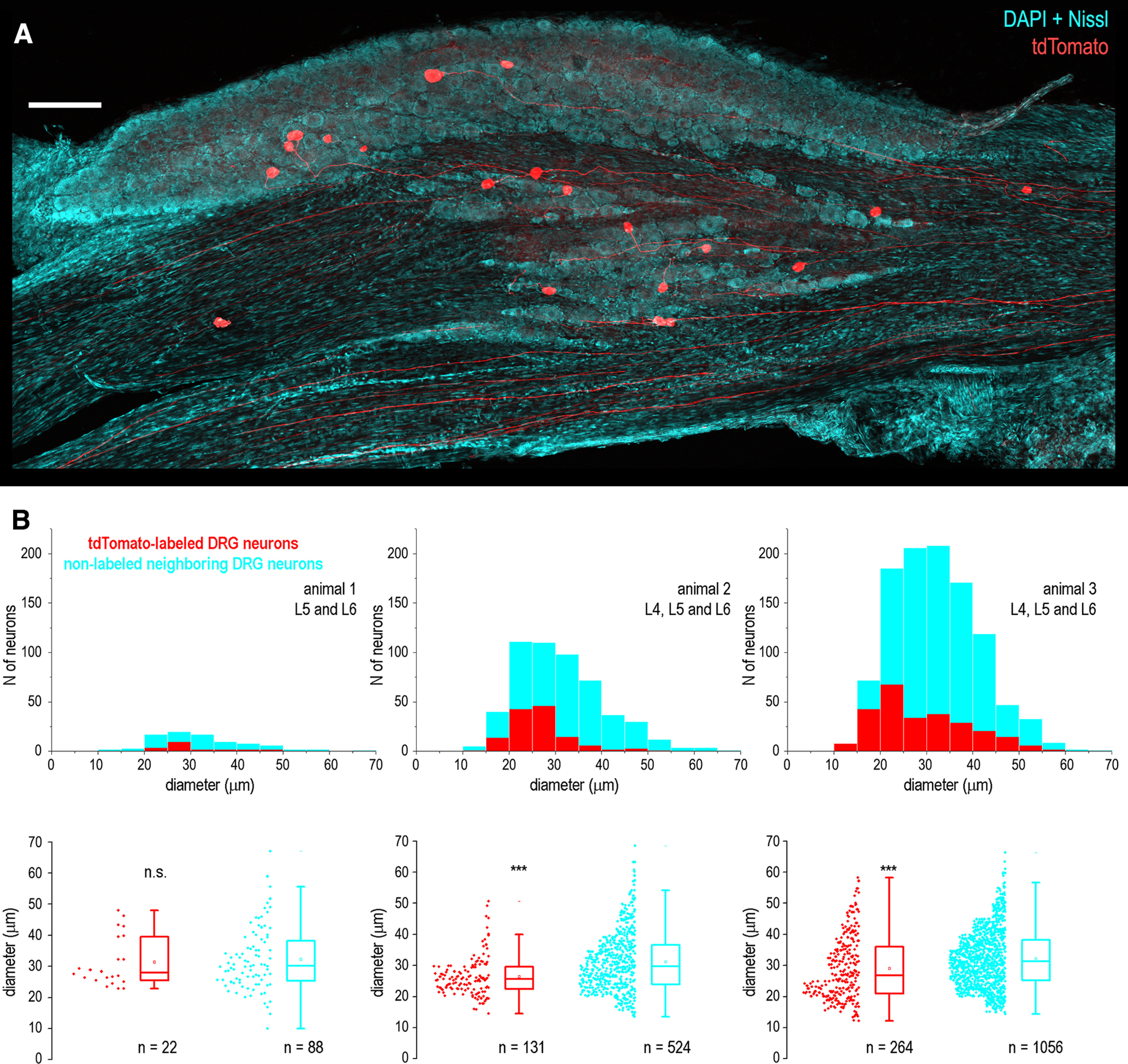
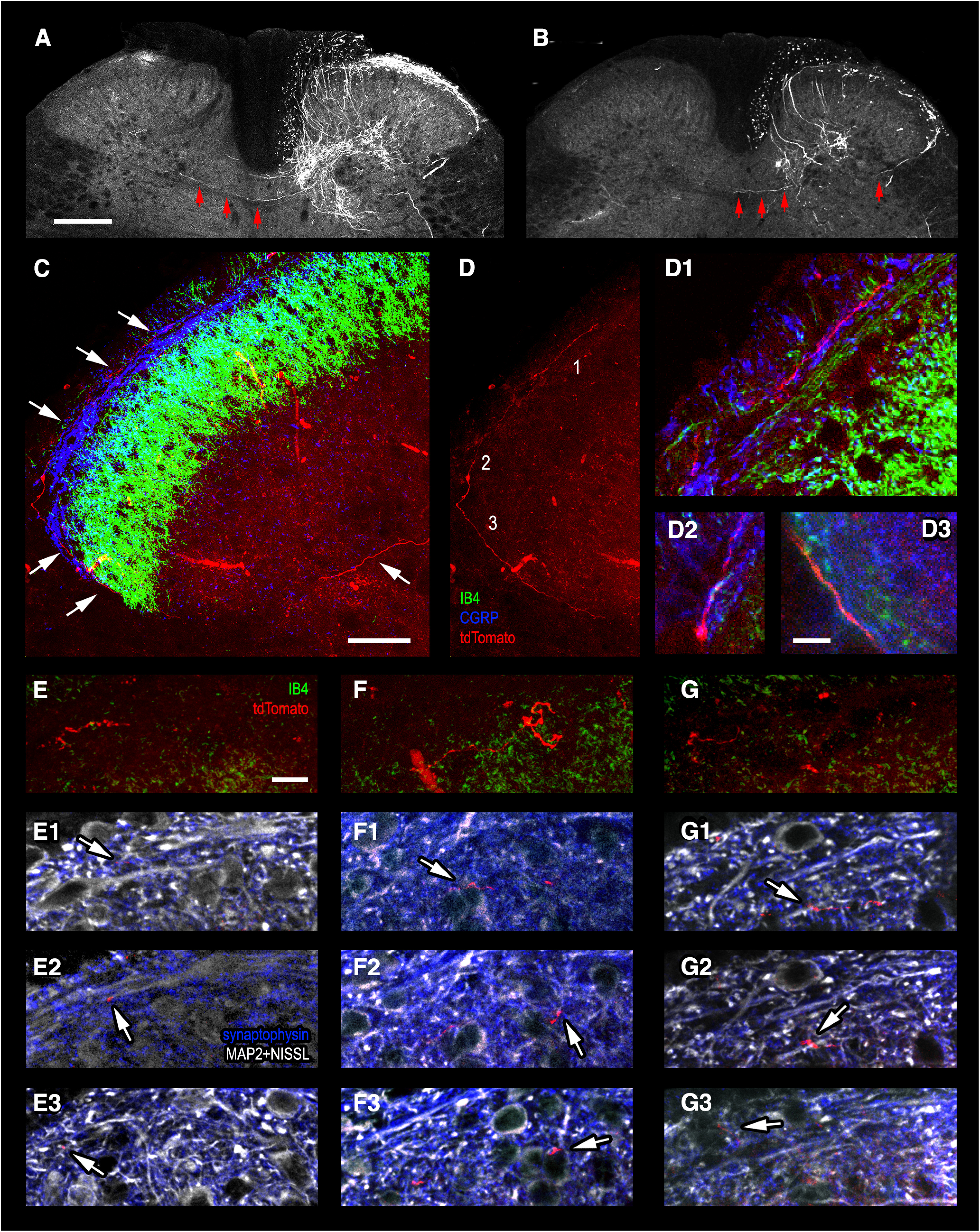
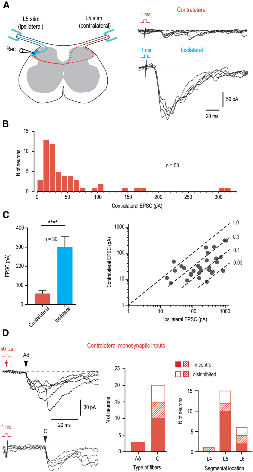
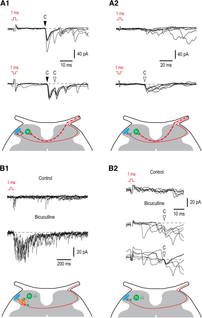
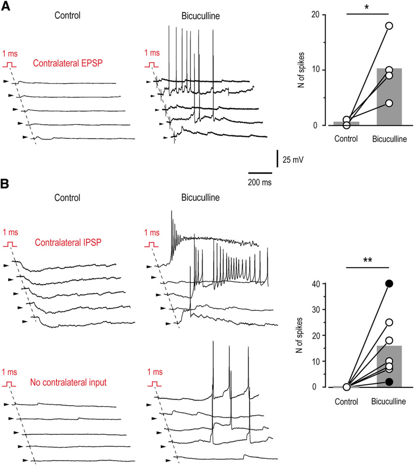
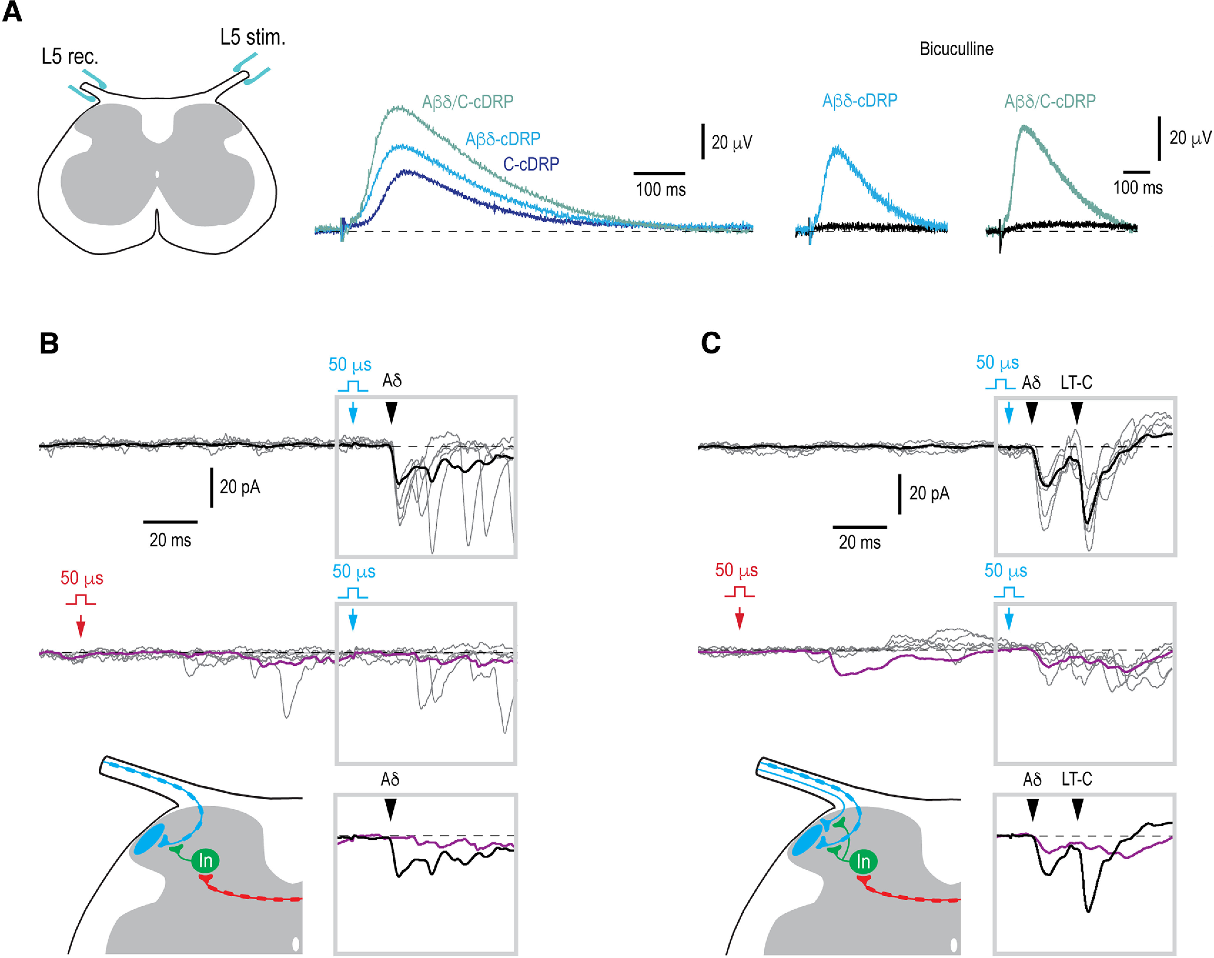
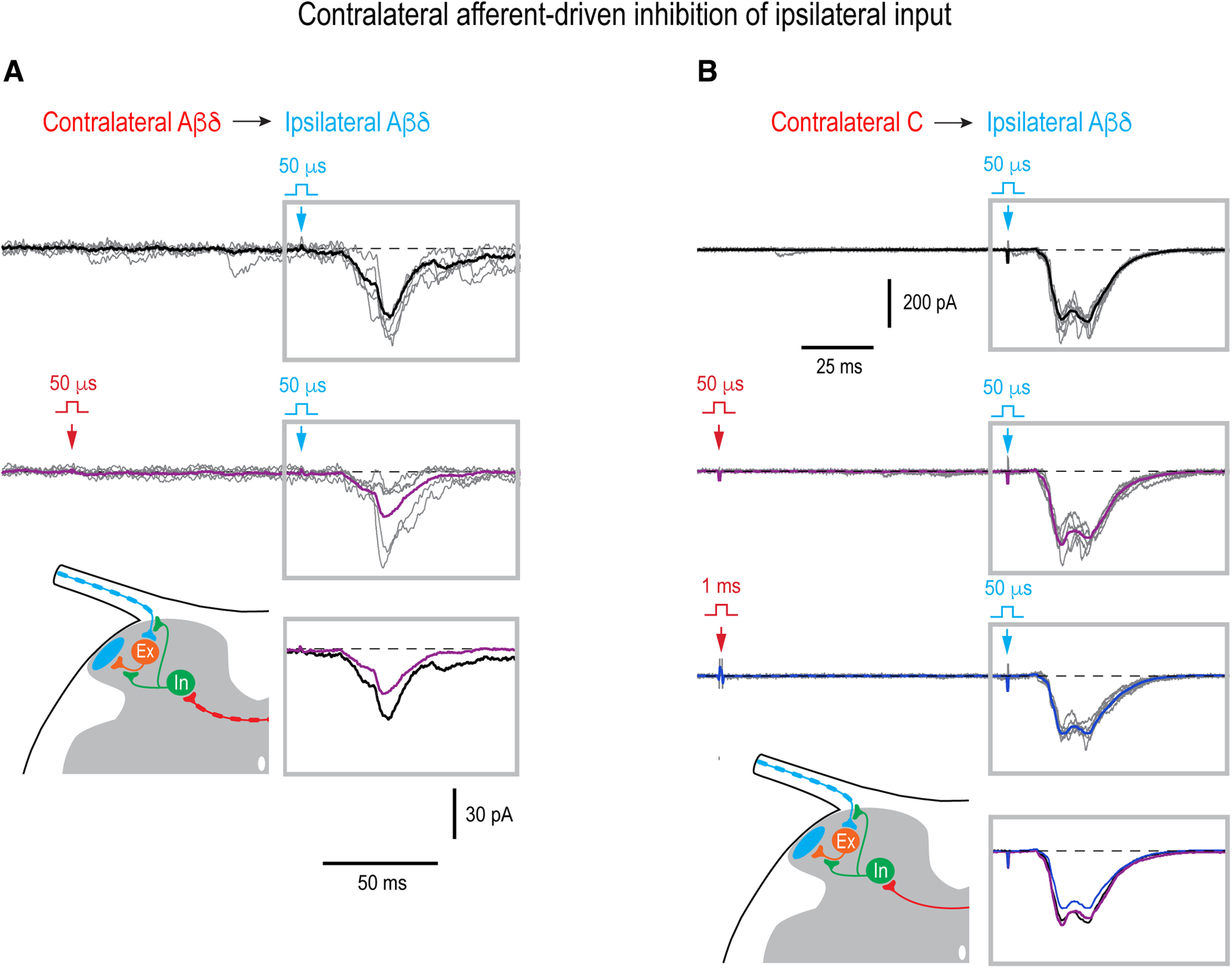
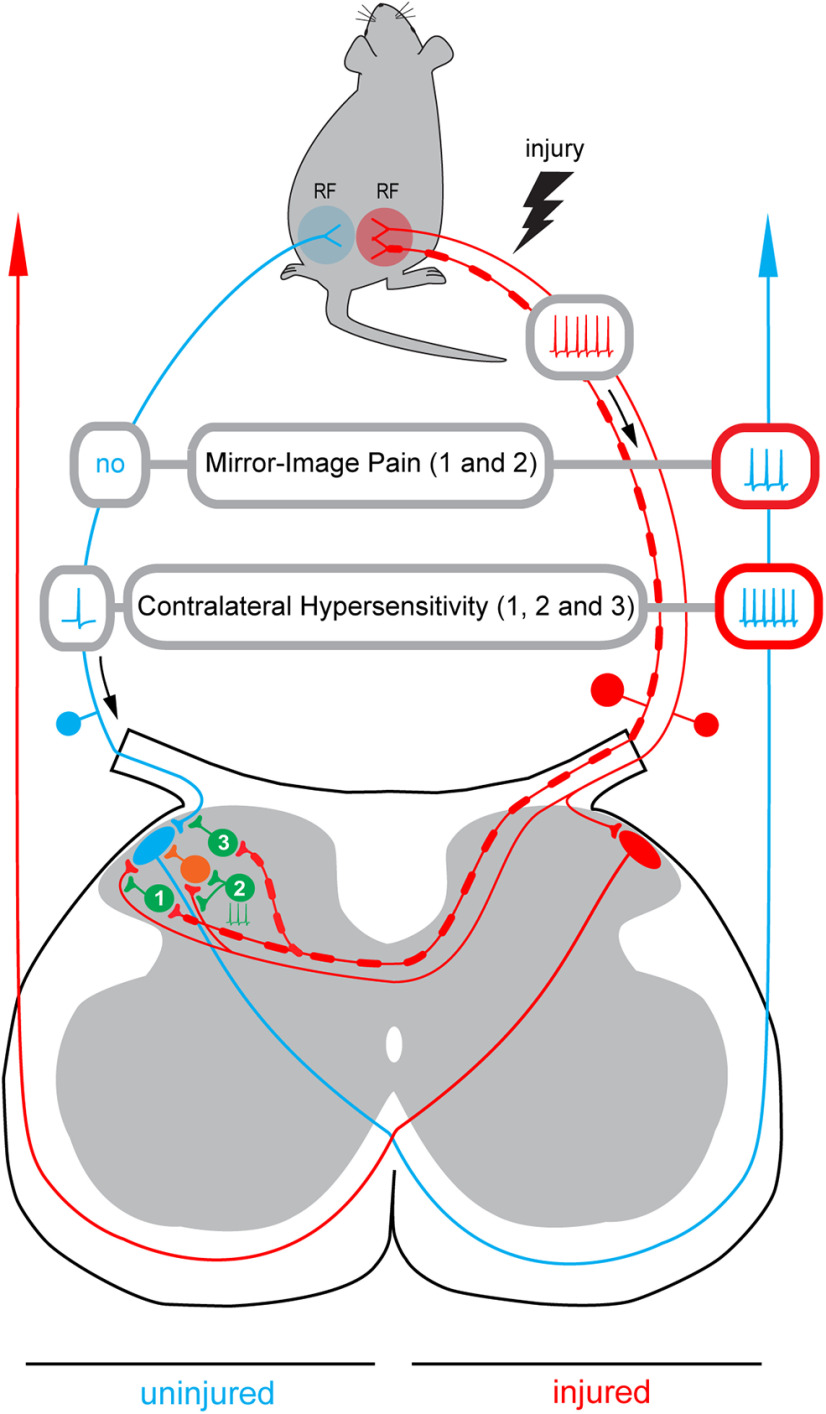
Mirror-image pain arises from pathologic alterations in the nociceptive processing network that controls functional lateralization of the primary afferent input. Although a number of clinical syndromes related to dysfunction of the lumbar afferent system are associated with the mirror-image pain, its morphophysiological substrate and mechanism of induction remain poorly understood. Therefore, we used ex vivo spinal cord preparation of young rats of both sexes to study organization and processing of the contralateral afferent input to the neurons in the major spinal nociceptive projection area Lamina I. We show that decussating primary afferent branches reach contralateral Lamina I, where 27% of neurons, including projection neurons, receive monosynaptic and/or polysynaptic excitatory drive from the contralateral Aδ-fibers and C-fibers. All these neurons also received ipsilateral input, implying their involvement in the bilateral information processing. Our data further show that the contralateral Aδ-fiber and C-fiber input is under diverse forms of inhibitory control. Attenuation of the afferent-driven presynaptic inhibition and/or disinhibition of the dorsal horn network increased the contralateral excitatory drive to Lamina I neurons and its ability to evoke action potentials. Furthermore, the contralateral Aβδ-fibers presynaptically control ipsilateral C-fiber input to Lamina I neurons. Thus, these results show that some lumbar Lamina I neurons are wired to the contralateral afferent system whose input, under normal conditions, is subject to inhibitory control. A pathologic disinhibition of the decussating pathways can open a gate controlling contralateral information flow to the nociceptive projection neurons and, thus, contribute to induction of hypersensitivity and mirror-image pain.SIGNIFICANCE STATEMENT We show that contralateral Aδ-afferents and C-afferents supply lumbar Lamina I neurons. The contralateral input is under diverse forms of inhibitory control and itself controls the ipsilateral input. Disinhibition of decussating pathways increases nociceptive drive to Lamina I neurons and may cause induction of contralateral hypersensitivity and mirror-image pain.

Keywords: dorsal horn; dorsal root potentials; marginal zone; nociception; presynaptic inhibition; primary afferents.


Figures

Figure 1.
Figure 1.
Labeling of primary sensory neurons after sciatic injection of AAV-CAG-tdTomato viral vector. A, DRG neurons expressing tdTomato (red) after the sciatic injection of viral vector. A low-magnification montage of 3 image tiles showing a Z-projection of 22 optical planes from a 100-µm-thick section of the L5 DRG. DAPI and Nissl staining (both cyan) revealed nuclei and cytoplasm of all DRG neurons. Scale bar, 200 µm. B, Distribution histograms of the largest cross-section diameters of all tdTomato-labeled and neighboring nonlabeled DRG neurons in three animals. Below, box-plots showing the largest cross-sectional diameter of all labeled and neighboring nonlabeled DRG neurons in the same three animals. The box indicates the 25th–75th percentile range, whiskers are set to outliers (coefficient, 1.5), square shows mean and the horizontal line indicates the median. n.s., not significant; ***p < 0.001.
Figure 2.
Figure 2.
Tracing projections of primary afferent fibers to the contralateral superficial dorsal horn. A, B, Low-magnification confocal images showing tdTomato-expressing primary afferent collaterals crossing the spinal cord midline in the dorsal commissure and running toward the contralateral dorsal horn (three arrows). A, Collateral originating from the medial aspect of the dorsal horn where tdTomato-expressing afferents enter the gray matter. B, Collateral arising from the lateral dorsal horn (single arrow). C, D, A thin primary afferent collateral (red) with bouton-like enlargements stretches along the dorsal surface of the contralateral dorsal horn (white arrows) in the termination zone of CGRP-positive (blue) and IB4-positive (green) afferents. The images are Z-projections of 81 single 0.5-µm-thick optical sections. D1–D3, Regions 1–3 from D are given at higher magnification to show close appositions of the tdTomato-expressing afferent (red) with CGRP-positive (blue) and IB4-positive (green) primary afferent terminals (Z-projections from 2–6 optical sections). E–G, tdTomato-expressing primary afferent collateral fragments with varicosities (red) near the termination zone of IB4-positive (green) primary afferents. Z-projections from 10–20 optical sections. E1–E3, F1–F3, G1–G3, Single optical sections showing tdTomato-expressing primary afferent varicosities contacting (arrows) elongated mediolaterally-oriented MAP2-positive dendrites of Lamina I neurons (white, E1–E3), as well as Nissl-stained somata (white, F1–F3) and MAP2-positive dendrites (white, G1–G3) of Lamina II neurons. Scale bars: 200 µm in A (the same in B), 50 µm in C (the same in D), 10 µm in D1–D3 and E–G (the same in E1–E3, F1–F3, and G1–G3).
Figure 3.
Figure 3.
Contralateral Aδ-fiber and C-fiber input to lumbar Lamina I neurons. A, Left panel, Schematic of the experimental design used to study ipsilateral (blue) and contralateral (red) primary afferent input to a Lamina I neuron. Note that decussating thin afferent collaterals can run via medial or lateral dorsal horn (dashed lines). Right, Overall mono- plus polysynaptic EPSCs evoked in an L5 Lamina I neuron after stimulating contralateral and ipsilateral L5 dorsal roots at intensity activating all Aβδ/C-fibers (5 traces each). Holding potential, −80 mV. B, Distribution of the amplitudes of the overall EPSCs evoked by the contralateral dorsal root stimulation in 53 Lamina I neurons. C, Comparison of the overall EPSCs evoked by contralateral and ipsilateral stimulation in neurons with bilateral input. Only Lamina I neurons with undistorted ipsilateral input were selected (n = 30). ****p < 0.0001, paired t test. Right, EPSC amplitudes for each of 30 neurons are plotted in double logarithmic coordinates. Dashed lines are shown for the ratios between the amplitudes of the contralateral and ipsilateral inputs of 1.0, 0.3, 0.1, and 0.03. D, Monosynaptic Aδ-EPSC and C-EPSC components (indicated by arrowheads) of the contralateral input (5 traces each). Holding potential, −70 mV. The neuron receiving the Aδ-input was from the segments L6. The neuron receiving monosynaptic C-fiber input was located in the segment L5 and identified as a PN. Right, Histograms show the number of neurons with Aδ-fiber and C-fiber input and segmental locations of Lamina I neurons with direct contralateral input. Filled bars, neurons showing monosynaptic input in control; open bars, neurons that showed monosynaptic components only after attenuation of the afferent-driven presynaptic inhibition or disinhibition of the dorsal horn network (Fig. 4); crosshatched bars, neurons showing both monosynaptic input in control and a new component appearing after removal of inhibition.
Figure 4.
Figure 4.
Inhibitory control of the contralateral input. A1, A2, Afferent-driven presynaptic inhibition of the monosynaptic C-fiber input to Lamina I neurons. A1, Recording from a neuron in which attenuation of the Aβδ-fiber-driven presynaptic inhibition by the inverted pulse stimulation resulted in an appearance of a new monosynaptic C-EPSC (open arrowhead) in addition to the component seen in control (filled arrowhead). Schematic shows a phasic Aβδ-fiber-mediated presynaptic inhibition at one of two contralateral C-fiber branches supplying Lamina I neuron. A2, Removal of the Aβδ-afferent-driven presynaptic inhibition unblocks a monosynaptic C-fiber input (open arrowhead). Schematic shows a phasic presynaptic inhibition at the contralateral C-fiber branch supplying Lamina I neuron. B1, B2, Network disinhibition by bicuculline (20 μm) increases the contralateral input to Lamina I neurons. B1, Disinhibition augments the polysynaptic input. Schematic, an increase in the polysynaptic input can be caused by removal of a tonic postsynaptic inhibition from excitatory interneuron or from the presynaptic terminal of the afferent supplying this interneuron. B2, Network disinhibition unblocks contralateral monosynaptic C-fiber input (open arrowhead) to an LCN. In bicuculline, the same family of traces is also shown below with membrane currents at the time point of the monosynaptic EPSC initiation shifted to the same level. Schematic, a tonic presynaptic inhibition at the contralateral C-fiber branch supplying Lamina I neuron. Holding potential was −70 mV in A1, B1, and B2, and −80 mV in A2. For each type of response, five traces are shown. Locations of inhibitory (In) and excitatory (Ex) interneurons are not known.
Figure 5.
Figure 5.
Network disinhibition increases efficacy of the contralateral afferent input. Current-clamp recordings done in control and in the presence of 20 μm bicuculline from Lamina I neurons receiving excitatory (EPSC of 16 pA; A), inhibitory or no (B) contralateral input. Right, Numbers of spikes evoked by 10 consecutive stimulations of the contralateral dorsal root are plotted for individual neurons. Gray bars indicate the mean value for each cell group. Neurons with inhibitory (filled symbols) or no (open symbols) contralateral input are presented as one group. In all traces, arrowheads indicate a potential of −70 mV. *p < 0.05, **p < 0.01; paired t test.
Figure 6.
Figure 6.
Contralateral control of the ipsilateral monosynaptic input. A, Recording of the cDRPs. Left panel shows a schematic of how the cDRPs were recorded in the isolated spinal cord preparation. The recording electrode was placed on the contralateral L5 dorsal root close to where it entered the spinal cord. Middle, cDRPs evoked by stimulating Aβδ-afferents (Aβδ-cDRP), C-afferents (C-cDRP), and Aβδ/C-afferents (Aβδ/C-cDRP). Right, Bicuculline (20 μm) effect on the Aβδ-cDRP and Aβδ/C-cDRP. B, Contralateral Aβδ-range conditioning abolished ipsilateral Aδ-EPSC component. Schematic, Contralateral Aβδ-afferent induces presynaptic inhibition of the ipsilateral Aδ-fiber input to a Lamina I neuron. C, Contralateral Aβδ-conditioning attenuated the ipsilateral Aδ-EPSC and abolished the low-threshold-(LT)-C-EPSC. Schematic, a contralateral Aβδ-afferent induces inhibition of the ipsilateral Aδ- and LT-C-afferents. In B, C, 50-µs stimuli were applied to activate Aβδ-afferents or LT-C-afferents. Monosynaptic EPSCs are indicated by filled arrowheads. Holding potential, −80 mV. The time moments when conditioning (contralateral) and test (ipsilateral) stimuli were applied are indicated by red and blue arrows, respectively. For each type of response, five individual traces are shown with the average of 10–15 traces. Averaged responses to the test stimuli are shown superimposed below. Location of an inhibitory interneuron (In) is not known.
Figure 7.
Figure 7.
Contralateral control of the ipsilateral overall input. A, Contralateral Aβδ-range conditioning attenuated the ipsilateral Aδ-fiber input in a PN (integral reduced by 55%, p = 0.03, unpaired t test). Holding potential, −70 mV. B, Contralateral Aβδ-conditioning did not change the ipsilateral Aδ-fiber input (p = 0.3, unpaired t test) that was however significantly attenuated by the contralateral Aβδ/C-conditioning (integral reduced by 21%, p < 0.001, unpaired t test). Thus, the effect was considered to be driven by the contralateral C-afferents. Holding potential, −80 mV. In A, B, 50-µs stimuli were applied to activate Aβδ-afferents and 1-ms stimuli to activate all Aβδ/C-afferents. The time interval between the contralateral conditioning stimulus (red arrow) and ipsilateral test stimulus (blue arrow) was 100 ms. For each type of response, five traces are shown with the average of 7–21 traces. Averaged responses to the test stimuli are shown superimposed below. Schematics show contralateral Aβδ-afferent-driven (A) and C-afferent-driven (B) presynaptic inhibition of the ipsilateral afferent supplying an intercalated excitatory neuron or/and of the axon terminal of the intercalated neuron. The presynaptic, rather than postsynaptic, mechanism of the contralateral inhibition of the ipsilateral polysynaptic input was assumed, since effect was observed 100 ms after conditioning stimulation when cDRP, and therefore PAD, reached its maximum (Fig. 6A), but most evoked IPSCs already terminated (Luz et al., 2019; Fernandes et al., 2022a). Locations of inhibitory (In) and excitatory (Ex) interneurons are not known.
Figure 8.
Figure 8.
Induction of the mirror-image pain and contralateral hypersensitivity. Schematic illustrating how disinhibition of the decussating pathways can cause induction of the mirror-image pain. Based on our data, three inhibitory pathways are considered. Pathway 1, the contralateral Aβδ-afferent-driven presynaptic inhibition of the contralateral C-fiber input (Fig. 4A1,A2). Pathway 2, tonic network inhibition of the contralateral input (Fig. 4B1). The tonic inhibition of the C-fiber directly supplying Lamina I neuron is not indicated. Pathway 3, the contralateral Aβδ-afferent-driven presynaptic inhibition of the ipsilateral input (Fig. 6B,C). Disinhibition of pathways 1 and 2 will allow the ongoing afferent barrage in the injured nerve (red) to reach a Lamina I PN on the uninjured side (blue). Its excitation by the contralateral afferent barrage will result in a perception of pain as arising from the ipsilateral receptive field (RF) even if there is no activity in the ipsilateral afferents (Mirror-Image Pain). Disinhibition of pathway 3 will increase the ipsilateral afferent drive reaching a PN on uninjured side after stimulation of its ipsilateral receptive field. Note that the ipsilateral input can be affected even in the neuron that does not receive contralateral supply. This can reduce the noxious threshold for the ipsilateral receptive field and increase the nociceptive afferent discharge reaching the PN, thereby contributing to development of allodynia and hyperalgesia on the uninjured side (Contralateral Hypersensitivity). The hyperalgesia can further be augmented by disinhibition of pathways 1 and 2 that will open a gate allowing injured afferent barrage to reach the contralateral PN. Inhibitory interneurons are shown in green, an excitatory neuron in orange.

References

    1. Al Ghamdi KS, Polgár E, Todd AJ (2009) Soma size distinguishes projection neurons from neurokinin 1 receptor-expressing interneurons in lamina I of the rat lumbar spinal dorsal horn. Neuroscience 164:1794–1804. 10.1016/j.neuroscience.2009.09.071 - DOI - PMC - PubMed
    1. Antal Z, Luz LL, Safronov BV, Antal M, Szücs P (2016) Neurons in the lateral part of the lumbar spinal cord show distinct novel axon trajectories and are excited by short propriospinal ascending inputs. Brain Struct Funct 221:2343–2360. 10.1007/s00429-015-1046-3 - DOI - PubMed
    1. Barron DH, Matthews BH (1938) The interpretation of potential changes in the spinal cord. J Physiol 92:276–321. 10.1113/jphysiol.1938.sp003603 - DOI - PMC - PubMed
    1. Brown AG (1982) The dorsal horn of the spinal cord. Q J Exp Physiol 67:193–212. 10.1113/expphysiol.1982.sp002630 - DOI - PubMed
    1. Brown AG, Kirk EJ, Martin HF 3rd (1973) Descending and segmental inhibition of transmission through the spinocervical tract. J Physiol 230:689–705. 10.1113/jphysiol.1973.sp010212 - DOI - PMC - PubMed

Publication types

LinkOut - more resources