

Differential Laminar Activation Dissociates Encoding and Retrieval in the Human Medial and Lateral Entorhinal Cortex
- PMID: 36948584
- PMCID: PMC10124959
- DOI: 10.1523/JNEUROSCI.1488-22.2023
Differential Laminar Activation Dissociates Encoding and Retrieval in the Human Medial and Lateral Entorhinal Cortex
Abstract
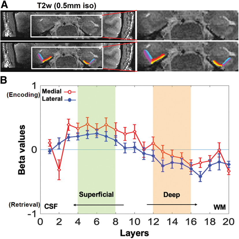
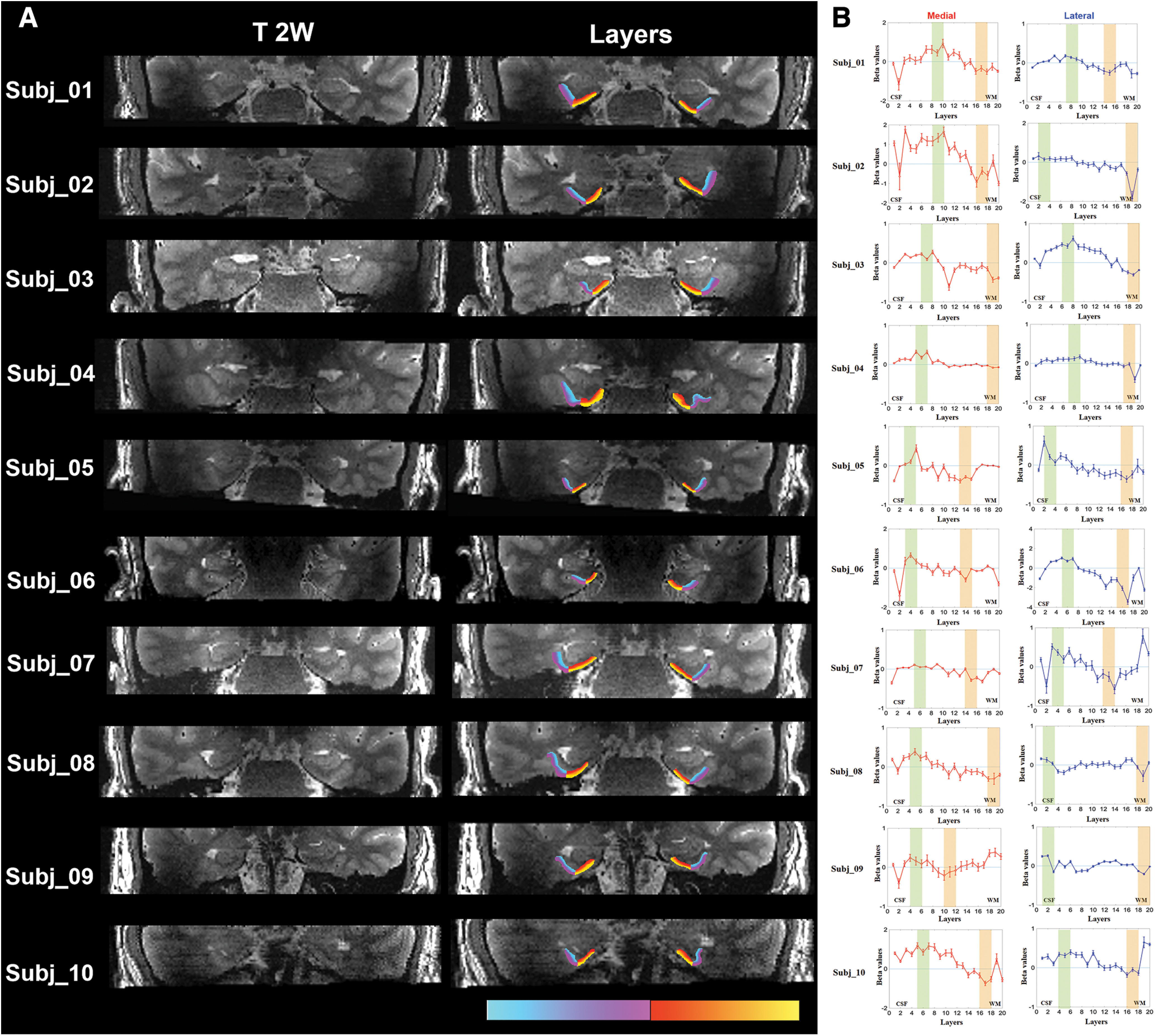
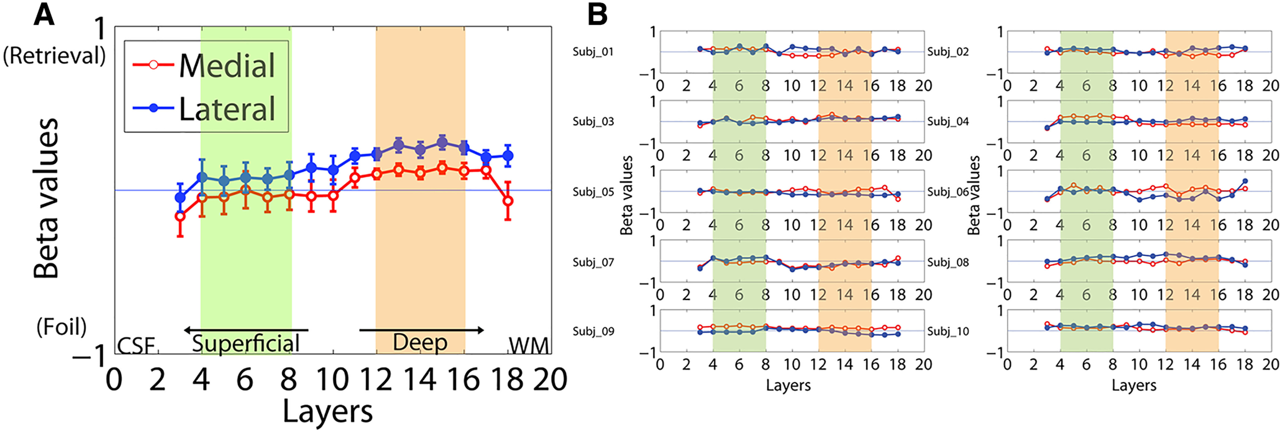
The hierarchically organized structures of the medial temporal lobe are critically important for episodic memory function. Accumulating evidence suggests dissociable information processing pathways are maintained throughout these structures including in the medial and lateral entorhinal cortex. Cortical layers provide an additional dimension of dissociation as the primary input to the hippocampus derives from layer 2 neurons in the entorhinal cortex, whereas the deeper layers primarily receive output from the hippocampus. Here, novel high-resolution T2-prepared functional MRI methods were successfully used to mitigate susceptibility artifacts typically affecting MRI signals in this region providing uniform sensitivity across the medial and lateral entorhinal cortex. During the performance of a memory task, healthy human subjects (age 25-33 years, mean age 28.2 ± 3.3 years, 4 female) showed differential functional activation in the superficial and deep layers of the entorhinal cortex associated with task-related encoding and retrieval conditions, respectively. The methods provided here offer an approach to probe layer-specific activation in normal cognition and conditions contributing to memory impairment.SIGNIFICANCE STATEMENT This study provides new evidence for differential neuronal activation in the superficial versus deep layers of the entorhinal cortex associated with encoding and retrieval memory processes, respectively, in cognitively normal adults. The study further shows that this dissociation can be observed in both the medial and the lateral entorhinal cortex. The study was achieved by using a novel functional MRI method allowing us to measure robust functional MRI signals in both the medial and lateral entorhinal cortex that was not possible in previous studies. The methodology established here in healthy human subjects lays a solid foundation for subsequent studies investigating layer-specific and region-specific changes in the entorhinal cortex associated with memory impairment in various conditions such as Alzheimer's disease.
Keywords: 7T; T2prep; cortical layer; encoding and retrieval; high field functional MRI; medial temporal lobe.
Copyright © 2023 the authors.
Figures
Similar articles
-
Age-related functional changes in domain-specific medial temporal lobe pathways.Neurobiol Aging. 2018 May;65:86-97. doi: 10.1016/j.neurobiolaging.2017.12.030. Epub 2018 Jan 31. Neurobiol Aging. 2018. PMID: 29454154
-
Object and spatial mnemonic interference differentially engage lateral and medial entorhinal cortex in humans.Proc Natl Acad Sci U S A. 2014 Oct 7;111(40):E4264-73. doi: 10.1073/pnas.1411250111. Epub 2014 Sep 22. Proc Natl Acad Sci U S A. 2014. PMID: 25246569 Free PMC article. Clinical Trial.
-
Laminar activity in the hippocampus and entorhinal cortex related to novelty and episodic encoding.Nat Commun. 2014 Nov 26;5:5547. doi: 10.1038/ncomms6547. Nat Commun. 2014. PMID: 25424131 Free PMC article. Clinical Trial.
-
Structural magnetic resonance imaging for the early diagnosis of dementia due to Alzheimer's disease in people with mild cognitive impairment.Cochrane Database Syst Rev. 2020 Mar 2;3(3):CD009628. doi: 10.1002/14651858.CD009628.pub2. Cochrane Database Syst Rev. 2020. PMID: 32119112 Free PMC article.
-
Deep entorhinal cortex: from circuit organization to spatial cognition and memory.Trends Neurosci. 2021 Nov;44(11):876-887. doi: 10.1016/j.tins.2021.08.003. Epub 2021 Sep 28. Trends Neurosci. 2021. PMID: 34593254 Review.
Cited by
-
Entorhinal cortex-hippocampal circuit connectivity in health and disease.Front Hum Neurosci. 2024 Sep 20;18:1448791. doi: 10.3389/fnhum.2024.1448791. eCollection 2024. Front Hum Neurosci. 2024. PMID: 39372192 Free PMC article. Review.
-
Communication of perceptual predictions from the hippocampus to the deep layers of the parahippocampal cortex.Sci Adv. 2025 May 23;11(21):eads4970. doi: 10.1126/sciadv.ads4970. Epub 2025 May 21. Sci Adv. 2025. PMID: 40397746 Free PMC article.
-
Short-term gradient imperfections in high-resolution EPI lead to Fuzzy Ripple artifacts.Magn Reson Med. 2025 Aug;94(2):571-587. doi: 10.1002/mrm.30489. Epub 2025 Apr 2. Magn Reson Med. 2025. PMID: 40173320 Free PMC article.
-
Fuzzy ripple artifact in high resolution fMRI: identification, cause, and mitigation.bioRxiv [Preprint]. 2024 Sep 9:2024.09.04.611294. doi: 10.1101/2024.09.04.611294. bioRxiv. 2024. Update in: Magn Reson Med. 2025 Aug;94(2):571-587. doi: 10.1002/mrm.30489. PMID: 39314458 Free PMC article. Updated. Preprint.
-
Heterogenous effect of early adulthood stress on cognitive aging and synaptic function in the dentate gyrus.Front Mol Neurosci. 2024 Apr 4;17:1344141. doi: 10.3389/fnmol.2024.1344141. eCollection 2024. Front Mol Neurosci. 2024. PMID: 38638601 Free PMC article.
References
Publication types
MeSH terms
Grants and funding
LinkOut - more resources
Full Text Sources
Medical