. 2023 Dec;17(1):2192457.
doi: 10.1080/19336934.2023.2192457.

Downregulation of Plasma Membrane Ca2+ ATPase driven by tyrosine hydroxylase-Gal4 reduces Drosophila lifespan independently of effects in neurons

Affiliations

Downregulation of Plasma Membrane Ca2+ ATPase driven by tyrosine hydroxylase-Gal4 reduces Drosophila lifespan independently of effects in neurons

Brenda Erhardt et al. Fly (Austin). 2023 Dec.

Abstract

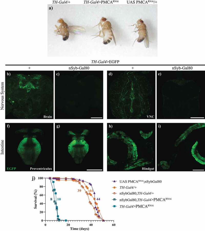
In Drosophila melanogaster, several Gal4 drivers are used to direct gene/RNAi expression to different dopaminergic neuronal clusters. We previously developed a fly model of Parkinson's disease, in which dopaminergic neurons had elevated cytosolic Ca2+ due to the expression of a Plasma Membrane Ca2+ ATPase (PMCA) RNAi under the thyroxine hydroxylase (TH)-Gal4 driver. Surprisingly, TH-Gal4>PMCARNAi flies died earlier compared to controls and showed swelling in the abdominal area. Flies expressing the PMCARNAi under other TH drivers also showed such swelling and shorter lifespan. Considering that TH-Gal4 is also expressed in the gut, we proposed to suppress the expression specifically in the nervous system, while maintaining the activation in the gut. Therefore, we expressed Gal80 under the direction of the panneuronal synaptobrevin (nSyb) promoter in the context of TH-Gal4. nSyb-Gal80; TH-Gal4>PMCARNAi flies showed the same reduction of survival as TH-Gal4>PMCARNAi flies, meaning that the phenotype of abdomen swelling and reduced survival could be due to the expression of the PMCARNAi in the gut. In perimortem stages TH-Gal4>PMCARNAi guts had alteration in the proventriculi and crops. The proventriculi appeared to lose cells and collapse on itself, and the crop increased its size several times with the appearance of cellular accumulations at its entrance. No altered expression or phenotype was observed in flies expressing PMCARNAi in the dopaminergic PAM cluster (PAM-Gal4>PMCARNAi). In this work we show the importance of checking the global expression of each promoter and the relevance of the inhibition of PMCA expression in the gut.

Keywords: Calcium; GAL4-UAS; GAL80; dopaminergic neurons; flies; gut.


Conflict of interest statement

No potential conflict of interest was reported by the authors.

Figures

Figure 1.
Figure 1.
The expression of PMCARNAi under dopaminergic drivers, with the exception of PAM-Gal4 decreased flies’ lifespan. Diagrams show the brain dopaminergic neurons in which each promoter is expressed. a, TH-Gal4 is active in several dopaminergic clusters in the anterior and posterior regions of the brain. The expression of TH-Gal4>PMCARNAi markedly reduced lifespan compared to controls. b and c, TH-F1-Gal4 and TH-D4-Gal4 are active in 28 and 22 neurons, respectively, and, when used to drive expression of PMCARNAi, also presented a lower survival rate, but higher than TH-Gal4>PMCARNAi flies. d, Flies expressing PMCARNAi under the PAM-Gal4 promoter showed only minor differences in lifespan compared to its controls (with a mean lifespan similar to most other controls). All curves were analysed with the Log-rank Mantel Cox test and each curve was replicated 2 or 3 times.
Figure 2.
Figure 2.
TH-Gal4 was expressed in the digestive tract. a, Schematic representation of the central nervous system and digestive tract of Drosophila melanogaster (VNC: ventral nerve cord). Microscopy images of the gut from TH-Gal4/+ and TH-Gal4>mcherry flies: b-c, Bright field; d-e, mCherry fluorescence; and f-g, merge. Red fluorescence was detected only in the proventriculus and hindgut (arrows, e and g) from TH-Gal4>mcherry flies. Scale bar: 250 µm.
Figure 3.
Figure 3.
TH-F1-Gal4 and TH-D4-Gal4, but not PAM-Gal4, also showed expression in the gut. Confocal images showing expression of EGFP driven by TH-Gal4, TH-F1-Gal4, TH-D4-Gal4 and PAM-Gal4 in brain (a-d), VNC (e-h), proventriculus (i-l), crop (m-p) and hindgut (q-t). VNC: central nervous cord. Scale bar: d, h and p, 100 µm; l, 50 µm and t, 200 µm.
Figure 4.
Figure 4.
TH-Gal4>PMCARNAi flies showed a perimortem swelling of the abdomen and suppressing neuronal expression of PMCARNAi did not prevent the shortening of their lifespan. a, Abdominal swelling phenotype in 10 day-old TH-Gal4>PMCARNAi flies, compared to TH-Gal4/+ and UAS PMCARNAi/+ controls. b-i, Confocal images showing that nSyb-Gal80 suppresses TH-Gal4 driven EGFP expression in the brain (b,c) and the VNC (d,e), but not in the proventriculus (f,g) and hindgut (h,i). j, the lifespan curve of nSyb-Gal80;TH-Gal4>PMCARNAi flies was similar to that of TH-Gal4>PMCARNAi flies. Scale bars: 100 µm.
Figure 5.
Figure 5.
10-days-old, but not 2-days-old, TH-Gal4>PMCARNAi flies presented an increase in crop size and abnormal accumulation of cells at the entrance of the crop. a-b, Bright field microscopy images of the crops from 2 (a) and 10-days-old (b) TH-Gal4/+ (control) and TH-Gal4>PMCARNAi flies. c, Quantification of crop sizes (mm2) showed no difference at 2 days of age, whereas at 10 days, those from TH-Gal4>PMCARNAi flies were about 10 times larger than those from TH-Gal4/+ controls. d-i, Confocal microscopy images of the crop entrance (dotted lines) from TH-Gal4>egfp/mCherry (d-f) and TH-Gal4>EGFP/PMCARNAi (g-i). The latter showed fragmented nuclei (g) and increased EGFP fluorescence (h, merge in i) compared to the former (control). Nuclei outside the dotted line belong to a different section of the intestine that was left next to the crop in the dissection. Two-Way ANOVA Tukey’s multiple comparisons test; 10-days-old TH-Gal4>PMCARNAi versus 10-days-old TH-Gal4/+, 2-days-old TH-Gal4/+ and 2-days-old TH-Gal4>PMCARNAi, ****p > 0,0001. Scale bar: 300 µm.
Figure 6.
Figure 6.
10-days-old TH-Gal4>PMCARNAi flies showed shrinking and cell loss in the proventriculus. a-c, Bright field microscopy images of proventriculi (arrows) from TH-Gal4/+ (a), TH-Gal4>egfp/mCherry (b) and TH-Gal4>EGFP/PMCARNAi (c) 10-days-old flies. The latter exhibited perimortem collapsing of the proventriculus, which was not evident in the controls. The diameter (indicated with a red line) of the proventriculus shown were 218,48 µm (a), 220.82 µm (b) and 131.93 µm (c). d-i, Confocal images of proventriculi from TH-Gal4>egfp/mCherry (d-f) and TH-Gal4>EGFP/PMCARNAi (g-i) flies, which presented a decreased number of nuclei and EGFP fluorescence. f and i show mCherry fluorescence, represented in magenta (no fluorescence shown in TH-Gal4>EGFP/PMCARNAi flies as they do not express mCherry). Scale bar: c, 500 µm; i, 50 µm.

References

    1. Mao Z, Davis RL.. Eight different types of dopaminergic neurons innervate the drosophila mushroom body neuropil: anatomical and physiological heterogeneity. Front Neural Circuits. 2009;3:5. - PMC - PubMed
    1. Brand AH, Perrimon N. Targeted gene expression as a means of altering cell fates and generating dominant phenotypes. Development. 1993;118(2):401–11. - PubMed
    1. Friggi-Grelin F, Coulom H, Meller M, et al. Targeted gene expression in drosophila dopaminergic cells using regulatory sequences from tyrosine hydroxylase. J Neurobiol. 2003;54(4):618–627. - PubMed
    1. Qili L, Liu S, Kodama L, et al. Two dopaminergic neurons signal to the dorsal fan-shaped body to promote wakefulness in drosophila. Curr Biol. 2012;22(22):2114–2123. - PMC - PubMed
    1. Liu C, Yves Plaaais P, Yamagata N, et al. A subset of dopamine neurons signals reward for odour memory in drosophila. Nature. 2012;488(7412):512–516. - PubMed

Publication types

MeSH terms

LinkOut - more resources