. 2023 Mar 23;8(1):45.
doi: 10.1038/s41541-023-00637-7.

Combination Ad26.RSV.preF/preF protein vaccine induces superior protective immunity compared with individual vaccine components in preclinical models

Affiliations

Combination Ad26.RSV.preF/preF protein vaccine induces superior protective immunity compared with individual vaccine components in preclinical models

Eirikur Saeland et al. NPJ Vaccines. .

Abstract

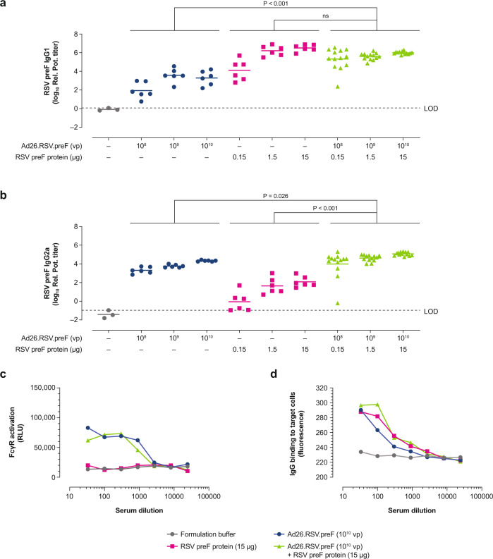
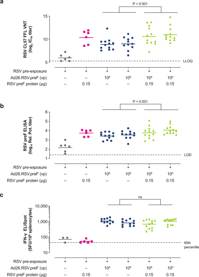
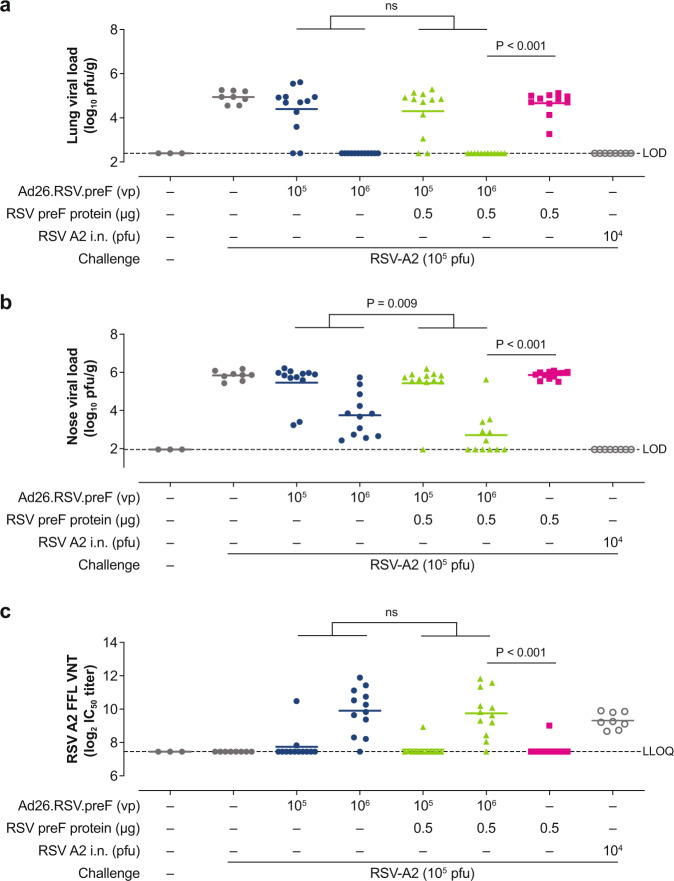
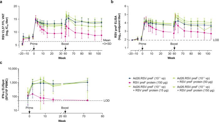
Respiratory syncytial virus (RSV) is a leading cause of severe respiratory disease for which no licensed vaccine is available. We have previously shown that a prefusion (preF) conformation-stabilized RSV F protein antigen and an adenoviral vector encoding RSV preF protein (Ad26.RSV.preF) are immunogenic and protective in animals when administered as single components. Here, we evaluated a combination of the 2 components, administered as a single injection. Strong induction of both humoral and cellular responses was shown in RSV-naïve and pre-exposed mice and pre-exposed African green monkeys (AGMs). Both components of the combination vaccine contributed to humoral immune responses, while the Ad26.RSV.preF component was the main contributor to cellular immune responses in both mice and AGMs. Immunization with the combination elicited superior protection against RSV A2 challenge in cotton rats. These results demonstrate the advantage of a combination vaccine and support further clinical development.


Conflict of interest statement

All authors except M.J.J. are employees of Janssen Infectious Diseases & Vaccines, and may own stock or stock options in Johnson & Johnson, its parent company. M.J.J. has no competing interests to report.

Figures

Fig. 1
Fig. 1. Humoral and cellular immunogenicity in naïve mice.
Naïve female BALB/c mice were immunized at Week 0 and Week 4 with Ad26.RSV.preF (1 × 108 or 1 × 109 vp), Ad26.RSV.preF (1 × 108 vp) combined with RSV preF protein (15, 1.5, 0.15 or 0.015 µg), or Ad26.RSV.preF (1 × 109 vp) combined with RSV preF protein (15 µg; n = 6 each group). VNT against RSV CL57, expressed as log2 IC90 titers, using an FFL virus neutralization assay (a) and RSV preF-binding antibody titers, expressed as log10 EC50 titers (b), were measured in serum at Week 6. Horizontal bars indicate mean titers per group, and dotted lines indicate the LLOQ or the LOD. Cellular responses were measured by IFN-γ ELISpot in splenocytes at Week 6 (c), and the responses are shown as SFU per million splenocytes. The horizontal lines indicate the geometric mean titer per group, and the dotted line indicates the 95th percentile of the response of unstimulated cells. Statistical comparisons were made by ANOVA for potentially censored measurements (Tobit model) and were conducted at an overall significance level of α = 0.05. Ad26, adenovirus type 26; EC50, 50% effective concentration; ELISA, enzyme-linked immunosorbent assay; ELISpot, enzyme-linked immune absorbent spot; FFL, firefly luciferase; IC90, 90% inhibitory concentration; IFN-γ, interferon-γ; LLOQ, lower limit of quantification; LOD, limit of detection; ns, not significant; preF, prefusion conformation-stabilized RSV F protein; RSV, respiratory syncytial virus; SFU, spot-forming units; VNT, virus neutralization titers; vp, viral particles.
Fig. 2
Fig. 2. IgG subclass responses and FcγR engagement of antibodies induced by Ad26.RSV.preF/RSV preF protein combinations in naïve mice.
Naïve female BALB/c mice were immunized at Week 0 and Week 6 with Ad26.RSV.preF (1 × 108, 1 × 109, or 1 × 1010 vp; n = 6 in each group), RSV preF protein (0.15, 1.5, or 15 µg; n = 6 each), or Ad26.RSV.preF combined with RSV preF protein at varying dose levels (n = 13 each). IgG1 (a) and IgG2a (b) preF-binding antibody titers, expressed as relative potency titer, were measured in serum by ELISA at Week 8. Horizontal lines indicate mean antibody responses per group, and the dotted line indicates the LOD, specified as the titer of the sample with the highest titer in the formulation buffer control group. Serum obtained at Week 8 from mice immunized with the highest vaccine concentration (Ad26.RSV.preF [1 × 1010 vp] and RSV preF protein [15 ug], alone or in combination) was pooled and used to measure FcγRIV activation in an ADCC reporter assay (c), expressed as RLU. Total IgG binding to RSV F-expressing target cells (d) was measured by fluorescence using AF488-conjugated goat anti-mouse IgG. Average of duplicate measurements is shown. Statistical comparisons were made across dose by ANOVA for potentially censored measurements (Tobit model) with Bonferroni correction for multiple comparisons (at an overall significance level of α = 0.05). Ad26, adenovirus type 26; ADCC, antibody-dependent cellular cytotoxicity; ELISA, enzyme- linked immunosorbent assay; FcγR, Fcγ receptor; FcγRIV, Fcγ receptor IV; IgG, immunoglobulin G; LOD, limit of detection; ns, not significant; preF, prefusion conformation-stabilized RSV F protein; Rel. Pot., relative potency; RLU, relative light units; RSV, respiratory syncytial virus; vp, viral particles.
Fig. 3
Fig. 3. Humoral and cellular immunogenicity in RSV pre-exposed mice.
Naïve female BALB/c mice were intranasally pre-exposed with RSV A2 (5 × 105 pfu) at Week –79 and immunized intramuscularly at Week 0 with Ad26.RSV.preF (1 × 108 or 1 × 109 vp; n = 12 in each group), RSV preF protein (0.15 µg; n = 6), or Ad26.RSV.preF (1 × 108 or 1 × 109 vp) combined with RSV preF protein (0.15 µg; n = 12 each group). Negative control animals received formulation buffer (n = 6). VNT against RSV CL57, expressed as log2 IC90 titers, were measured in serum at Week 5 (a). Horizontal bars indicate mean VNT per group, and the dotted line indicates the LLOQ. RSV preF total IgG-binding antibody titers (b), expressed as log10 relative potency titers, were also measured in serum at Week 5. Horizontal bars indicate mean titers per group, and the dotted line indicates the LOD. Cellular responses were measured by IFN-γ ELISpot in splenocytes at Week 5 (c), and the responses are expressed as SFU per million splenocytes. The horizontal lines indicate the geometric mean response per group, and the dotted line indicates the 95th percentile of the response of unstimulated cells. Statistical comparisons were made across Ad26.RSV.preF dose by ANOVA for potentially censored measurements (Tobit model) with Bonferroni correction for multiple comparisons (at an overall significance level of α = 0.05). Ad26, adenovirus type 26; ELISA, enzyme-linked immunosorbent assay; ELISpot, enzyme-linked immune absorbent spot; FFL, firefly luciferase; IC90, 90% inhibitory concentration; IFN- γ, interferon-γ; IgG, immunoglobulin G; LLOQ, lower limit of quantification; LOD, limit of detection; ns, not significant; pfu, plaque-forming units; preF, prefusion conformation-stabilized RSV F protein; Rel. Pot., relative potency; RSV, respiratory syncytial virus; SFU, spot-forming units; VNT, virus neutralization titers; vp, viral particles.
Fig. 4
Fig. 4. Immunogenicity and protective efficacy in cotton rats.
Naïve female cotton rats were immunized intramuscularly at Week 0 with Ad26.RSV.preF (1 × 105 or 1 × 106 vp; n = 12 in each group), RSV preF protein (0.5 µg; n = 12), or Ad26.RSV.preF (1 × 105 or 1 × 106 vp) combined with RSV preF protein (0.5 µg; n = 13 in each group). Negative control groups received phosphate-buffered saline (n = 3 and n = 8, assay control and challenge control, respectively) and the positive control group received i.n. RSV A2 (1 × 104 pfu; n = 8). Cotton rats were challenged i.n. with RSV A2 (1 × 105 pfu) at Day 49 and Day 54. Viral load was measured in lung (a) and nose homogenates (b) and expressed as log10 pfu/g tissue. Horizontal lines indicate the mean viral load per group, and the dotted line indicates the LOD. RSV A2 VNT, expressed as log2 IC50, were measured in prechallenge serum samples obtained at Day 49 (c). Horizontal lines indicate mean VNT per group, and the dotted line indicates the LLOQ. Statistical comparisons between groups receiving Ad26.RSV.preF alone versus those receiving Ad26.RSV.preF/RSV preF protein combinations were made using a Cochran–Mantel–Haenszel test based on rank scores with the Ad26.RSV.preF dose as the stratification variable. Comparisons between animals receiving Ad26.RSV.preF/RSV preF protein combinations versus those receiving RSV preF protein alone were made using a Wilcoxon rank sum test with Bonferroni correction. Tests were conducted at an overall significance level of α = 0.05. Ad26, adenovirus type 26; FFL, firefly luciferase; IC50, half-maximum inhibitory concentration; i.n., intranasally; LLOQ, lower limit of quantification; LOD, limit of detection; ns, not significant; pfu, plaque-forming units; preF, prefusion conformation-stabilized RSV F protein; RSV, respiratory syncytial virus; VNT, virus neutralization titers; vp, viral particles.
Fig. 5
Fig. 5. Humoral and cellular immunogenicity measured in African green monkey serum.
Female African green monkeys were pre-exposed i.n. with RSV Memphis 37 (7.5 × 105 pfu) at Week –19 and immunized intramuscularly with Ad26.RSV.preF (1 × 1011 vp; n = 11); RSV preF protein (150 µg; n = 4); or a combination of Ad26.RSV.preF (1 × 1011 vp) with RSV preF protein (15, 50, or 150 µg; n = 7 in each group) at Week 0 with homologous boost immunization at Week 60. RSV CL57 VNT, expressed as log2 IC50, were measured in serum samples; dots represent mean VNT per group; error bars indicate SD (a). LOD was determined as 3×SD above mean VNT of Week –19 serum samples and indicated by a dotted line. RSV preF IgG-binding titers, expressed as log10 endpoint titers, were measured by ELISA in serum samples (b). Dots represent mean titers per group; error bars indicate SD. LOD was determined as log10 of the inverse of the lowest serum dilution. PBMCs were obtained before immunization (but after pre-exposure) and after immunization at Weeks 2, 7, 9, 15, and 72 for measurements of cellular immune responses by IFN-γ ELISpot (c), expressed as SFU/106 PBMCs. Each dot represents geometric mean responses per group, and error bars indicate 95% CIs. LOD was set at 50 SFU/106 PBMCs. Measurement of the animal showing the highest SFU counts at Weeks 2, 7, and 9 (in the group receiving Ad26.RSV.preF/RSV preF protein [15 μg]) is missing at Week 15 due to too many spots. Ad26, adenovirus type 26; CI, confidence interval; EC50, 50% effective concentration; ELISA, enzyme-linked immunosorbent assay; ELISpot, enzyme-linked immune absorbent spot; FFL, firefly luciferase; IC50, half-maximum inhibitory concentration; IFN-γ, interferon-γ; IgG, immunoglobulin G; i.n., intranasally; LOD, limit of detection; PBMC, peripheral blood mononuclear cell; pfu, plaque-forming units; preF, prefusion conformation-stabilized RSV F protein; RSV, respiratory syncytial virus; SD, standard deviation; SFU, spot-forming units; VNT, virus neutralization titers; vp, viral particles.
Fig. 6
Fig. 6. RSV preF IgA and IgG titers measured in nasal swabs and BAL of African green monkeys.
African green monkeys were immunized as described in Fig. 5. RSV preF IgA (a) and IgG (b) antibody-binding titers, expressed as endpoint titer (ELISA) divided by total protein concentration measured at OD280, in BAL samples obtained before immunization and after immunization at Week 2. No BAL samples were collected after boost immunization. RSV preF IgA ELISA titers (c) were measured in nasal swabs obtained before immunization and at Weeks 2, 60, 61, and 104. Error bars indicated SD. Ad26, adenovirus type 26; BAL, bronchoalveolar lavage; ELISA, enzyme-linked immunosorbent assay; IgA, immunoglobulin A; IgG, immunoglobulin G; preF, prefusion conformation-stabilized RSV F protein; RSV, respiratory syncytial virus; SD, standard deviation; vp, viral particles.

References

    1. Falsey AR, Hennessey PA, Formica MA, Cox C, Walsh EE. Respiratory syncytial virus infection in elderly and high-risk adults. N. Engl. J. Med. 2005;352:1749–1759. doi: 10.1056/NEJMoa043951. - DOI - PubMed
    1. Stockman LJ, Curns AT, Anderson LJ, Fischer-Langley G. Respiratory syncytial virus-associated hospitalizations among infants and young children in the United States, 1997-2006. Pediatr. Infect. Dis. J. 2012;31:5–9. doi: 10.1097/INF.0b013e31822e68e6. - DOI - PubMed
    1. Falsey AR, Walsh EE. Respiratory syncytial virus infection in adults. Clin. Microbiol. Rev. 2000;13:371–384. doi: 10.1128/CMR.13.3.371. - DOI - PMC - PubMed
    1. National Institutes of Health & National Institute of Allergy and Infectious Diseases. Respiratory syncytial virus (RSV). https://www.niaid.nih.gov/diseases-conditions/respiratory-syncytial-viru... (2008). Accessed 28 March 2022.
    1. Falsey AR, et al. Risk factors and medical resource utilization of respiratory syncytial virus, human metapneumovirus and influenza related hospitalizations in adults—a global study during the 2017–2019 epidemic seasons (Hospitalized Acute Respiratory Tract Infection [HARTI] study) Open Forum Infect. Dis. 2021;8:ofab491. doi: 10.1093/ofid/ofab491. - DOI - PMC - PubMed