. 2023 Mar 22;14(1):1592.
doi: 10.1038/s41467-023-36846-w.

Androgens show sex-dependent differences in myelination in immune and non-immune murine models of CNS demyelination

Affiliations

Androgens show sex-dependent differences in myelination in immune and non-immune murine models of CNS demyelination

Amina Zahaf et al. Nat Commun. .

Abstract

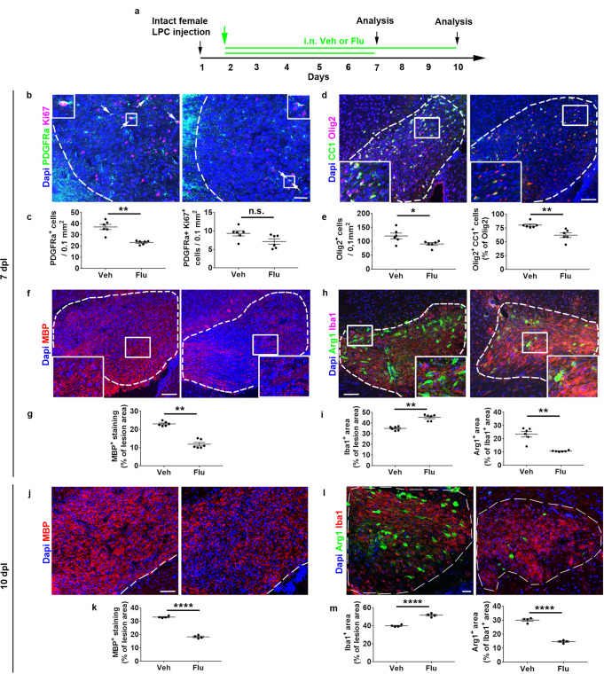
Neuroprotective, anti-inflammatory, and remyelinating properties of androgens are well-characterized in demyelinated male mice and men suffering from multiple sclerosis. However, androgen effects mediated by the androgen receptor (AR), have been only poorly studied in females who make low androgen levels. Here, we show a predominant microglial AR expression in demyelinated lesions from female mice and women with multiple sclerosis, but virtually undetectable AR expression in lesions from male animals and men with multiple sclerosis. In female mice, androgens and estrogens act in a synergistic way while androgens drive microglia response towards regeneration. Transcriptomic comparisons of demyelinated mouse spinal cords indicate that, regardless of the sex, androgens up-regulate genes related to neuronal function integrity and myelin production. Depending on the sex, androgens down-regulate genes related to the immune system in females and lipid catabolism in males. Thus, androgens are required for proper myelin regeneration in females and therapeutic approaches of demyelinating diseases need to consider male-female differences.


Conflict of interest statement

The authors declare no competing interests.

Figures

Fig. 1
Fig. 1. The androgen receptor is strongly up-regulated in the LPC-demyelinated corpus callosum from female but not male mice.
a Scheme of the experimental protocol. b, c Differential detection of AR transcripts in the corpus callosum (cc), but not cortex (Cx) from LPC-injected females or males. Dashed lines delineate the demyelinated area. d AR signal quantification in the lesions. e Double AR ISH (left) and Iba1, GFAP or Olig2 immunostainings (middle) and, merge images (right) in the female demyelinated lesions. The white arrows show a high number of AR-expressing microglial cells compared to a more restricted number of AR+GFAP+ astrocytes or AR+Olig2+ oligodendroglia. The white arrowheads indicate AR-expressing cells clearly devoid of GFAP or Olig2 markers. The boxed areas are magnified in the insets. f, g Visualization of cells co-expressing either the AR protein or the DHT ligand with Iba1 marker. The white arrows show microglia co-expressing nuclear/perinuclear AR f and nuclear DHT staining g. In g, microglial or non-microglial (white and yellow arrowheads, respectively) cells displaying a perinuclear DHT labeling or cells expressing none of the markers (yellow arrow) are shown. h Quantification of the percentage of Iba1+ cells displaying a nuclear (Nuc) or perinuclear (Perinuc) labeling. i Co-vizualization of AR transcripts with Iba1 or GFAP immunostainings in the demyelinated corpus callosum (dotted line) from LPC-injected male mice. The boxes are cortical (yellow) and callosal (white) areas magnified in the corresponding inset. j, k Triple labeling of female or male lesions using Iba1 immunostaining with Esr1 and Esr2 ISH. The lesions are delineated by the dashed lines. The boxed areas are magnified in the insets. Esr2 can be observed colocalized or not with Esr1. l, m Quantification of Esr1 and Esr2 signals. Scale bars (µm): 100 b, c, ik, 50 e, 10 f, g. Data are mean values±SEM from n = 4 d, l, m or n = 5 h animals/condition examined over two independent experiments. P values d, h, l, m were calculated using the unpaired two-tailed t-test with Welch’s correction d, l; **p = 0.001 d, p = 0.003 l; ****p < 0.0001 h. Source data are provided as a Source Data file.
Fig. 2
Fig. 2. AR mRNA expression in IBA1+ microglia/macrophages is up-regulated in the human female MS brain.
Post-mortem white matter samples from MS and non-neurological control donors were used to detect AR mRNA expression in fluorescently labeled IBA1+ microglia/macrophages. a Representative images showing AR mRNA expression (red) within Iba1+ microglia/macrophages (cyan) in white matter from a female control donor and in active white matter demyelinated lesions from a female and male MS donor. Sections were counterstained with DAPI (blue). Scale bar 50 μm. Magnified regions in inserts show AR+ (white arrowheads) and AR (yellow arrowheads) microglia/macrophages. Scale bar 20 μm. b Quantification of RNAscope experiment shows a significantly greater proportion of Iba1+ cells expressing AR mRNA in MS samples compared to controls (F 1, 18 = 8.778, p = 0.00821; post-hoc pairwise comparison: t = 2.963, p = 0.0083; control: median 34.70 (CI 30.00, 37.47), 25% percentile 32.50, 75% percentile 35.98, minimum 25.00, maximum 40.10; MS: median 46.60 (CI 31.92, 52.00), 25% percentile 33.33, 75% percentile 51.43, minimum 25.77, maximum 66.00), with a significant effect of sex, as significantly more Iba1+ cells express AR in females compared to males (linear mixed-effect model: F 1, 18 = 10.411, p = 0.00459; post-hoc pairwise comparison: t = 3.231, p = 0.0046). c Within MS donors, female samples show a significantly greater proportion of AR+Iba1+ cells compared to males (F 1, 8 = 28.579, p = 0.000494). No significant differences were detected among different lesion types (F 3, 97 = 2.027, p = 0.1151; WM: median 34.70 (CI 30.00, 37.47), 25% percentile 32.50, 75% percentile 35.98, minimum 25.00, maximum 40.10; CIL: median 45.41 (CI 22.94, 56.00), 25% percentile 32.89, 75% percentile 52.28, minimum 22.94, maximum 56.00; AL: median 46.60 (CI 25.00, 72.03), 25% percentile 31.54, 75% percentile 59.94, minimum 25.00, maximum 72.03; NAWM: median (43.33 (CI 28.26, 52.00), 25% percentile 29.41, 75% percentile 51.36, minimum 27.78, maximum 64.00; CAL: median 40.54 (CI 26.53, 59.21), 25% percentile 32.64, 75% percentile 54.57, minimum 26.53, maximum 59.21). Data were analyzed by linear mixed-effects models followed by ANOVA to determine main effects and Tukey post-hoc pairwise comparisons. Each data point represents the average quantification of 5–10 different regions of interest from the same case (n = 10 controls (5 M, 5 F), n = 11 MS (5 M, 6 F)). AL active lesion, CAL chronic active lesion, CIL chronic inactive lesion, NAWM normal-appearing white matter, WM white matter from control donors. Source data are provided as a Source Data file.
Fig. 3
Fig. 3. Testosterone and DHT induce a potent regeneration of myelin in female mice.
a Scheme of the experimental protocol. bg Visualization of OPC proliferation in (b, c), OPC differentiation in (d, e) and MBP expression in (f, g) evaluated 7 days after LPC injection into the corpus callosum of ovariectomized females daily treated with the drug vehicle (Veh), testosterone (T) or dihydrotestosterone (DHT). In (b), the white arrows indicate Ki67+ PDGFRα+ proliferating OPCs. hk Immunostaining of local inflammatory cells using Iba1 and Arg-1 antibodies for the detection of the microglial population and the cell subset expressing the anti-inflammatory marker Arg-1 in (h, i) as well as GFAP and STAT3 antibodies, as markers of astrocytes and their reactive state in (j, k). The dashed lines in (d, f, h, j) indicate the lesion. The boxed area in (f, j) is magnified in the inset. Scale bars: 50 µm unless indicated. Data in (c, e, g, i, k) are presented as mean values ± SEM from n = 8 mice/group examined over 2 independent experiments (3–5 slices/animal). lo Scheme of the experimental protocol in (l). Electron microscopy analysis of the spinal cords from Vehicle and DHT-treated EAE females in (m) and determination of the g-ratio values plotted according to axon diameter in (n; 100 axons per animal, n = 3/group) as well as the mean value of g-ratios in each group in (o; 100 axons per animal, n = 3 mice/group). The upper, middle and lower horizontal lines of the boxplots represent the upper, median and lower quartile, respectively. Whiskers depict the smallest or largest values within 1.5-fold of the interquartile range. P values were calculated by using the one-way ANOVA test together with Tukey’s (c, i, k) or Holm-Sidak’s (g) multiple comparisons test, Kruskal-Wallis test together with Dunn’s.multiple comparisons test e, two-tailed Mann-Whitney test (o). Brown-Forsythe correction was used for (e left, i left, k right). **p ≤ 0.01; ***p ≤ 0.001; ****p ≤ 0.0001 compared to the control (Veh). ##p = 0.0049 compared to the indicated condition. Source data are provided as a Source Data file.
Fig. 4
Fig. 4. The combination of androgens and estrogens in LPC-demyelinated female animals leads to a regeneration process more efficient than the one induced by each molecule used alone.
a Scheme of the experimental paradigm. bi Visualization of OPC proliferation in b, c, OPC differentiation d, e and MBP immunostaining f, g as well as quantifications carried out 7 days after stereotaxic injection of LPC into the corpus callosum of ovariectomized female mice daily treated with the drug vehicle (Veh), dihydrotestosterone (DHT), estradiol (E2) or the combination of these molecules (DHT + E2). h, i Immunostaining of microglial cells by Iba1 and Arg-1 antibodies for the detection of the whole microglial population and the cell subset expressing the anti-inflammatory marker Arg-1. jn Scheme of the protocol used for pharmacologically inhibiting the conversion of testosterone to estradiol by using the aromatase inhibitor, fadrozole (Fad) in (j). MBP in (k, l) and Iba1/Arg-1 in (m, n) immunostaining experiments were performed and quantified on slices from the different groups of LPC-demyelinated animals. In (b), the white arrows indicate Ki67+ PDGFRα+ proliferating OPCs. The dashed lines in (d, f, h, k, m) delineate the lesion. Scale bars (µm): 50 in (b), 100 in (d, f, h, k, m). Data are presented as mean values ± SEM from n = 8 mice/group in (c, e, g, i) examined over two independent experiments and n = 4 mice/group in (l, n) examined in a single experiment (3–4 slices/per animal). P values were calculated by using the one-way ANOVA test together with Tukey’s (c) or Holm-Sidak’s (e, g, l, n) multiple comparisons test or Kruskal-Wallis test together with Dunn’s.multiple comparisons test (i). Brown-Forsythe correction was used for (e left, l). *p ≤ 0.05; **p ≤ 0.01; ***p ≤ 0.001; ****p ≤ 0.0001 compared to the control (Veh). #p ≤ 0.05; ##p ≤ 0.01; ####p ≤ 0.0001 compared to the indicated condition. Source data are provided as a Source Data file.
Fig. 5
Fig. 5. AR blockade alters spontaneous regeneration in female mice.
a Scheme of the experimental paradigm. Visualization and quantification of OPC proliferation in (b, c), OPC differentiation in (d, e) and MBP immunostaining in (f, g) at 7 days after stereotaxic injection of LPC into the corpus callosum of ovariectomized female mice daily treated with the drug vehicle (Veh) or the AR antagonist flutamide (Flu). In (b), the white arrows indicate Ki67+ PDGFRα+ proliferating OPCs. h, i Immunostaining of microglial cells by using Iba1 and Arg-1 antibodies for the detection of the whole microglial population and the cell subset expressing the anti-inflammatory marker Arg-1. The dashed lines delineate the lesions. The boxed areas are magnified in the insets. (jm) Visualization and quantification of MBP in (j, k) and Iba1/Arg-1 in (l, m) immunostaining at 10 dpl. Scale bars (µm): 50 in (b, j), 100 in (d, f, h, l). Data are presented as mean values ± SEM from n = 6 mice/group in (c, e, g, i) and n = 4 mice/group in (k, m) (3–4 slices/per animal). P values were calculated by using the unpaired two-tailed t-test (c, k, m) or two-tailed Mann-Whitney (e, g, i). Welch’s correction was used for (c left, i right). *p ≤ 0.05; **p ≤ 0.01; ***p ≤ 0.001; ****p ≤ 0.0001 compared to the control (Veh); n.s., non-significant. Source data are provided as a Source Data file.
Fig. 6
Fig. 6. Microglial AR is required for DHT-induced control of microglia response to demyelination.
a Scheme of the experimental paradigm. bg Visualization and quantification of Arg-1 expression in Iba1-expressing microglial cells in b, c, OPC differentiation in d, e and GFAP immunostaining of astrocytes in f, g at 7 days after stereotaxic injection of LPC into the corpus callosum of ovariectomized female mice expressing (AR wt/wt) or not (AR fl/fl) AR in microglia and treated with the drug vehicle (Veh) or DHT. The dashed lines delineate the lesions. Scale bars: 50 µm. Data are presented as mean values ± SEM from n = 6 mice/condition examined in two independent experiments (3 slices/per animal). P values (c, e, g,) were calculated by using the two-way ANOVA test together with Tukey’s multiple comparisons test. **p = 0.0069 (e left), p = 0.0061 (e right); ****p < 0.0001 c, p = 0.0001 g; n.s. not significant. Source data are provided as a Source Data file.
Fig. 7
Fig. 7. Therapeutic administration of androgens mitigates the course of EAE in female mice.
a Functional scores derived from EAE ovariectomized female mice treated with the drug vehicle (Veh), testosterone (T) or dihydrotestosterone (DHT) at onset of the first neurological symptoms (day 1) for 30 days (two-way ANOVA: treatment: F(2, 1307) = 298.2, p < 0.0001; time: F(29, 1307) = 33.23, p < 0.0001). b, c Smi-32 and MBP IHF performed on spinal cord slices derived from animals of each group. The boxed areas are magnified in the bottom panels. Determination of the fluorescent area is shown in the histograms on the right. d, e Electron microscopy analysis of the spinal cords and determination of the g-ratios plotted according to axon diameter or represented by their mean value. f, g Visualization and quantification of microglia immunostained with Iba1 (red) and Arg-1 (green) as markers of the whole microglia population and the cell subset that express the anti-inflammatory molecule Arg-1, respectively. h, i Visualization and quantification of astrocytes by using GFAP. j, k Detection of the tight junction protein Claudin-5 in each animal group. The boxed areas are magnified in the insets. Data are the mean ± SEM from n = 12 animals/condition in (a) or from n = 8 animals/condition in g, i, k examined in a single experiment (3–4 slices/per animal). 600–900 axons from n = 3 mice in (e) were evaluated. P values were calculated by using the Kruskal-Wallis test together with Dunn’s.multiple comparisons test (c, g) or one-way ANOVA test together with Tukey’s (e, k) or Holm-Sidak’s i multiple comparisons test. Brown-Forsythe correction was used in (k). *p < 0.05; **p < 0.01; ***p < 0.001 ****p < 0.0001 versus the control condition. Scale bars (µm): 200 in (b top), 50 f, h, j, 25 (b bottom). Source data are provided as a Source Data file.
Fig. 8
Fig. 8. The immune response triggered by DHT in EAE animals is strikingly different in female compared to male mice.
a Scheme of the experimental protocol. b, c Scoring of neurological disabilities in EAE female and male mice (n = 8/group) daily treated with DHT or the drug vehicle (Veh) at onset of neurological symptoms (day 1) for 8 days. df The spleen (n = 8 females, 5 males) in d, lymph nodes in e and spinal cord in f (n = 7 females, 5 males) from each animal group examined in a single experiment were harvested in order to perform flow cytometry analysis and dosage of cytokines. Data are presented as mean values ± SEM. The gating strategies for flow cytometry analysis are shown in Supplementary Figs. 4–7. Only the cell types regulated by DHT are shown. In the lymphoid organs and spinal cord, immune cell types are expressed in percentage of all cells and CD45+ leukocytes, respectively. Similarly, a panel of 9 cytokines has been assessed (as described in Methods). Only cytokines regulated by DHT are shown in red boxes. P values (d, e, f) were calculated by using the unpaired two-tailed t-test or Mann-Whitney tests. Welch’s correction was used for IFNγ e and IL1-β f in females. *p ≤ 0.05; **p ≤ 0.01; ***p ≤ 0.001 compared to the control (Veh); ns non-significant. Source data are provided as a Source Data file.
Fig. 9
Fig. 9. DHT controls differently the local inflammatory cells in EAE female and male mice.
a Scheme of the experimental protocol. Immunostaining of microglial and astroglial cells in the spinal cord from vehicle or DHT-treated female in (bh) or male in (io) mice. b, c Visualization and quantification of microglia in the whole white matter of vehicle-treated females indicate numerous spots of Arg-1+ cells extending deeply into the white matter (white arrows in b) strongly reduced under DHT treatment. d, e Visualization and quantification of Iba1 and Arg-1 staining at the level of an individual lesion indicating that Iba1 staining is still decreased whereas Arg-1+ area is significantly higher under DHT treatment. f, g GFAP+ astrogliosis is shown in whole spinal cord slices co-labeled by MBP antibody aimed at visualizing myelin. Numerous spots of demyelinated tissue are shown (white arrows) in the vehicle- compared to the DHT condition. Magnifications of the boxed areas show that DHT treatment is accompanied by the decrease of GFAP staining in the gray matter (GM) and conversely its increase in the white matter (WM). h Quantification of MBP+ area in the white matter. il Visualization and quantification of Iba1 and Arg-1 staining in the spinal cord from male in the whole white matter in (i, j) and at the level of individual lesions in k, l. mo Visualization of GFAP and MBP staining in m. Quantification of GFAP+ fluorescence in the white (WM) and gray (GM) matter in n and of MBP in the white matter in o. The boxed areas in b, i are magnified in d, k. Data are presented as mean values ± SEM from n = 8 mice/group examined in a single experiment (3–4 slices/per animal). P values (c, e, g, h, j, l, n, o) were calculated by using the unpaired two-tailed t-test or Mann-Whitney test. Welch’s correction was used for (e left, j, h). **p ≤ 0.01; ***p ≤ 0.001; ****p ≤ 0.0001; n.s, non-significant. Scale bars (µm): 200 in (b, f top, i), 50 in m, 25 (d, f bottom, k). Source data are provided as a Source Data file.
Fig. 10
Fig. 10. RNA-Seq analysis of the spinal cord derived from EAE mice therapeutically treated with DHT reveal major differences between female and male animals.
a Scheme of the experimental protocol. b, c PCA plot of two first components with their contribution to the variance depicturing clear differences between DHT-treated (DHT, n = 3) and control (CT, n = 4) samples in the first PCA component, which contributes for more than half of the variance of the experiment, in females in (b) and in males in (c) examined in two independent experiments. The size of the sample name and the circle indicate the relative contribution to the total variance. d, e Barplots and tables showing the contribution of oligodendroglial curated DEGs genes to promote (positive) or inhibit (negative) each process of oligodendrogenesis in females in d and in males in e. Note that DHT mainly promotes (re)myelination. f, g Dotplot representating the top 7 biological processes enriched in up-regulated genes in DHT-treated females in f and DHT-treated males in g compared to their respective controls, showing similar up-regulation of synaptic and neuronal associated processes in both sexes. h, i Dotplot representating the top 7 biological processes enriched in down-regulated genes in DHT-treated females in h and DHT-treated males in i. Note that while down-regulated genes are implicated in immune processes in females in h, they are implicated in catabolism in males in i. jl Histograms visualizing the deregulation of genes characterizing homeostatic in j, Disease-Associated in k and White matter-Associated in l microglia by DHT in EAE females or males. Multiple testing correction aimed at controling the false discovery rate (FDR, p-adjust) was performed by using the Benjamini-Hochberg method fi. Fisher test was used as well as multiple testing correction aimed at controling the false discovery rate (FDR, p-adjust) performed by using the Benjamini–Hochberg method jl. *FDR < 0.05; **FDR < 0.01; ***FDR < 0.001.

Similar articles

Cited by

References

    1. Thompson AJ, Baranzini SE, Geurts J, Hemmer B, Ciccarelli O. Multiple sclerosis. Lancet. 2018;391:1622–1636. doi: 10.1016/S0140-6736(18)30481-1. - DOI - PubMed
    1. Orton SM, et al. Sex ratio of multiple sclerosis in Canada: a longitudinal study. Lancet Neurol. 2006;5:932–936. doi: 10.1016/S1474-4422(06)70581-6. - DOI - PubMed
    1. Schwendimann RN, Alekseeva N. Gender issues in multiple sclerosis. Int Rev. Neurobiol. 2007;79:377–392. doi: 10.1016/S0074-7742(07)79017-7. - DOI - PubMed
    1. Bove R, Chitnis T. The role of gender and sex hormones in determining the onset and outcome of multiple sclerosis. Mult. Scler. 2014;20:520–526. doi: 10.1177/1352458513519181. - DOI - PubMed
    1. Vukusic S, et al. Pregnancy and multiple sclerosis (the PRIMS study): clinical predictors of post-partum relapse. Brain: a J. Neurol. 2004;127:1353–1360. doi: 10.1093/brain/awh152. - DOI - PubMed

Publication types