. 2023 Mar 22;6(1):304.
doi: 10.1038/s42003-023-04583-x.

Learning to predict RNA sequence expressions from whole slide images with applications for search and classification

Affiliations

Learning to predict RNA sequence expressions from whole slide images with applications for search and classification

Areej Alsaafin et al. Commun Biol. .

Abstract

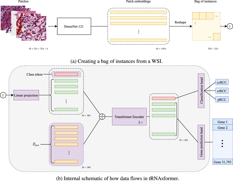
Deep learning methods are widely applied in digital pathology to address clinical challenges such as prognosis and diagnosis. As one of the most recent applications, deep models have also been used to extract molecular features from whole slide images. Although molecular tests carry rich information, they are often expensive, time-consuming, and require additional tissue to sample. In this paper, we propose tRNAsformer, an attention-based topology that can learn both to predict the bulk RNA-seq from an image and represent the whole slide image of a glass slide simultaneously. The tRNAsformer uses multiple instance learning to solve a weakly supervised problem while the pixel-level annotation is not available for an image. We conducted several experiments and achieved better performance and faster convergence in comparison to the state-of-the-art algorithms. The proposed tRNAsformer can assist as a computational pathology tool to facilitate a new generation of search and classification methods by combining the tissue morphology and the molecular fingerprint of the biopsy samples.


Conflict of interest statement

The authors declare no competing interests.

Figures

Fig. 1
Fig. 1. A diagram showing how tRNAsformer works.
a 49 tiles of size 224 × 224 × 3 selected from 49 spatial clusters in a WSI are embedded with a DenseNet-121. The outcome is a matrix of size 49 × 1024 as DenseNet-121 has 1024 deep features after the last pooling. Then the matrix is reshaped and rearranged to 224 × 224 matrix in which each 32 × 32 block corresponds to a tile embedding 1 × 1024. b Applying a 2D convolution with kernel 32, stride 32, and 384 kernels, each 32 × 32 block has linearly mapped a vector of 384 dimensional. Next, a class token is concatenated with the rest of the tile embeddings, and Epos is added to the matrix before entering L Encoder layers. The first row of the outcome, which is associated with the class token, is fed to the classification head. The rest of the internal embeddings that are associated with all tile embeddings are passed to the gene prediction head. All parts with learnable variables are shown in purple.
Fig. 2
Fig. 2. The distribution of the correlation coefficients between 31,793 genes predicted and their true value for the TCGA test set.
The violin diagrams depict the distribution, min, max, and mean values of the correlation coefficients. a Violin diagrams for Pearson correlation coefficients and b violin diagrams for Spearman’s correlation coefficients. The violin diagrams are plotted for tRNAsformerL for L = (1, 2, 4, 8, 12) and HE2RNAbb1024. The mean and standard deviation of the correlation coefficients are included in the legend for violins from left to right.
Fig. 3
Fig. 3. ROC Curves for TCGA and External Dataset.
The micro ROC curve of different models applied on a the TCGA test set and b the external dataset. The AUC is reported in the legend for all models.
Fig. 4
Fig. 4. An example of clustering for creating bag of tiles from a WSI.
a Shows a thumbnail of a WSI, b shows the tissue mask obtained by segmenting the WSI, and c shows the clustered WSI using k-means.

References

    1. Hou, L. et al. Patch-based convolutional neural network for whole slide tissue image classification. in Proceedings of the IEEE Conference on Computervision and Pattern Recognition 2424–2433 (2016). - PMC - PubMed
    1. Kalra S, et al. Pan-cancer diagnostic consensus through searching archival histopathology images using artificial intelligence. NPJ Digit. Med. 2020;3:1–15. doi: 10.1038/s41746-020-0238-2. - DOI - PMC - PubMed
    1. Wang H, et al. Mitosis detection in breast cancer pathology images by combining handcrafted and convolutional neural network features. J. Med. Imaging. 2014;1:034003. doi: 10.1117/1.JMI.1.3.034003. - DOI - PMC - PubMed
    1. Bulten W, et al. Automated deep-learning system for gleason grading of prostate cancer using biopsies: a diagnostic study. Lancet Oncol. 2020;21:233–241. doi: 10.1016/S1470-2045(19)30739-9. - DOI - PubMed
    1. Schmauch B, et al. A deep learning model to predict rna-seq expression of tumours from whole slide images. Nat. Commun. 2020;11:1–15. doi: 10.1038/s41467-020-17678-4. - DOI - PMC - PubMed

Publication types