. 2023 Jun;112(6):666-674.
doi: 10.1007/s00223-023-01076-1. Epub 2023 Mar 22.

Hip Labral Morphological Changes in Patients with Femoroacetabular Impingement Speed Up the Onset of Early Osteoarthritis

Affiliations

Hip Labral Morphological Changes in Patients with Femoroacetabular Impingement Speed Up the Onset of Early Osteoarthritis

Michela Battistelli et al. Calcif Tissue Int. 2023 Jun.

Abstract

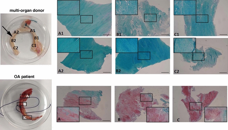
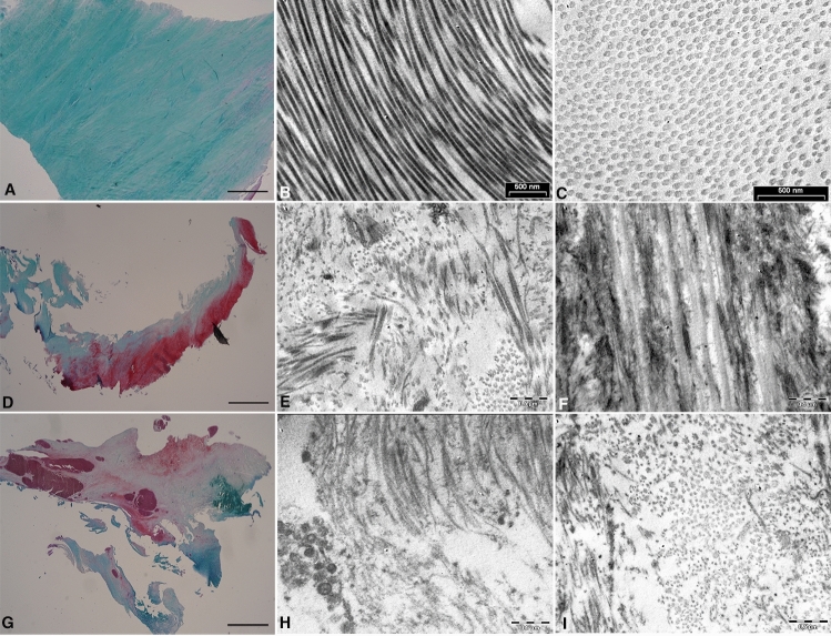
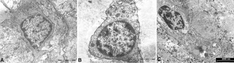
Over the last decade, evidence has mounted for a prominent etiologic role of femoroacetabular impingement (FAI) in the development of early hip osteoarthritis (OA). The aim of this study was to compare the ultrastructure and tissue composition of the hip labrum in healthy and pathological conditions, as FAI and OA, to provide understanding of structural changes which might be helpful in the future to design targeted therapies and improve treatment indications. We analyzed labral tissue samples from five healthy multi-organ donors (MCDs) (median age, 38 years), five FAI patients (median age, 37 years) and five late-stage OA patients undergoing total hip replacement (median age, 56 years). We evaluated morpho-functional by histology and transmission electron microscopy. Extracellular matrix (ECM) structure changes were similar in specimens from FAI compared to those from patients with OA (more severe in the latter) showing disorganization of collagen fibers and increased proteoglycan content. In FAI and in OA nuclei the chromatin was condensed, organelle degenerated and cytoplasm vacuolized. Areas of calcification were mainly observed in FAI and OA labrum, as well as apoptotic-like features. We showed that labral tissue of patients with FAI had similar pathological alterations of tissue obtained from OA patients, suggesting that FAI patients might have high susceptibility to develop OA.

Keywords: Calcification; Extracellular matrix degeneration; Femoroacetabular impingement; Labrum; Osteoarthritis.


Conflict of interest statement

Michela Battistelli, Enrico Tassinari, Giovanni Trisolino, Marco Govoni, Gianluca Ruspaggiari, Lucia De Franceschi, Dante Dallari, Debora Burini, Roberta Ramonda, Marta Favero, Francesco Traina, Brunella Grigolo, Eleonora Olivotto have declair that they don’t have conflict of interest.

Figures

Fig. 1
Fig. 1
Labral tissue samples. Upper side: samples from one representative multi-organ donor (the arrow indicates the area where the biopsies were collected in FAI patients). Lower side: samples from one representative patient with end-stage OA. For both samples, LEFT: high resolution digital photograph of labral tissue; RIGHT: Safranin-O-Fast Green matrix staining for the histological grading score. A = anterior, B = middle and C = posterior region of each labral sample cut perpendicular relative to the sagittal plane (Magnification 2x, bar 1000 μm; insert 10x, bar 100 μm)
Fig. 2
Fig. 2
Labral tissue morphology. Labral samples collected from one representative multi-organ donor showed strong stain for Collagen and virtually no staining for proteoglycans observed by Safranin-O/Fast green staining (A). The collagen fibers appeared with homogenous distribution, regular size and orientation by transmission electron microscopy (TEM) (BC). In samples from one representative patient with FAI (DF) and with end-stage OA (G and H), there was a strong staining for proteoglycans and the TEM revealed collagen fibers with a structural disorganization of the ECM (Magnification: A, D, G = 2 x, bar 1000 μm; B, C, E, F, H, I = bar 0.5 µm)
Fig. 3
Fig. 3
Calcium deposition. Labrum samples collected from one representative multi-organ donor showed small calcium deposits, evaluated by alizarin red (AR) staining (A and insert) and rarely observed with TEM (D). In FAI samples big calcium deposits were visible with AR (B and insert) also detected by TEM (E). Extensive calcified areas were observed in one representative OA patient (C and F) (White arrows = calcium deposits. Magnification panel A, B, C = 2x, bar 1000 μm; insert 10x; panel D = bar 2 µm, E = bar 0.5 µm, F = bar 1 µm)
Fig. 4
Fig. 4
Cell morphology. In labrum from MCDs, the cells embedded in the ECM were round and showed a healthy morphology, consistent with viable and metabolically active cells. The nuclear chromatin appeared diffuse with small condensed areas near the nuclear membrane (A). In patient with FAI cells contained condensed nuclear chromatin, with a specific pattern, called ‘‘chondroptosis’’. Cytoplasmic vacuoles were present with scarcity of organelles (B). In the representative OA patient large areas of condensed chromatin (C) and vacuoles within the cytoplasm were observed (C). TEM A, C bar 2 µm, B bar 1 µm

References

    1. Smith E, Hoy D, Cross M, Merriman TR, Vos T, Buchbinder R, Woolf A, March L. The global burden of gout: estimates from the global burden of disease 2010 study. Ann Rheum Dis. 2014;73:1470–1476. doi: 10.1136/annrheumdis-2013-204647. - DOI - PubMed
    1. Guillemin F, Rat AC, Mazieres B, Pouchot J, Fautrel B, Euller-Ziegler L, Fardellone P, Morvan J, Roux CH, Verrouil E, Saraux A, Coste J, Osteoarthritis g, Prevalence of symptomatic hip and knee osteoarthritis: a two-phase population-based survey. Osteoarthritis Cartil. 2011;19:1314–1322. doi: 10.1016/j.joca.2011.08.004. - DOI - PubMed
    1. Melugin HP, Hale RF, Zhou J, LaPrade M, Bernard C, Leland D, Levy BA, Krych AJ. Risk factors for long-term hip osteoarthritis in patients with femoroacetabular impingement without surgical intervention. Am J Sports Med. 2020;48:2881–2886. doi: 10.1177/0363546520949179. - DOI - PMC - PubMed
    1. Agricola R, Waarsing JH, Arden NK, Carr AJ, Bierma-Zeinstra SM, Thomas GE, Weinans H, Glyn-Jones S. Cam impingement of the hip: a risk factor for hip osteoarthritis. Nat Rev Rheumatol. 2013;9:630–634. doi: 10.1038/nrrheum.2013.114. - DOI - PubMed
    1. Ganz R, Parvizi J, Beck M, Leunig M, Notzli H, Siebenrock KA. Femoroacetabular impingement: a cause for osteoarthritis of the hip. Clin Orthop Relat Res. 2003 doi: 10.1097/01.blo.0000096804.78689.c2. - DOI - PubMed