. 2023 Apr;616(7955):183-189.
doi: 10.1038/s41586-023-05836-9. Epub 2023 Mar 22.

From primordial clocks to circadian oscillators

Affiliations

From primordial clocks to circadian oscillators

Warintra Pitsawong et al. Nature. 2023 Apr.

Abstract

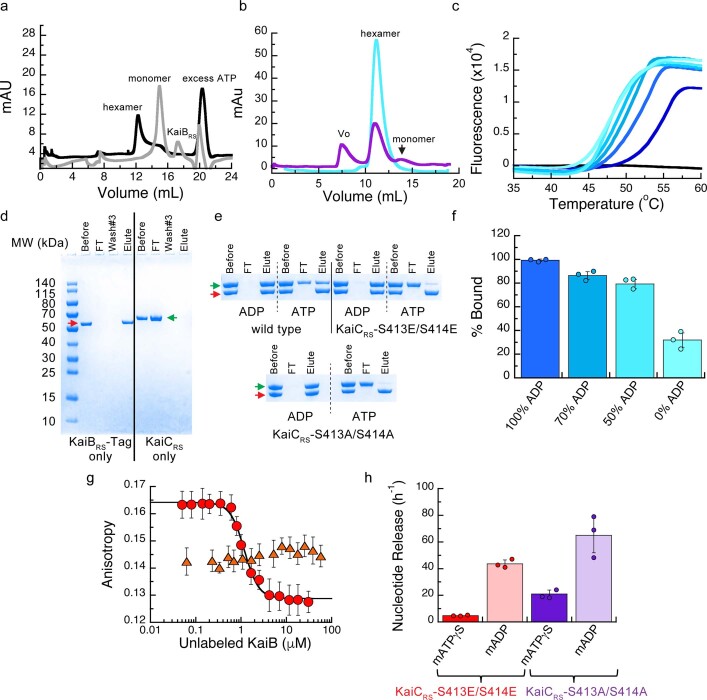
Circadian rhythms play an essential part in many biological processes, and only three prokaryotic proteins are required to constitute a true post-translational circadian oscillator1. The evolutionary history of the three Kai proteins indicates that KaiC is the oldest member and a central component of the clock2. Subsequent additions of KaiB and KaiA regulate the phosphorylation state of KaiC for time synchronization. The canonical KaiABC system in cyanobacteria is well understood3-6, but little is known about more ancient systems that only possess KaiBC. However, there are reports that they might exhibit a basic, hourglass-like timekeeping mechanism7-9. Here we investigate the primordial circadian clock in Rhodobacter sphaeroides, which contains only KaiBC, to elucidate its inner workings despite missing KaiA. Using a combination of X-ray crystallography and cryogenic electron microscopy, we find a new dodecameric fold for KaiC, in which two hexamers are held together by a coiled-coil bundle of 12 helices. This interaction is formed by the carboxy-terminal extension of KaiC and serves as an ancient regulatory moiety that is later superseded by KaiA. A coiled-coil register shift between daytime and night-time conformations is connected to phosphorylation sites through a long-range allosteric network that spans over 140 Å. Our kinetic data identify the difference in the ATP-to-ADP ratio between day and night as the environmental cue that drives the clock. They also unravel mechanistic details that shed light on the evolution of self-sustained oscillators.


Conflict of interest statement

D.K. is co-founder of Relay Therapeutics and MOMA Therapeutics. The remaining authors declare no competing interests.

Figures

Fig. 1
Fig. 1. The extended C-terminal tail of KaiCRS forms a coiled-coil interaction with an exposed A loop for KaiA-independent phosphorylation of KaiC.
a, Schematic of the phylogenetic tree of kaiC showing the appearance of kaiB and kaiA during evolution. The kaiC clade with an approximately 50-amino-acid C-terminal extension is labelled in red, and a timeline was predicted as previously reported. Ga, billion years ago. b, Phosphorylation rate over time of KaiCRS (6.5 ± 1.0 h−1) and KaiCSE in the presence (0.40 ± 0.02 h−1) or absence of KaiASE at 30 °C. The s.d. in reported parameters were obtained from the fitting. c, Nucleotide exchange between ATP and mant-ATP in KaiCRS alone (18.0 ± 1.5 h–1) compared with KaiCSE in the presence (4.7 ± 0.3 h–1) and absence of KaiASE (0.08 ± 0.04 h–1) measured at 30 °C. Representative traces are shown, and the fitted parameters (mean ± s.d.) were obtained from three replicate measurements. d, X-ray structure of dodecameric KaiCRS (PDB: 8DBA) coloured by hexamer A (light green) and hexamer B (dark green). The CI, CII and coiled-coil domains are indicated, and the A loop is labelled in e. e, Superposition based on an alignment of the CII domain of KaiCRS (green; PDB: 8DBA, chain B), KaiCSE (purple; PDB: 1TF7, chain B) and KaiCSE-S431E/T432A (yellow; PDB: 7S65, chain A) shows that KaiCRS has an extended A loop orientation that no longer forms the inhibitory interaction with the 422 loop (KaiCSE numbering). The conformation of the 422 loop in KaiCRS resembles the one seen in the cryo-EM structure of the phosphomimetic KaiCSE-S431E/T432A (yellow; PDB: 7S65). No electron density is observed for the C-terminal part of wild-type KaiCSE and the S431E/T432A mutant owing to flexibility, and the missing 22 residues for wild-type KaiCSE (46 for S431E/T432A) are represented by a dashed line (not shown for the mutant).
Fig. 2
Fig. 2. A coiled-coil partner switch coupled to an allosteric network in the CII domain promotes autodephosphorylation.
a, X-ray structure of KaiCRS-Δcoil was solved in the C2221 space group and contained three monomers in the asymmetric unit, with ADP present in all active sites. The obtained electron density map allowed for model building up to Pro463, which indicated that the truncation at position 490 results in enhanced flexibility beyond Pro463. Phosphorylation of Ser414 (pS414) was observed in chain B (cyan) as shown by the electron density mFoDFc polder map (green mesh, 3σ contour level). b, Assembly analysis using the PISA software revealed a hexamer as the most probable quaternary structure (top view). c, Structural comparison of the coiled-coil domain for unphosphorylated KaiCRS (dark and light green; X-ray structure) and the KaiCRS-S413E/S414E phosphomimetic mutant (dark and light blue; cryo-EM structure). d, Overlay of interacting dimers of the structures in c using the CII domain of chain A as a reference (dark shades; bottom). Unphosphorylated KaiCRS (dark green) interacts with the opposite partner on the right (light green), whereas KaiCRS-S413E/S414E (dark blue) interacts with the partner on the left (light blue). The hydrophobic packing in the coiled-coil domain is mediated by only the Cβ atoms of alanine and arginine residues in unphosphorylated KaiCRS, but involves the entire side chain of leucine and isoleucine residues in the phosphomimetic structure. e, Allosteric network in the phosphomimetic state (blue) from the coil (light blue) propagating through the KaiCRS CII domain to the active site (dark blue) compared with the unphosphorylated state (dark green) (Supplementary Video 1). f, Autodephosphorylation of KaiCRS and KaiCRS-Δcoil over time in the presence of 4 mM ADP at 30 °C. The phosphorylated (P) and unphosphorylated (U) proteins were separated by Zn2+ Phos-tag SDS–PAGE (for gel source data, see Supplementary Fig. 1).
Fig. 3
Fig. 3. The regulatory role of KaiBRS in the phosphorylation–dephosphorylation cycle of KaiCRS.
a, SDS–PAGE gel of 3.5 μM KaiCRS and 4 mM ATP in the absence (top) and presence (bottom) of 3.5 μM KaiBRS at 35 °C, with the percentage of ATP indicated at specific time points. b, Phosphorylation (single and double) of KaiCRS during the reaction in the absence (grey circles) or presence (red diamonds) of KaiBRS. c, Phosphorylation–dephosphorylation cycle of 3.5 μM phosphorylated KaiCRS in the absence and presence of 3.5 μM KaiBRS in a constant ATP-to-ADP ratio of high ATP (4 mM) to mimic daytime and about 25% ATP to mimic the night time (exact percentage of ATP indicated at specific time points) at 30 °C. U, S and D in a and c represent the unphosphorylated, single phosphorylated (at Ser413 or Ser414) and double phosphorylated state of KaiCRS, respectively (for gel source data, see Supplementary Fig. 1). d, ATPase activity of wild-type KaiCRS in the absence and presence of KaiBRS, KaiCRS-E62Q/E63Q in the absence and presence of KaiBRS, and KaiCRS-E302Q/E303Q in the absence and presence of KaiBRS at 30 °C. Bar graphs show mean ± s.d. from three replicates. e, Time-dependent autodephosphorylation of 32P-labelled KaiCRS bound with ADP in the presence of 20 μM KaiBRS and 4 mM ADP at 30 °C showing phosphorylated 32P-KaiCRS, 32P-ATP and free 32Pi. The reaction products were separated by thin layer chromatography. f, The decay of phosphorylated 32P-KaiCRS bound with 4 mM ADP in the absence (grey circles) and presence (red diamonds) of KaiBRS at 30 °C is obtained from autoradiography quantification (Extended Data Fig. 7). g, The nucleotide exchange of 3.5 μM KaiCRS (grey trace) and 3.5 μM KaiCRS in complex with 30 μM KaiBRS (red dotted trace) in the presence of ATP with mant-ATP. Representative traces are shown, and the fitted parameters (mean ± s.d.) were obtained from three replicate measurements.
Fig. 4
Fig. 4. KaiBRS binds to the post-hydrolysis state and accelerates the ATPase activity of KaiCRS.
a, Cryo-EM structure of KaiCRS-S413E/S414E (yellow) in complex with KaiBRS (blue) (PDB: 8FWJ). b, Superposition of KaiCRS-S413E/S414E (yellow) bound to KaiBRS (blue) (PDB: 8FWJ) and KaiCTE-S413E (dark grey) bound to fsKaiBTE (fold-switch, binding-competent state of KaiBTE; light grey) (PDB: 5JWQ). c,d, Binding of KaiBRS (blue) creates a tunnel (grey mesh) that enables water to reach the catalytic position (red sphere) for ATP hydrolysis in the CI domain. e, Binding of wild-type and mutant forms of KaiCRS to His-tagged KaiBRS in the presence of ADP or an ATP-recycling system at 25 °C. Bar graphs show mean ± s.d. from three replicates. f, Fluorescence anisotropy of unlabelled KaiBRS competitively displacing KaiBRS–6IAF (where 6IAF is the fluorophore) from unphosphorylated KaiCRS in the presence of ADP (dark green circles) and phosphorylated KaiCRS in the presence of the ATP-recycling system (light green triangles) at 30 °C. The average anisotropy and standard error were calculated from ten replicate measurements. g, Schematic of the uncovered mechanism of KaiCRS regulated by coiled-coil interactions and KaiBRS in the CI and CII domains.
Extended Data Fig. 1
Extended Data Fig. 1. Evolution of kaiC and sequence alignment of kaiC subgroups.
(a) Phylogenetic tree of kaiC homologs, where kaiC genes that have an approximately 50 amino acids C-terminal extension are labeled in red. Rhodobacter sphaeroides strain KD131 studied here and Synechococcus elongatus PCC 7942 (widely studied in the literature) are highlighted in green and pink, respectively. The accession code and organism are shown at the tip of the branches, the numbers at each node represent the aBayes bootstrap values, and the legend for branch length is shown (see also Supplementary Datasets 1 and 2). (b) A sequence alignment of the CII domain of the kaiC subgroups annotated with its sequence similarity. Residue Glu490, the position where the stop codon was introduced in the truncated KaiCRS-Δcoil construct, is shown in red and marked with an arrow.
Extended Data Fig. 2
Extended Data Fig. 2. Auto-phosphorylation and nucleotide exchange rates of KaiC.
(a) 10% SDS-PAGE gel of 3.5 μM KaiCRS in the presence of 4 mM ATP and using an ATP-recycling system at 30 °C. U, S, and D represent unphosphorylated, singly, and doubly phosphorylated KaiCRS, respectively. (b) Densitometric analysis of auto-phosphorylation (single + double phosphorylation) from panel (a) over time yields a rate of 6.5 ± 1.0 h−1. (c) 6.5% SDS-PAGE gel of 3.5 μM KaiCSE in the presence of 1.2 μM KaiASE and 4 mM ATP at 30 °C. U and P represent unphosphorylated and phosphorylated KaiCSE, respectively. (d) Densitometric analysis of auto-phosphorylation of KaiCSE activated by KaiASE (panel (c)) shows a rate of 0.40 ± 0.02 h−1 and is substantially slower than for KaiCRS. The standard deviation for parameters in (b) and (d) were obtained from data fitting. (e) 10% SDS-PAGE gels for experiments with 3.5 μM KaiCRS in the presence of 4 mM ATP and using an ATP-recycling system between 20 and 35 °C show that the level of phosphorylation increases with temperature. U, S, and D represent unphosphorylated, singly, and doubly phosphorylated KaiCRS, respectively. For gel source data in (a), (c), and (e), see Supplementary Figure 2. (f) Bar graphs indicating the nucleotide exchange rate in the CII domain of KaiCRS incubated with 50 μM ATP in the presence of an ATP-recycling system, and then mixed with 250 μM mant-ATP. An increase in fluorescence intensity at 440 nm was recorded and the single-exponential time traces were fitted to obtain the exchange rate constants: 3.6 ± 0.8 h−1 (20 °C), 12.2 ± 1.0 h−1 (25 °C), 18.5 ± 1.5 h−1 (30 °C), and 25.2 ± 0.2 h−1 (35 °C). Experiments were performed in triplicate and data are presented as mean values ± s.d.
Extended Data Fig. 3
Extended Data Fig. 3. Oligomeric states of KaiCRS and effect of coiled-coil domain on rates of nucleotide exchange and auto-phosphorylation.
(a) Oligomerization analysis of KaiCRS (dodecamer, green line) and truncated KaiCRS-Δcoil (hexamer, cyan line) by analytical gel-filtration chromatography. The protein size markers are indicated at the top. (b) Comparison of the elution profiles of KaiCRS-Δcoil (cyan line) and KaiCSE (purple line) from size-exclusion chromatography shows a hexameric state for both KaiCRS-Δcoil and KaiCSE. (c) Oligomeric states of KaiCRS (dodecamer, green line), KaiCRS-Δcoil (hexamer, cyan line), and KaiCSE (hexamer, purple line) were also measured by analytical ultracentrifugation (sedimentation velocity at 30,000 rpm and 20 °C) and the results agree with the data shown in panels (a) and (b). The graph in panel (c) represents the sedimentation coefficient distribution [c(s)]. (d) The change in fluorescence at 440 nm (ΔF440nm) represents the nucleotide exchange between ATP and mant-ATP at 30 °C for KaiCRS (green trace, 18.0 ± 1.5 h−1) and KaiCRS-Δcoil (cyan trace, 19.1 ± 0.8 h−1). Representative traces are shown and the fitted parameters (mean ± s.d.) were obtained from three replicate measurements. (e) Zn2+ Phos-tagTM SDS-PAGE gel shows the level of phosphorylation over time of KaiCRS (upper gel) and KaiCRS-Δcoil (lower gel) at 35 °C. P and U represent phosphorylated and unphosphorylated protein, respectively. (f) Phosphorylation level over time of KaiCRS (green circles, 7.4 ± 0.3 h−1) and KaiCRS-Δcoil (cyan circles, 5.5 ± 0.4 h−1) analyzed by densitometric analysis of Zn2+ Phos-tagTM SDS-PAGE gel in (e). (g) First derivative of thermal-stability curves measured for unphosphorylated KaiCRS bound with ADP (brown line) and phosphorylated KaiCRS bound with ATP (green line). The extracted temperatures of denaturation are 50 °C (unphosphorylated KaiCRS in the presence of 1 mM ADP) and 58 °C (phosphorylated KaiCRS in the presence of 1 mM ATP), respectively. (h) Dodecameric state of unphosphorylated KaiCRS (40 μM) bound with ADP measured by size-exclusion chromatography. (i) SDS-PAGE gel shows dephosphorylation of Ser413 over time at 30 °C in the presence of 4 mM ADP (U and pSer413 represent unphosphorylated and Ser413-phosphorylated KaiCRS, respectively) with the corresponding kinetics shown in the right panel (confirmed by MS/MS) with a rate constant of 11.5 ± 0.8 h−1. This result suggests that the coiled-coil domain promotes KaiCRS dephosphorylation. For gel source data in (e) and (i), see Supplementary Figure 2. The standard deviation for parameters in (f) and (i) were obtained from data fitting.
Extended Data Fig. 4
Extended Data Fig. 4. Graphical description of the cryo-EM processing workflow and validation of the final dodecamer structures.
The workflow (see description in Methods section) demonstrates a typical image (scale bar: 60 nm) and representative good class averages. The ab initio and final reconstructions are shown. Shown alongside the final reconstruction is the angular plot demonstrating the distribution of particle views and the Fourier shell correlation curve used for the global resolution estimation (a) KaiCRS-S413E/S414E alone and (b) KaiCRS-S413E/S414E:KaiBRS complex. To validate the final combined dodecamer structures, the data were reprocessed for the full dodecamer. The figure shows representative good class averages, the final reconstruction, angular distribution and Fourier shell correlation curve for the (c) KaiCRS-S413E/S414E alone and (d) KaiCRS-S413E/S414E:KaiBRS complex dodecamers. (e) Comparison of the C1 and D6 reconstructions of KaiCRS-S413E/S414E alone and KaiCRS-S413E/S414E:KaiBRS, and Fourier shell correlation curves for the C1 reconstructions. The C1/D6 comparisons do not reveal discernable differences, suggesting that these complexes have D6 symmetry.
Extended Data Fig. 5
Extended Data Fig. 5. Correlation between the coiled-coil register shift and phosphorylation, and model for consecutive phosphorylation/dephosphorylation events in CII domain of KaiCRS.
(a) Structural comparison between KaiCRS (green) and KaiCRS-S413E/S414E (orange, single-chain for clarity) reveals that the coiled-coil in the phosphomimetic structure points outwards, with an angle of about 20° relative to the KaiCRS coiled-coil. (b) The conformational change in the coiled-coil domain affects the dimer interface due to partner swaps with the opposite hexamer (see also Fig. 2). From an “outside perspective”: the C-terminal helix in KaiCRS interacts with the right chain from the opposite hexamer, whereas in KaiCRS-S413E/S414E the interaction is with the chain on the left. (c) Coiled-coil diagrams describe the heptad register shift that accompanies this structural rearrangement. (d) Based on the overlay of our structures, we propose the following model for the phosphorylation/dephosphorylation events. First, the phosphorylation cycle starts with the transfer of the γ-phosphate of ATP to the hydroxyl group of Ser414 (1; green arrow) in unphosphorylated KaiCRS (green) or KaiCRS-Δcoil (cyan). Secondly, pSer414 of KaiCRS-Δcoil (purple, singly phosphorylated) moves away from the active site placing the hydroxyl group of Ser413 closer to the γ-phosphate of ATP for the second phosphorylation (2; purple arrow). Thirdly, the doubly phosphomimetic state (KaiCRS-S413E/S414E, orange) reveals that the phosphoryl group of pSer414 moves back towards the active site for dephosphorylation (3; orange arrow). Lastly, we hypothesize that the indole group of Trp419 “pushes” pSer413 into the active site for the second dephosphorylation event (dashed arrow), in agreement with the slower dephosphorylation rate observed in the KaiCRS-Δcoil construct (cf. Fig. 2d).
Extended Data Fig. 6
Extended Data Fig. 6. Effect of ATP-to-ADP ratio on KaiCRS auto-dephosphorylation and the dependence of temperature and KaiBRS binding on the ATPase activity of KaiCRS.
(a) 10% SDS-PAGE gel of 3.5 μM KaiCRS and 3.5 μM KaiBRS in the presence of 4 mM ATP and 10 mM 2-phosphoenolpyruvate at 30 °C shows that the auto-phosphorylation cycle restarts upon regeneration of ATP by the addition of 2 U ml−1 pyruvate kinase at the 24-hour time mark. (b) 10% SDS-PAGE gel of 3.5 μM KaiCRS without (upper panel) and with (lower panel) 3.5 μM KaiBRS in the presence of 4 mM ATP with an ATP-recycling system added from the beginning showing that under these conditions KaiB does not accelerate dephosphorylation. For gel source data in (a) and (b), see Supplementary Figure 2. (c) Representative curves for ADP production of KaiCRS (3.5 μM) alone and (d) in the presence of KaiBRS (3.5 μM) in 4 mM ATP measured by HPLC. The data were analysed as described in the Methods section and result in ATPase activities of 108 ± 10 day−1 KaiC−1 (with KaiBRS = 176 ± 29 day−1 KaiC−1) at 20 °C, 163 ± 16 day−1 KaiC−1 (with KaiBRS = 1052 ± 143 day−1 KaiC−1) at 25 °C, 208 ± 19 day−1 KaiC−1 (with KaiBRS = 1557 ± 172 day−1 KaiC−1) at 30 °C, and 300 ± 21 day−1 KaiC−1 (with KaiBRS = 2584 ± 245 day−1 KaiC−1) at 35 °C. The temperature coefficient, Q10, was calculated using the data obtained at 25 °C and 35 °C and yields a value of ~ 1.9. The standard deviations of ATPase activity at each temperature (right panels) were obtained from three replicate measurements and data are presented as mean values ± s.d. (e) The comparison of ADP production of KaiCRS in the absence (orange circles, data from panel (c)) and presence (orange diamonds, data from panel (d)) of KaiBRS at 30 °C indicate a 7.5-fold increase in ATPase activity for the complex. The binding of KaiBRS accelerates the ATPase activity of KaiCRS in both the CI and CII domains (see also Extended Data Fig. 8b, c). (f) The SDS-PAGE gel of KaiCRS (10 μg) and KaiBRS (10 μg) shows that both proteins were purified to homogeneity and the measured ATPase activity is, therefore, not due to impurities. (g) ADP production of KaiBRS in 4 mM ATP at 30 °C shows, as expected, no ATPase activity for KaiBRS alone and confirms the increase in ATPase activity shown in panel (d) is due to complex formation. The standard deviation for the representative curves shown in panels (c-d, left), e, and g was set to 6% assuming the largest systematic error originates from the injector.
Extended Data Fig. 7
Extended Data Fig. 7. Dephosphorylation of KaiCRS occurs via an ATP-synthase mechanism and the phosphoryl-transfer step is unaffected by the binding of KaiBRS.
(a) Possible mechanisms how KaiBRS could accelerate KaiCRS dephosphorylation at nighttime. Binding of KaiBRS on the CIRS domain directly accelerates the phosphoryl transfer from pSer to bound ADP to generate transiently bound ATP. The cartoon represents the interface between two monomers in the CIIRS domain. (b) Autoradiograph of separation of 32P-KaiCRS at Ser413, transiently formed 32P-ATP, and free 32Pi via thin-layer chromatography (TLC) with 4 mM ADP at 30 °C, with the corresponding kinetics shown in (c) where gray circle, purple triangle, and cyan diamonds represent the relative concentrations of phosphorylated 32P-KaiCRS, 32P-ATP, and free 32Pi, respectively. (d) Comparison of transient 32P-ATP formation and decay in the absence (open triangle) and presence (solid triangle) of KaiBRS and free 32P formation in the absence (open circles) and presence of KaiBRS (solid circles). Faster decay of transient 32P-ATP together with higher free 32P production in the presence of KaiBRS indicated that KaiBRS accelerates hydrolysis in KaiCRS. (e) SDS-PAGE gel (10%) of dephosphorylation of phosphorylated 3.5 μM KaiCRS at Ser413 without (upper gel) and with (lower gel) 3.5 μM KaiBRS in the presence of 4 mM ADP at 30 °C (for gel source data, see Supplementary Figure 2). (f) Densitometric analysis of data in panel (e) shows the decay of total KaiCRS phosphorylation in the absence (gray circles) and presence (red diamonds) of KaiBRS and yields rates of 11.5 ± 0.8 h−1 and 11.0 ± 0.8 h−1, respectively. This result indicates that binding of KaiBRS does not accelerate the phosphoryl-transfer step in KaiCRS.
Extended Data Fig. 8
Extended Data Fig. 8. Effect of KaiBRS binding on ATPase activity and nucleotide exchange in the CII domain of KaiCRS.
(a) Second possible mechanism to explain how KaiBRS accelerates KaiCRS dephosphorylation at nighttime: binding of KaiBRS to the CIRS domain could increase the hydrolysis rate in the CIIRS domain and, thereby, prevent the phosphoryl transfer back from transiently formed or external ATP back to serine residues. (b) Representative curves for ADP production of phosphorylated KaiCRS with catalytic mutations in the CI domain (KaiCRS-E62Q/E63Q, 3.5 μM) in the absence (dark green circles) and presence (light green circles) of 3.5 μM KaiBRS at 30 °C with 4 mM ATP was quantified using HPLC. From these data an ATPase activity in the CII domain of 112 ± 8 day−1 KaiC−1 and 195 ± 16 day−1 KaiC−1 in the presence of KaiBRS was determined. (c) Representative curves for ADP production measured by HPLC as in panel (b) of KaiCRS but with catalytic mutations in the CII domain (KaiCRS-E302Q/E303Q, 3.5 μM) in the absence (dark pink circles) and presence (light pink diamonds) of 3.5 μM KaiBRS. The corresponding ATPase activities in the CI domain are 110 ± 12 day−1 KaiC−1 in the absence and 320 ± 22 day−1 KaiC−1 in the presence of KaiBRS. (d) Representative curves for ADP production of KaiCIRS-E62Q/E63Q (construct of only CI domain with catalytic mutations) in 4 mM ATP at 30 °C shows no ATPase activity indicating that Glu62 and Glu63 are the only two residues that are responsible for ATPase activity in CI domain of KaiCRS and confirms the ATPase activity shown in panel b is due to ATPase activity in CII domain of KaiCRS. The standard deviation for the representative curves shown in panels (b-d) was set to 6% assuming the largest systematic error originates from the injector. The experiments in panel (b-d) were performed in triplicate and ATPase rate given in the legend for panels (b) and (c) are presented as mean values ± s.d. (e) Third possible mechanism to explain how KaiBRS accelerates KaiCRS dephosphorylation at nighttime: binding of KaiBRS to the CIRS domain could promote faster nucleotide exchange in the CIIRS domain to displace transient ATP by ADP. (f) Time course of fluorescence intensity at 440 nm due to binding of mant-ATP to KaiCRS-S413E bound with ATP in the absence (solid blue trace) and presence (red dotted trace) of KaiBRS. KaiCRS-S413E (3.5 μM) was pre-incubated with 3.5 μM KaiBRS for 16 h at 20, 25, 30, and 35 °C in the presence of 50 μM ATP and an ATP-recycling system and then mixed with 250 μM mant-ATP. The observed exchange rates at each temperature are listed in the table (g). (h) Nucleotide exchange of KaiCIRS (i.e., only CIRS domain) cannot be measured since there is no tryptophan residue in close proximity of the nucleotide binding site. In summary, KaiBRS accelerates KaiCRS dephosphorylation by increasing the hydrolysis rate in the CI and CII domains and does not affect the nucleotide exchange rate. Representative traces are shown in (f) and (h) and the fitted parameters (g; mean ± s.d.) were obtained from three replicate measurements.
Extended Data Fig. 9
Extended Data Fig. 9. KaiB-KaiB interface in the KaiBCRS complex affects the solvent accessibility into the active site of KaiCRS-CI.
(a) Size-exclusion chromatography of KaiBRS (blue) shows that it is monomeric in solution in contrast to KaiBSE (gray), which elutes as a tetramer. Molecular-weight standards are shown above the chromatogram. (b) Structural comparison of KaiBTE (gray, PDB 5jwq) and KaiBRS (blue) when bound to their corresponding KaiC hexamers. The PISA software package determines that for the KaiBCTE complex the interface between the KaiBTE monomers is 255 Å2, whereas the average interface between KaiBRS monomers is only 45 Å2 in the KaiBCRS complex. (c) To understand how KaiBRS binding to KaiCRS-CI domain increases the hydrolysis rate, we investigated whether conformational changes modulated substrate access to the active site. The CAVER software was used to calculate tunnels (gray mesh) leading into the active site of the CI domain of KaiCRS-S413E/S414E alone (orange) and the KaiCRS-S413E/S414E:KaiBRS complex (yellow:blue) with varying probe radii. In both structures, the active site was occupied by ADP:Mg2+ (sticks and green sphere, respectively). The crystal structure of bovine F1-ATPase in complex with a transition-state analogue (PDB 1w0j, chain D) was used as a reference to determine the position of the catalytic water molecule in the active site (shown as a red sphere). The calculated tunnels connect bulk solvent to the catalytic water when KaiBRS is bound for probe radii larger than the default value of 0.9 Å, but never in its absence. These results suggest that KaiBRS facilitates the access of water into the active site of KaiCRS-CI via long-range conformational changes and thus enhances ATP hydrolysis.
Extended Data Fig. 10
Extended Data Fig. 10. KaiBRS binds preferentially to the post-hydrolysis state of KaiCRS and affects its stability.
(a) Size-exclusion chromatography of 50 μM KaiCRS-CI (CIRS domain) in the absence (black line, hexamer) and presence (gray line, monomer) of 50 μM KaiBRS in 1 mM ATP buffer. (b) Size-exclusion chromatography of 50 μM KaiCRS-Δcoil in the presence of 50 μM KaiBRS (purple). The reference sample (50 μM KaiCRS-Δcoil) is a hexamer in solution (cyan) and after the addition of 50 μM KaiBRS the mixture was incubated at 30 °C for 3.5 h (purple) before running the samples again on a Superdex-200 10/300 GL column at 4 °C. These data show that binding of KaiBRS results in (i) disassembly of the hexameric KaiCRS-Δcoil structure into its monomers and (ii) aggregation as detected by the elution in the void volume of the column (v0). (c) Thermal denaturation profiles for KaiCRS-S413E/S414E in the presence of 1 mM ADP are shown from dark to light blue for increasing concentrations of KaiBRS (between 0 – 4 μM). The black line represents KaiBRS alone (15 μM), which shows no fluorescence signal as it does not bind to SYPRO Orange due to a lack of a hydrophobic core. The Tm decreases upon the addition of KaiBRS, indicating that binding of KaiBRS destabilizes the KaiCRS dodecamer. Likely due to loosening up of interface and the KaiCRS structure, thereby allowing for the formation of a tunnel that connects bulk solvent to the position of the hydrolytic water in the active site (see Extended Data Fig. 9). (d) SDS-PAGE analysis showing the control experiment for pull-down assay. The first four lanes after the molecular weight marker are KaiBRS-Tag samples (red arrow) and show that KaiBRS-Tag binds tightly to the column. The last four lanes are control pull-down assay experiments for KaiCRS (green arrow) and show that KaiCRS alone is unable to bind to the column. The lanes represent the initial sample used in pull-down assay (Before), flow-through after loading sample onto the column (FT), flow-through after washing the column three times with the binding buffer (Wash #3), and sample after elution with imidazole (Elute). (e) SDS-PAGE analysis of pull-down assay to measure the complex formation between KaiBRS-Tag and wild-type KaiCRS, KaiCRS-S413E/S414E, or KaiCRS-S413A/S414A in the presence of 4 mM ADP or ATP (with an ATP-recycling system). For gel source data, see Supplementary Figure 2. (f) Percentage of wild-type KaiCRS bound to KaiBRS-Tag protein for different ATP-to-ADP ratios (4 mM total nucleotide concentration) at 25 °C as measured from pull-down assays. (g) Fluorescence anisotropy at 30 °C of unlabeled KaiBRS competitively replacing the fluorophore-labeled KaiBRS (KaiBRS-6IAF) from KaiCRS-S413E/S414E in the presence of 4 mM ADP (red circles, KD value of 0.79 ± 0.06 μM) and 4 mM ATP with an ATP-recycling system (orange triangles). In the latter experiment no change in anisotropy is observed, indicating that only a small fraction of KaiBRS-6IAF is bound under these conditions. The average anisotropy and standard error were calculated from ten replicate measurements. (h) The mant-ATPγS or mant-ADP release is shown as bar graphs with observed rates of 4.8 ± 0.2 h−1 and 43.6 ± 3.0 h−1 for mant-ATPγS and mant-ADP releasing from KaiCRS-S413E/S414E, respectively, and 21.0 ± 3.0 h−1 and 65 ± 15 h−1 for mant-ATPγS and mant-ADP releasing from KaiCRS-S413A/S414A, respectively. The result shows that CII domain of KaiCRS prefers binding of ATP over ADP. Experiments in panels (f) and (h) were performed in triplicate and data are presented as mean values ± s.d.

References

    1. Ishiura M, et al. Expression of a gene cluster kaiABC as a circadian feedback process in cyanobacteria. Science. 1998;281:1519–1523. doi: 10.1126/science.281.5382.1519. - DOI - PubMed
    1. Dvornyk V, Vinogradova O, Nevo E. Origin and evolution of circadian clock genes in prokaryotes. Proc. Natl Acad. Sci. USA. 2003;100:2495–2500. doi: 10.1073/pnas.0130099100. - DOI - PMC - PubMed
    1. Cohen SE, Golden SS. Circadian rhythms in cyanobacteria. Microbiol. Mol. Biol. Rev. 2015;79:373–385. doi: 10.1128/MMBR.00036-15. - DOI - PMC - PubMed
    1. Golden SS. Principles of rhythmicity emerging from cyanobacteria. Eur. J. Neurosci. 2020;51:13–18. doi: 10.1111/ejn.14434. - DOI - PMC - PubMed
    1. Partch CL. Orchestration of circadian timing by macromolecular protein assemblies. J. Mol. Biol. 2020;432:3426–3448. doi: 10.1016/j.jmb.2019.12.046. - DOI - PMC - PubMed

Publication types