. 2023 Jul;61(3-4):e23515.
doi: 10.1002/dvg.23515. Epub 2023 Mar 22.

A CRISPR/Cas9-engineered mouse carrying a conditional knockout allele for the early growth response-1 transcription factor

Affiliations

A CRISPR/Cas9-engineered mouse carrying a conditional knockout allele for the early growth response-1 transcription factor

Vineet K Maurya et al. Genesis. 2023 Jul.

Abstract

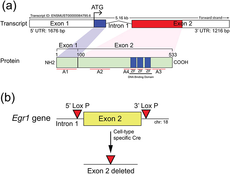
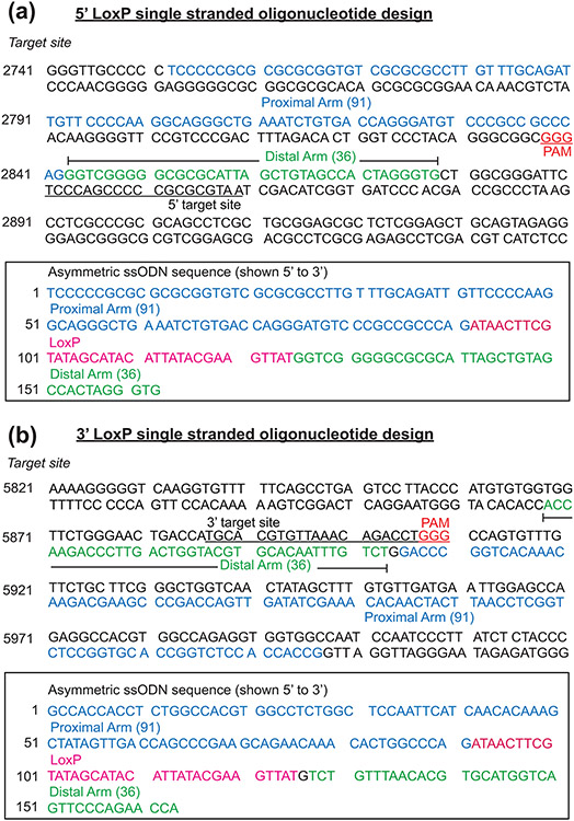
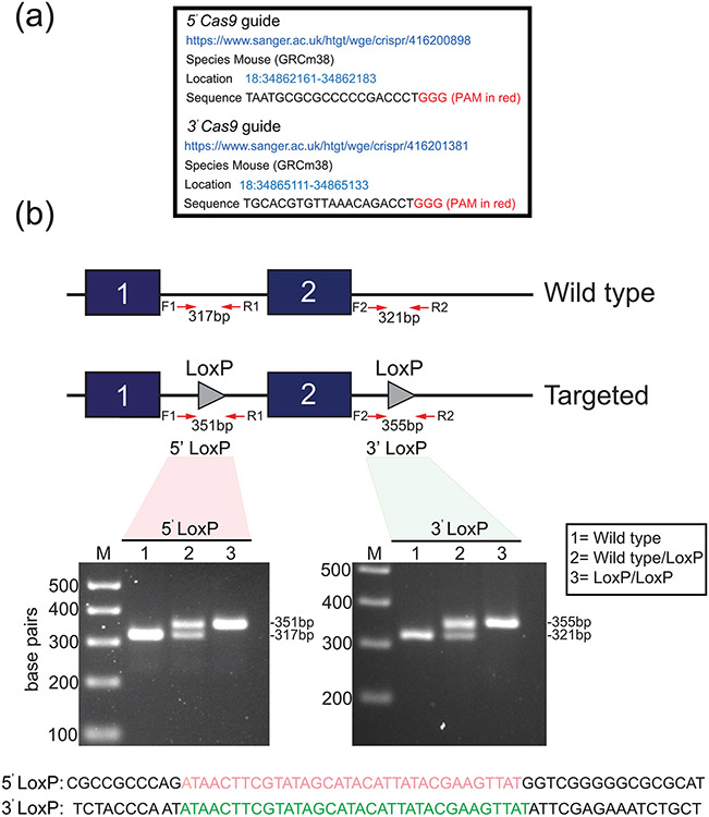
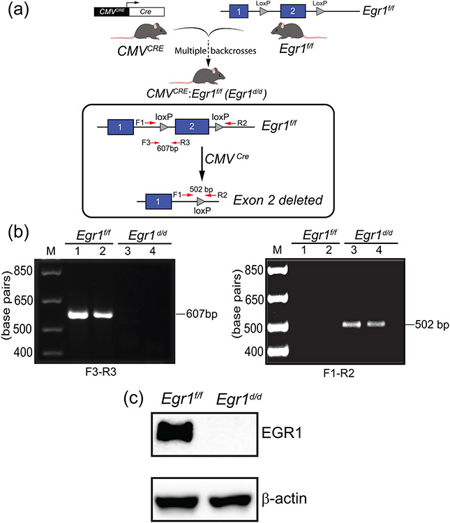
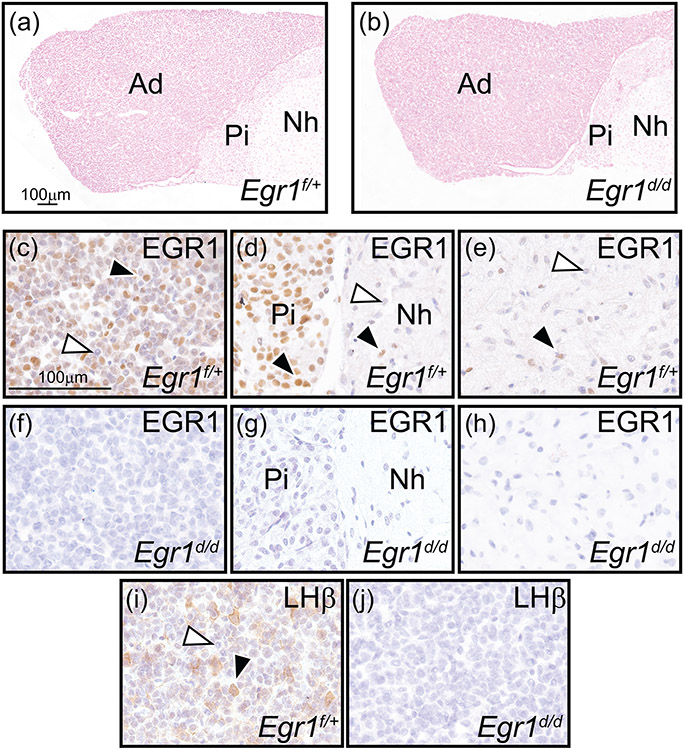
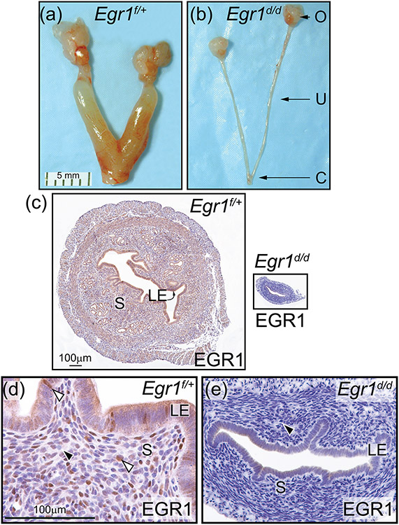
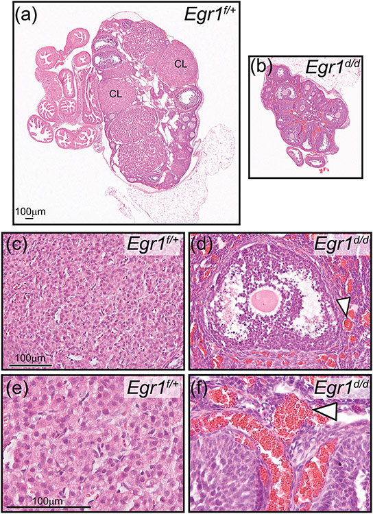
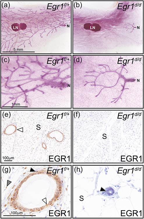
Early growth response 1 (EGR1) mediates transcriptional programs that are indispensable for cell division, differentiation, and apoptosis in numerous physiologies and pathophysiologies. Whole-body EGR1 knockouts in mice (Egr1KO ) have advanced our understanding of EGR1 function in an in vivo context. To extend the utility of the mouse to investigate EGR1 responses in a tissue- and/or cell-type-specific manner, we generated a mouse model in which exon 2 of the mouse Egr1 gene is floxed by CRISPR/Cas9 engineering. The floxed Egr1 alleles (Egr1f/f ) are designed to enable spatiotemporal control of Cre-mediated EGR1 ablation in the mouse. To confirm that the Egr1f/f alleles can be abrogated using a Cre driver, we crossed the Egr1f/f mouse with a global Cre driver to generate the Egr1 conditional knockout (Egr1d/d ) mouse in which EGR1 expression is ablated in all tissues. Genetic and protein analysis confirmed the absence of exon 2 and loss of EGR1 expression in the Egr1d/d mouse, respectively. Moreover, the Egr1d/d female exhibits overt reproductive phenotypes previously reported for the Egr1KO mouse. Therefore, studies described in this short technical report underscore the potential utility of the murine Egr1 floxed allele to further resolve EGR1 function at a tissue- and/or cell-type-specific level.

Keywords: CRISPR/Cas9; early growth response-1; floxed; mouse; ovary; pituitary; uterus.


Figures

FIGURE 1
FIGURE 1
General targeting strategy to flox exon 2 of the murine Egr1 gene. (a) The schematic displays the overall structural organization of the mouse Egr1 gene and EGR1 protein (http://useast.ensembl.org and https://www.uniprot.org/uniprotkb/Q544D6/entry). Located on mouse chromosome 18, the Egr1 gene is comprised of two exons, in which exon 1 encodes ~30% of the protein that includes the initiating ATG and the activation 1 (A1) domain. Exon 2 encodes the remainder of the protein, which contains activation domains 2-4 (A2-4) as well as the DNA binding domain with its three zinc finger (ZF) motifs. (b) For effective ablation of EGR1 protein expression by cre-mediated excision in the mouse, exon 2 was chosen to be floxed by 5’ and 3’ LoxP sites using a CRISPR/Cas9 mediated targeting strategy; note: exon 2 of the Egr1 gene in the Egr1KO mouse was ablated by the Milbrandt group (Lee et al., 1996; Lee et al., 1995).
FIGURE 2
FIGURE 2
The design and position of the 5’ and 3’ ssODNs that were used to create the mouse Egr1 floxed allele. (a) Shown is the sequence of the 5’ target site in intron 1 of the mouse Egr1 allele. Sequences in blue (91 base pairs (bp)) and green (36 bp) represent the asymmetric homology arms that are proximal and distal respectively relative to the location of the 3’ protospacer adjacent motif (PAM (red)); the ssODN sequence is complementary to the non-targeted strand. The sequence of the 5’ target site is underlined whereas the 3’ PAM (GGG) site is highlighted in red. The box contains the sequence (in the 5’ to 3’ direction) of the ssODNs that were used for CRISPR/Cas9 mediated homology directed repair. The LoxP sequence is highlighted in red, the underlined sequence shows the target sequence disrupted by the LoxP sequence in the ssODN donor. The proximal and distal homology arm sequences are highlighted in blue and green respectively. (b) The sequence of the 3’ LoxP target site located 3’ to the mouse Egr1 gene. The sequences of the proximal and distal asymmetric homology arms are displayed blue and green respectively. The location and sequence of the 3’ target site is underlined with the 3’ PAM shown in red (GGG). The box encloses the sequence of the ssODN donor that was used to insert the LoxP site in the 3’ location relative to the mouse Egr1 gene. The underlined target sequence is disrupted by the LoxP sequence (highlighted in red), which is flanked by the proximal and distal homology arms that are highlighted in blue and green respectively.
FIGURE 3
FIGURE 3
Generation of the murine Egr1 floxed allele. (a) The location information of the 5’ and 3’ Cas9 guide sequences (sense strand) that were used to generate the murine Egr1 floxed allele by CRISPR/Cas9 genome editing is shown. Note: in vitro transcribed sgRNAs were microinjected into C57BL/6NJ zygotes. (b) Schematic of the CRISPR/Cas9-mediated targeting strategy to insert the LoxP sites (grey triangles) into intron 1 of and 3’ from the Egr1 gene to flox exon 2. The location of the forward and reverse PCR primers to amplify the 5’ and 3’ LoxP sites is indicated. Using forward and reverse PCR primers (F1 and R1 primers to amplify the 5’ LoxP site (351 bp); F2 and R2 primers to amplify the 3’ LoxP site (355 bp)), the gel shows a typical genotype result for wild type (lane 1 (no LoxP sites (317/321 bp)), wild type/LoxP (lane 2 (heterozygote (Egr1f/+ (317/321 and 351/355 bp)), and LoxP/LoxP (lane 3 (homozygous (Egr1f/f) for the 5’ and 3’ LoxP insertion (351/355 bp). The PCR genotyping result shown was performed on tail biopsy genomic DNA from F2 generation mice. The sequences for the 5’ (red) and 3’ (green) LoxP sites that were carried by these mice are shown.
FIGURE 4
FIGURE 4
Generation of the Egr1d/d bigenic mouse. (a) The schematic summarizes the breeding scheme to generate the Egr1d/d bigenic mouse. The CMVcre transgenic mouse was crossed with Egr1f/f mice to generate the CMVcre: Egr1f/+ heterozygote, which is then intercrossed to generate CMVcre: Egr1f/f bigenic mouse (abbreviated Egr1d/d). The location of the F1, R2, F3, and R3 PCR primers to detect loss of the Egr1 exon 2 in the Egr1d/d bigenic mouse is shown. (b) Using the F1 and F3 forward and R2 and R3 reverse primers, the PCR genotyping result confirms the absence of exon 2 in the Egr1d/d bigenic mouse. (c) Western analysis using brain tissue protein isolates confirms that EGR1 protein is not produced in the Egr1d/d mouse. Lanes 1 and 2 represent protein isolated from Egr1f/f and Egr1d/d brain tissue respectively. Note the absence of EGR1 protein in the Egr1d/d lane. The experiment was performed in triplicate and β-actin served as a loading control.
FIGURE 5
FIGURE 5
Absence of EGR1 and LH-β subunit expression in the female Egr1d/d pituitary gland. (a) and (b) show representative nuclear-fast red stained sections of Egr1f/+ and Egr1d/d pituitary gland tissue respectively; representative of four adult (nine weeks old) mice per genotype. Pituitary gland size and histomorphology were equivalent for both genotypes (adenohypophysis (or the anterior pituitary gland or pars distalis), pars intermedia (or intermediate gland), and neurohypophysis (or posterior pituitary gland or pars nervosa) are indicated by Ad, Pi, and Nh respectively). Scale bar in (a) also applies to (b). (c-e) Immunohistochemical analysis of EGR1 expression in the Ad, Pi, and Nh of the Egr1f/+ pituitary gland respectively. Note in (c) the presence of numerous EGR1 positive cells in the Ad (black arrowhead); a subgroup of Ad cells is EGR1 negative (white arrowhead). (d) The majority of cells in the Pi region is immunopositive for EGR1 (black arrowhead), with few cells EGR1 positive in the Nh region ((d) and (e) (black arrowhead)); EGR1 negative cells in the Nh are indicated by a white arrowhead ((d) and (e)). (f-h) Expression of EGR1 is absent in all regions of the Egr1d/d pituitary gland. (i) Immunohistochemical detection of the LH β subunit in the Ad region of the Egr1f/+ pituitary gland. Note a subset of Ad cells is positive for LH β expression (black arrowhead); the white arrowhead indicates an Ad cell that is LH β negative. (j) Expression of the LH β subunit is absent in the Ad region of the Egr1d/d pituitary gland. Scale bar in (c) applies to (d-j).
Figure 6
Figure 6
Abrogation of EGR1 results in a hypoplastic uterus in the Egr1d/d mouse. (a) and (b) show the gross morphology of the Egr1f/+ and Egr1d/d female reproductive tract respectively; representative of five adult mice per genotype. Compared with the Egr1f/+ uterus (a), note the thin Egr1d/d uterine horn (b); ovary, uterus, and cervix are denoted by O, U, and C respectively. Scale bar in (a) applies to (b). (c) Transverse section of the mid-region of the Egr1f/+ and Egr1d/d uterine horn (left and right panels respectively) is shown; luminal epithelium and stroma are indicated by LE and S respectively. Both tissue sections were immunohistochemically stained for EGR1 expression. Note EGR1 expression in S and LE compartments of the Egr1f/+ uterus whereas EGR1 expression is not detected in the Egr1d/d uterus. Scale bar in the left panel applies to right panel. (d) and (e) are higher magnification images of the micrographs shown in the left and right panels in (c) respectively. (d) Expression of EGR1 is detected in subsets of cells in the S and LE compartment of the Egr1f/+ uterus (white arrowhead). (e) Expression of EGR1 is absent in the Egr1d/d uterus (black arrowhead). Scale bar in (d) applies to (e).
Figure 7
Figure 7
Absence of corpora lutea in the atrophied Egr1d/d ovary. (a) Hematoxylin and eosin stained section of the Egr1f/+ ovary; a corpus luteum is indicated by CL. (b) Similarly stained Egr1d/d ovarian tissue section; note the absence of CLs. Also note the diminished size of the Egr1d/d ovary compared with the Egr1f/+ ovary shown in (a). Scale bar in (a) applies to (b). The histological data are representative of five adult mice per genotype. (c) Field of view shows luteal cells within the CL of an Egr1f/+ ovary. (d) Image shows an antral follicle within the Egr1d/d ovary; note the juxtaposed hemorrhagic area indicated by the white arrowhead. Scale bar in (c) applies to (d). (e) and (f) represent higher magnification images of regions in (c) and (d) respectively. Again, note the hemorrhagic areas in the Egr1d/d ovary (white arrowhead). Scale bar in (e) applies to (f).
Figure 8
Figure 8
A prepubescent mammary gland phenotype in the adult Egr1d/d mouse. (a) Whole mount of an inguinal mammary gland from an adult Egr1f/+ mouse, note the extensive epithelial ductal branching from the nipple (N) to the periphery of the fat pad. The lymph node is indicated by LN, which is used as a structural reference point in the inguinal gland. (b) Absence of epithelial ductal elongation and dichotomous branching in the mammary of the adult Egr1d/d mouse. Note that the majority of the Egr1d/d mammary gland fat pad is devoid of the epithelial compartment. Mammary gland results are representative of five adult mice per genotype. Scale bar in (a) applies to (b). (c) and (d) represent higher magnification images shown in (a) and (b) respectively; scale bar in (c) applies to (d). (e) Immunohistochemical detection of EGR1 expression in a section derived from Egr1f/+ mammary gland tissue. Immunopositivity in the mammary epithelium is indicated by white arrowhead; mammary stroma is denoted as S. (f) Immunopositivity for EGR1 is absent in the mammary gland of the Egr1d/d mouse. As indicated in panel (b), the majority of the mammary gland is devoid of the epithelial compartment in the Egr1d/d mouse; scale bar in (e) applies to (f). (g) Higher magnification image clearly showing EGR1 expression in the basal epithelium (black arrowhead), a subset of luminal epithelial cells (white arrowhead), and stromal cells (gray arrowhead). (h) Immunopositivity for EGR1 is not detected in cells of vestigial epithelial ducts that are localized to the nipple region of the adult Egr1d/d mammary gland; the stromal compartment is also negative for EGR1 immunopositivity. Scale bar in (g) applies to (h).

References

    1. Adamson ED, & Mercola D (2002). Egr1 transcription factor: multiple roles in prostate tumor cell growth and survival. Tumour Biol, 23(2), 93–102. doi:10.1159/000059711 - DOI - PubMed
    1. Bhattacharyya S, Wu M, Fang F, Tourtellotte W, Feghali-Bostwick C, & Varga J (2011). Early growth response transcription factors: key mediators of fibrosis and novel targets for anti-fibrotic therapy. Matrix Biol, 30(4), 235–242. doi:10.1016/j.matbio.2011.03.005 - DOI - PMC - PubMed
    1. Blaschke F, Bruemmer D, & Law RE (2004). Egr-1 is a major vascular pathogenic transcription factor in atherosclerosis and restenosis. Rev Endocr Metab Disord, 5(3), 249–254. doi:10.1023/B:REMD.0000032413.88756.ee - DOI - PubMed
    1. Bocchinfuso WP, & Korach KS (1997). Mammary gland development and tumorigenesis in estrogen receptor knockout mice. J Mammary Gland Biol Neoplasia, 2(4), 323–334. doi:10.1023/a:1026339111278 - DOI - PubMed
    1. Davis BJ, Dixon D, & Herbert RA (1999). Ovary, oviduct, uterus, cervix, and vagina. In Maronpot RR, Boorman GA, & Gaul BW (Eds.), Pathology of the Mouse: Reference and atlas (pp. 409–444). Vienna, IL: Cache River Press.

Publication types

Substances

LinkOut - more resources