. 2023 Mar 23;24(1):90.
doi: 10.1186/s12931-023-02395-5.

Group 3 innate lymphoid cells secret neutrophil chemoattractants and are insensitive to glucocorticoid via aberrant GR phosphorylation

Affiliations

Group 3 innate lymphoid cells secret neutrophil chemoattractants and are insensitive to glucocorticoid via aberrant GR phosphorylation

Li Xiu He et al. Respir Res. .

Abstract

Background: Patients with neutrophil-mediated asthma have poor response to glucocorticoids. The roles and mechanisms of group 3 innate lymphoid cells (ILC3s) in inducing neutrophilic airway inflammation and glucocorticoid resistance in asthma have not been fully clarified.

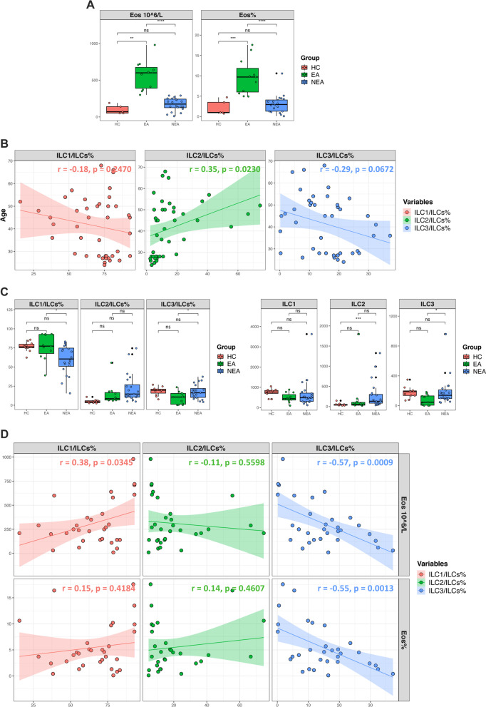
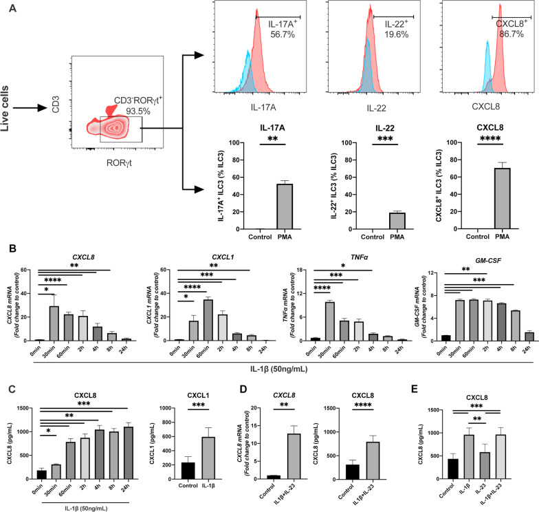
Methods: ILC3s in peripheral blood were measured by flow cytometry in patients with eosinophilic asthma (EA) and non-eosinophilic asthma (NEA). ILC3s were sorted and cultured in vitro for RNA sequencing. Cytokines production and signaling pathways in ILC3s after IL-1β stimulation and dexamethasone treatment were determined by real-time PCR, flow cytometry, ELISA and western blot.

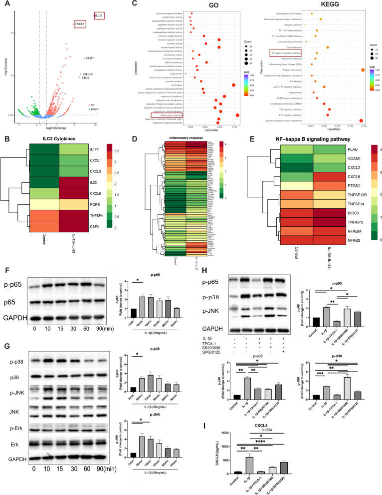
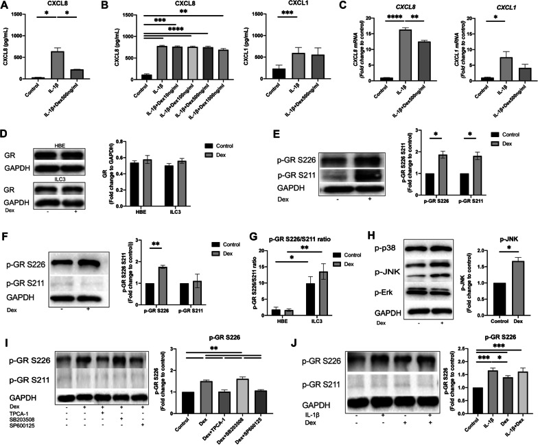
Results: The percentage and numbers of ILC3s in peripheral blood was higher in patients with NEA compared with EA, and negatively correlated with blood eosinophils. IL-1β stimulation significantly enhanced CXCL8 and CXCL1 production in ILC3s via activation of p65 NF-κB and p38/JNK MAPK signaling pathways. The expression of neutrophil chemoattractants from ILC3s was insensitive to dexamethasone treatment. Dexamethasone significantly increased phosphorylation of glucocorticoid receptor (GR) at Ser226 but only with a weak induction at Ser211 residues in ILC3s. Compared to human bronchial epithelial cell line (16HBE cells), the ratio of p-GR S226 to p-GR S211 (p-GR S226/S211) was significantly higher in ILC3s at baseline and after dexamethasone treatment. In addition, IL-1β could induce Ser226 phosphorylation and had a crosstalk effect to dexamethasone via NF-κB pathway.

Conclusions: ILC3s were elevated in patients with NEA, and associated with neutrophil inflammation by release of neutrophil chemoattractants and were glucocorticoid (GC) resistant. This paper provides a novel cellular and molecular mechanisms of neutrophil inflammation and GC-resistance in asthma. Trial registration The study has been prospectively registered in the World Health Organization International Clinical Trials Registry Platform (ChiCTR1900027125).

Keywords: Asthma; Glucocorticoid resistant; Group 3 innate lymphoid cells; Neutrophil chemoattractant; Neutrophilic inflammation.


Conflict of interest statement

The authors declare that they have no competing interests.

Figures

Fig. 1
Fig. 1
ILC3s elevated in patients with NEA compared to EA. A Blood eosinophils count and percentage among healthy control (HC) and patients with NEA and EA. B Correlation between subsets of ILCs and age. C Percentage and numbers (in every 50 × 105 recorded lymphocyte) of ILC1, ILC2 and ILC3 in HC, NEA and EA patients. D Correlation between ILC subsets and blood eosinophils count and percentage. ****p < 0.0001, ***p < 0.001, **p < 0.01, *p < 0.05, ns p > 0.05
Fig. 2
Fig. 2
Flow cytometry gating strategy for ILC3 identification and cytokines production profiles of ILC3s. A ILC3s were identified by transcription factor RORγt and the secretion of IL-17, IL-22 and CXCL8. Cytokines-producing cells of ILC3s were detected by Flow cytometry. Blue, ILC3s without stimulation. Red, ILC3s stimulated with PMA + ionomycin. Numbers represent percentages of cells producing IL-17A or IL-22 or CXCL8 in ILC3s. B Time course of CXCL8, CXCL1, TNF-α and GM-CSF mRNA expression in ILC3s after IL-1β (50 ng/mL) stimulation. C Protein level of CXCL8 and CXCL1 in culture supernatant of ILC3s with or without IL-1β (50 ng/mL) stimulation after 24 h. D, E Effect of IL-1β and IL-23 on CXCL8 expression in ILC3s
Fig. 3
Fig. 3
RNA sequencing of in vitro cultured ILC3s with or without IL-1β + IL-23 stimulation. A Volcano plots of differential expression genes (DEGs) in ILC3s stimulated by IL-1β + IL-23. The top five differential expressed genes were IL-22, CCR7, IGFBP4, CD22 and CXCL8. B Heatmap analysis of effector cytokines in ILC3s. In the heatmap, the redder the color is, the higher the expression is, and the greener the expression is, the lower the expression is. C Function terms of DEGs by Gene Ontology (GO) enrichment analysis and KEGG pathway analysis of DEGs in ILC3s upon IL-1β + IL-23(50 ng/mL) stimulation. D Heatmap analysis of DEGs in inflammation response. E The clustering heat map of DEGs involved in NF-κB signal pathway. F, G Western blot analysis by antiphospho-p65 Ab, antiphospho-p38 Ab, antiphospho-JNK Ab and antiphospho-Erk Ab. Cell extracts were prepared after stimulation with IL-1β (50 ng/mL) for indicated times. H Phosphorylation and inhibition of p65, p38 and JNK by specific inhibitors in ILC3s. Western blot analysis of p-p65, p-p38 and p-JNK were performed with cell extracts from ILC3s, preincubated for 1 h with p65 NF-κB inhibitor TPCA-1, p38-MAPK inhibitor SB203580 or JNK-MAPK inhibitor SP600125 and then stimulated with IL-1β (50 ng/mL) for 10 min. I Expression of CXCL8 protein in culture supernatant of ILC3 cells after IL-1β stimulation with or without corresponding inhibitors. ILC3s were preincubated for 1 h with indicated inhibitors and then stimulated with IL-1β (50 ng/mL) for 24 h
Fig. 4
Fig. 4
Effect of dexamethasone (Dex) on IL-1β-induced CXCL8 and CXCL1 production and phosphorylation of glucocorticoids receptor (GR) at S226 and S211 in ILC3s. A Expression of CXCL8 in 16HBEs with and without Dex treatment. B, C, Expression of CXCL8 and CXCL1 in ILC3s with Dex treatment by indicated concentrations. Cells were pre-treated with Dex (10 to 1000 ng/mL) for 1 h before 24 h stimulation with IL-1β (50 ng/mL), supernatants were collected and assayed for CXCL8 and CXCL1 release by ELISA, and ILC3 cells were extracted for qPCR analysis. D Western blotting analysis of GR in 16HBEs and ILC3s after stimulation with Dex (1000 ng/mL) for 15 min. E, F Western blotting analysis of phosphorylation of GR S226 and S211 in 16HBEs (E) and ILC3s (F) after stimulation with Dex (1000 ng/mL) for 15 min. G Ratio of p-GR S226/S211 in 16HBEs and ILC3s at baseline and after dexamethasone treatment. H Western blotting analysis of phosphorylation of MAPK in ILC3s with Dex stimulation (1000 ng/mL) for 15 min. I Effects of Dex and corresponding inhibitors on phosphorylated GR at S226 and S211 in ILC3s. J GR phosphorylation at S226 and S211 in ILC3s with indicated stimulation. Cells were stimulated with IL-1β (50 ng/mL) or dexamethasone (1000 ng/mL) alone or combination for 15 min. ILC3s were preincubated for 1 h with corresponding inhibitors then stimulated with dexamethasone (1000 ng/mL) for 15 min, then lysed and assessed for phosphorylation of GR S226 or GR S211 by Western blotting

References

    1. Spits H, Artis D, Colonna M, et al. Innate lymphoid cells–a proposal for uniform nomenclature. Nat Rev Immunol. 2013;13(2):145–149. doi: 10.1038/nri3365. - DOI - PubMed
    1. Vivier E, Artis D, Colonna M, et al. Innate Lymphoid Cells: 10 Years On. Cell. 2018;174(5):1054–1066. doi: 10.1016/j.cell.2018.07.017. - DOI - PubMed
    1. Kim CH, Hashimoto-Hill S, Kim M. Migration and tissue tropism of innate lymphoid cells. Trends Immunol. 2016;37(1):68–79. doi: 10.1016/j.it.2015.11.003. - DOI - PMC - PubMed
    1. Buonocore S, Ahern PP, Uhlig HH, et al. Innate lymphoid cells drive interleukin-23-dependent innate intestinal pathology. Nature. 2010;464(7293):1371–1375. doi: 10.1038/nature08949. - DOI - PMC - PubMed
    1. Ardain A, Porterfield JZ, Kløverpris HN, et al. Type 3 ILCs in lung disease. Front Immunol. 2019;10:92. doi: 10.3389/fimmu.2019.00092. - DOI - PMC - PubMed

Supplementary concepts