

Trichofolliculoma: A Case Series
- PMID: 36950043
- PMCID: PMC10027329
Trichofolliculoma: A Case Series
Abstract
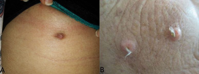
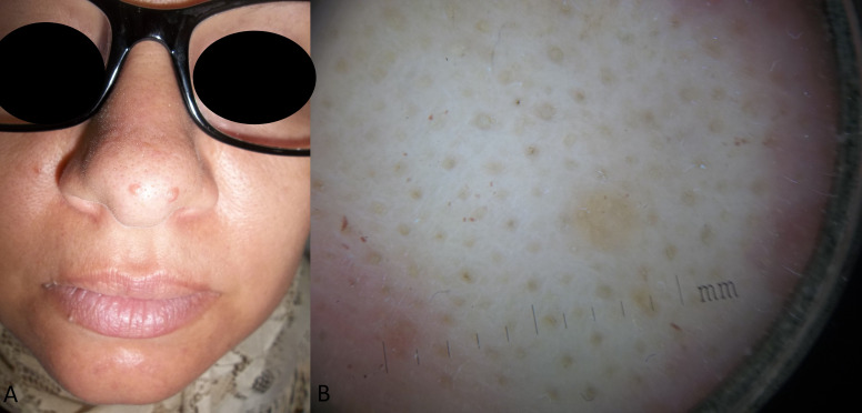
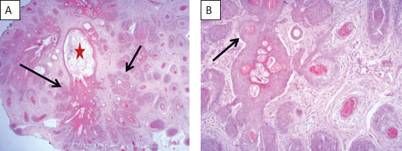
Trichofolliculoma (TF) is a rare benign adnexal follicular tumor, described as hamartoma with follicular differentiation according to some authors. It typically appears during adulthood on the face or scalp as an isolated nodule with protrusion of central tufted hairs. We present a retrospective series of eleven patients with histologically confirmed TF to evaluate epidemiological, clinical, and histopathologic characteristics. The mean age at excision was 46 years with extremes ranging from 20 to 75 years. The sex ratio M/F was 0.37. Clinical presentation was a papule or nodule with an average diameter of 6,7mm (2-15 mm), firm with central pit in 54 percent and visible emerging vellus hairs in 18 percent of cases only. The localization was on the face in seven cases (63.6%) and only four cases were located outside the face (scalp [n=2], sub mammary fold [n=1] and shoulder [n=1]). Histologically, a cystically dilated hair follicle containing keratinous material with several mature and immature branched follicular structures is described in all cases. According to our series, TF occurs predominantly in women without age predilection, in the face. Central tufted hairs are only found in a minority of cases corresponding histologically to many secondary vellus hair follicles. In fact, histopathological examination is the gold standard for the diagnosis because clinical diagnosis could be challenging. Histology and dermoscopy may vary according to the age of the lesion. To date, only few case series have been published.
Keywords: Trichofolliculoma; adnexal tumor; dermatoscopy.
Copyright © 2023. Matrix Medical Communications. All rights reserved.
Conflict of interest statement
FUNDING: No funding was provided for this article DISCLOSURES: The authors report no conflicts on interest relevant to the content of this article.
Figures
References
-
- El-Komy MH, Abdelkader HA. Congenital trichofolliculoma: a very rare presentation. Dermatol Online J. 2020;26(7):13030/qt4g69d9zc. Jul 15. - PubMed
-
- Lee HY, Kim EK, Choi HS Trichofolliculoma in the Auricle. Ear Nose Throat J. 2021. p. 145561321995599. Feb. 15. - PubMed
-
- Choi CM, Lew BL, Sim WY. Multiple trichofolliculomas on unusual sites: a case report and review of the literature. Int J Dermatol. 2013;52(1):87–89. Jan. - PubMed
-
- Al-Ghadeer H, Edward DP. Congenital Sebaceous Trichofolliculoma of the Upper Eyelid. Ophthalmic Plast Reconstr Surg. 2017;33(3):S60, S61. May/Jun; Suppl 1. - PubMed
Publication types
LinkOut - more resources
Full Text Sources
Miscellaneous