. 2023 Mar 20;9(1):E2-E10.
doi: 10.1055/a-1999-7818. eCollection 2023 Jan.

Quantification of Portal Vein Vascularization Using an Automated Post-Processing Video Analysis Tool

Affiliations

Quantification of Portal Vein Vascularization Using an Automated Post-Processing Video Analysis Tool

Valentin Blank et al. Ultrasound Int Open. .

Abstract

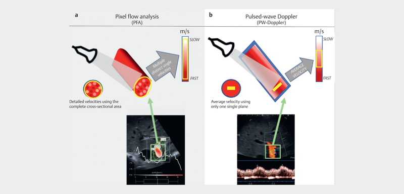
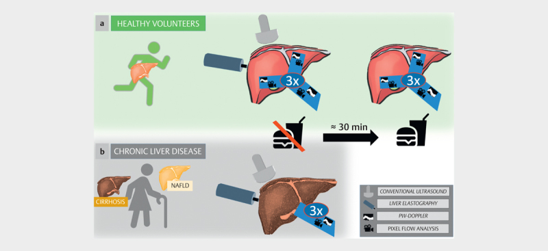
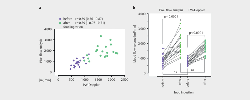
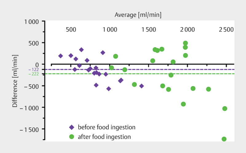
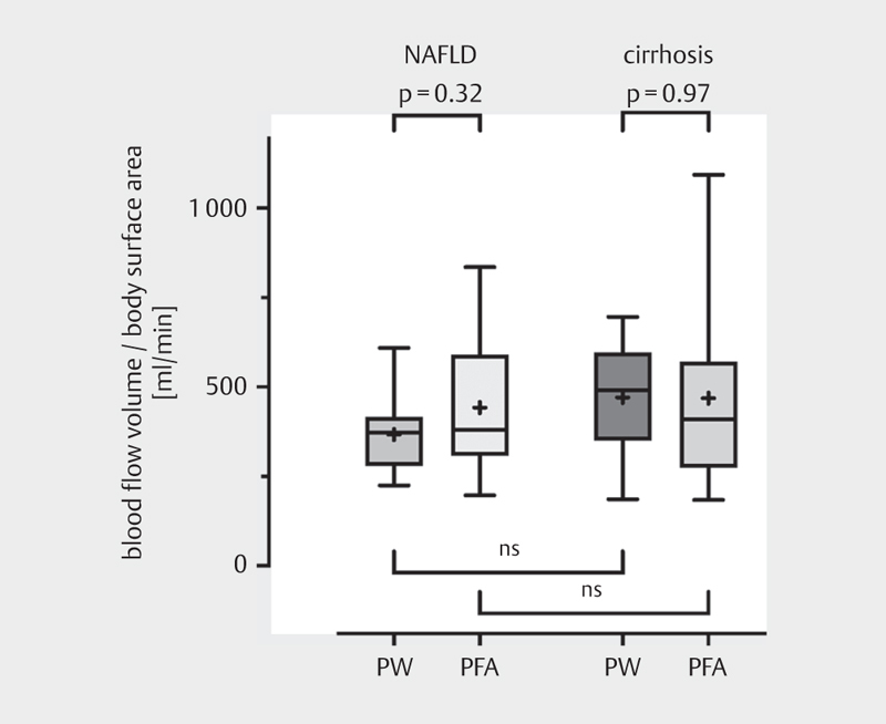
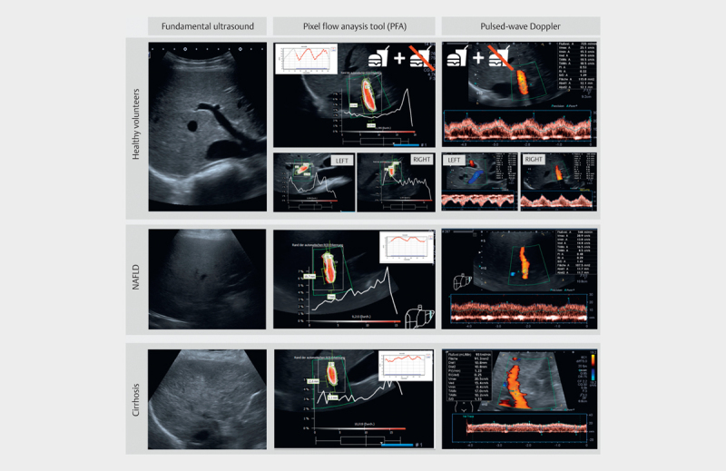
Purpose Blood flow dynamics represent a diagnostic criterion for many diseases. However, no established reference standard is available. In clinical practice, ultrasound pulsed-wave Doppler (PW-Doppler) is frequently used to assess visceral blood flow, despite its well-known limitations. A quantitative analysis of conventional color Doppler patterns can be performed using an innovative ultrasound-based algorithm (pixel flow analysis, PFA). This tool already shows promising results in obstetrics, but the technique has not yet been evaluated for portal venous blood flow assessment. Methods This prospective exploratory research study evaluated the applicability of PFA in the portal venous system. Measurements of portal venous flow using PFA and PW-Doppler were compared in healthy volunteers (n=20) and in patients with hepatic steatosis (n=10) and liver cirrhosis (n=10). Results In healthy volunteers (60% female, mean age 23 years, BMI 21.5 kg/m 2 [20.4-23.8]), PFA and PW-Doppler showed a strong positive correlation in fasting conditions (r=0.69; 95% CI 0.36-0.87), recording a median blood flow of 834 ml/min (624-1066) and 718 ml/min (620-811), respectively. PFA was also applicable in patients with chronic liver diseases (55% female, age 65 years (55-72); BMI 27.8 kg/m 2 (25.4-30.8)), but the correlation between PFA and PW-Doppler was poor (r=- 0.09) in the subgroup with steatosis. A better correlation (r=0.61) was observed in patients with liver cirrhosis. Conclusion PFA and PW-Doppler assessment of portal venous vascularization showed high agreement in healthy volunteers and patients with liver cirrhosis. Therefore, PFA represents a possible alternative to conventional PW-Doppler sonography for visceral blood flow diagnostics and merits further evaluation.

Keywords: PixelFlux; blood flow velocity; pixel flow analysis; portal venous blood flow; pulsed-wave Doppler.


Conflict of interest statement

Conflict of Interest The authors declare that they have no conflict of interest.

Figures

Fig. 1
Fig. 1
Principles of blood flow measurements. a ) Pixel flow analysis (PFA) is a post-processing tool that evaluates the blood flow velocities of each individual pixel in color Doppler visualization, with respect to the total cross-sectional area of the vessel using an ellipsoid plane. b ) Pulsed-wave Doppler (PW-Doppler) uses the mean vessel velocity and only considers a single plane. The blood flow is calculated using the manually measured diameter of the vessel.
Fig. 2
Fig. 2
Protocol for blood flow analyses and overview of all study investigations. In the cohort of healthy volunteers ( a ), the blood flow measurements were performed in the main portal trunk and both subsequent branches before and after food ingestion using both pixel flow analysis (PFA) and pulsed-wave Doppler (PW-Doppler). In patients with chronic liver disease ( b ), the blood flow analysis was restricted to the portal vein trunk in fasting conditions. All study participants underwent liver elastography and conventional ultrasound for study inclusion.
Fig. 3
Fig. 3
Comparison of blood flow analysis in the portal vein in healthy volunteers using both pixel flow analysis (PFA) and pulsed-wave Doppler (PW-Doppler). Panel a shows the Pearson correlation of both applied methods before and after food ingestion. Panel b illustrates the blood flow alterations after food ingestion for both measurements with PFA and PW-Doppler in the portal vein. Entries are in ml/min.
Fig. 4
Fig. 4
Bland–Altman plots for inter-method agreement of pixel flow analysis (PFA) and pulsed-wave Doppler (PW-Doppler) in healthy volunteers before (purple) and after (green) food ingestion.
Fig. 5
Fig. 5
Blood flow in the portal vein trunk in patients with chronic liver diseases in fasting conditions. The blood flow is adapted to the total surface area of the patients. PW=pulsed-wave Doppler; PFA=pixel flow analysis. NAFLD non-alcoholic fatty liver disease.
Fig. 6
Fig. 6
Examples of performed blood flow measurements for all cohorts showing conventional ultrasound (left), pixel flow analysis (PFA) (middle), and pulsed-wave Doppler (PW-Doppler) (right). In the cohort of healthy volunteers, the blood flow measurements were performed in the main portal trunk and both subsequent branches before and after food ingestion. In patients with chronic liver disease (NAFLD and cirrhosis), the blood flow analysis was restricted to the portal vein trunk in fasting conditions.

Similar articles

References

    1. et al.[S2k Guideline Gastrointestinal Bleeding – Guideline of the German Society of Gastroenterology DGVS] Z Gastroenterol. 2017;55:883–963. doi: 10.1055/S-0043-116856. - DOI - PubMed
    1. de RF. Expanding consensus in portal hypertension: Report of the Baveno VI Consensus Workshop: Stratifying risk and individualizing care for portal hypertension. J Hepatol. 2015;63:543–545. doi: 10.1016/J.JHEP.2015.05.022. - DOI - PubMed
    1. Farid WR R, De Jonge J, Slieker J C et al.The Importance of Portal Venous Blood Flow in Ischemic-Type Biliary Lesions after Liver Transplantation. Am J Transplant. 2011;11:857–862. doi: 10.1111/j.1600-6143.2011.03438.x. - DOI - PubMed
    1. Bosch J, Abraldes J G, Berzigotti A et al.The clinical use of HVPG measurements in chronic liver disease. Nat Rev Gastroenterol Hepatol. 2009;6:573–582. doi: 10.1038/NRGASTRO.2009.149. - DOI - PubMed
    1. Zoli M, Magalotti D, Giampaolo B et al.Functional hepatic flow and Doppler-assessed total hepatic flow in control subjects and in patients with cirrhosis. J Hepatol. 1995;23:129–134. doi: 10.1016/0168-8278(95)80326-2. - DOI - PubMed