

Optimizing MRI-based brain tumor classification and detection using AI: A comparative analysis of neural networks, transfer learning, data augmentation, and the cross-transformer network
- PMID: 36950474
- PMCID: PMC10027502
- DOI: 10.1016/j.ejro.2023.100484
Optimizing MRI-based brain tumor classification and detection using AI: A comparative analysis of neural networks, transfer learning, data augmentation, and the cross-transformer network
Abstract
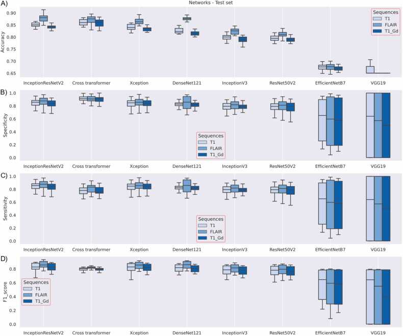
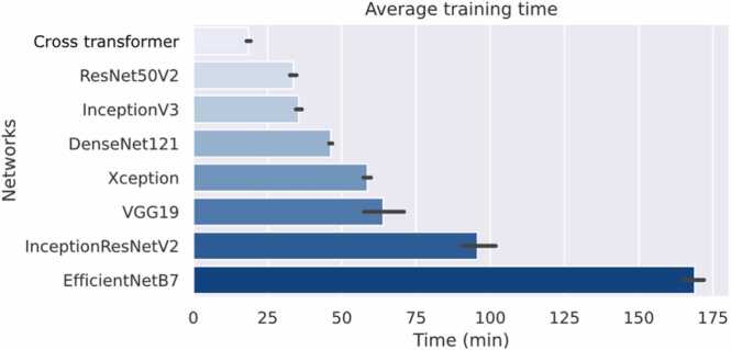
Early detection and diagnosis of brain tumors are crucial to taking adequate preventive measures, as with most cancers. On the other hand, artificial intelligence (AI) has grown exponentially, even in such complex environments as medicine. Here it's proposed a framework to explore state-of-the-art deep learning architectures for brain tumor classification and detection. An own development called Cross-Transformer is also included, which consists of three scalar products that combine self-care model keys, queries, and values. Initially, we focused on the classification of three types of tumors: glioma, meningioma, and pituitary. With the Figshare brain tumor dataset was trained the InceptionResNetV2, InceptionV3, DenseNet121, Xception, ResNet50V2, VGG19, and EfficientNetB7 networks. Over 97 % of classifications were accurate in this experiment, which provided a network's performance overview. Subsequently, we focused on tumor detection using the Brain MRI Images for Brain Tumor Detection and The Cancer Genome Atlas Low-Grade Glioma database. The development encompasses learning transfer, data augmentation, as well as image acquisition sequences; T1-weighted images (T1WI), T1-weighted post-gadolinium (T1-Gd), and Fluid-Attenuated Inversion Recovery (FLAIR). Based on the results, using learning transfer and data augmentation increased accuracy by up to 6 %, with a p-value below the significance level of 0.05. As well, the FLAIR sequence was the most efficient for detection. As an alternative, our proposed model proved to be the most effective in terms of training time, using approximately half the time of the second fastest network.
Keywords: Artificial intelligence; Cancer detection; Machine learning; Magnetic resonance imaging; Transformers; Tumors.
© 2023 The Authors. Published by Elsevier Ltd.
Conflict of interest statement
The authors declare that they have no known competing financial interests or personal relationships that could have appeared to influence the work reported in this paper.
Figures


References
-
-
American Cancer Society, Cancer Facts & Figures 2020, 1–76, 2020.
-
-
-
Cancer, https://www.who.int/en/news-room/fact-sheets/detail/cancer (accedido 17 de noviembre de 2020).
-
-
- Mack T.M. Cancers in the Urban Environment. Elsevier; 2021. What a cancer is; pp. 5–8. - DOI
-
- Ray S.D., Yang N., Pandey S., Bello N.T., Gray Y.J.P. en Reference Module in Biomedical Sciences. Elsevier; 2019. Apoptosis. - DOI
-
- Foster J.R. Comprehensive Toxicology. Elsevier; 2018. Introduction to Neoplasia; pp. 1–10. - DOI
LinkOut - more resources
Full Text Sources
Research Materials
