

A pathomic approach for tumor-infiltrating lymphocytes classification on breast cancer digital pathology images
- PMID: 36950640
- PMCID: PMC10025040
- DOI: 10.1016/j.heliyon.2023.e14371
A pathomic approach for tumor-infiltrating lymphocytes classification on breast cancer digital pathology images
Abstract
Background and objectives: The detection of tumor-infiltrating lymphocytes (TILs) could aid in the development of objective measures of the infiltration grade and can support decision-making in breast cancer (BC). However, manual quantification of TILs in BC histopathological whole slide images (WSI) is currently based on a visual assessment, thus resulting not standardized, not reproducible, and time-consuming for pathologists. In this work, a novel pathomic approach, aimed to apply high-throughput image feature extraction techniques to analyze the microscopic patterns in WSI, is proposed. In fact, pathomic features provide additional information concerning the underlying biological processes compared to the WSI visual interpretation, thus providing more easily interpretable and explainable results than the most frequently investigated Deep Learning based methods in the literature.
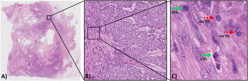
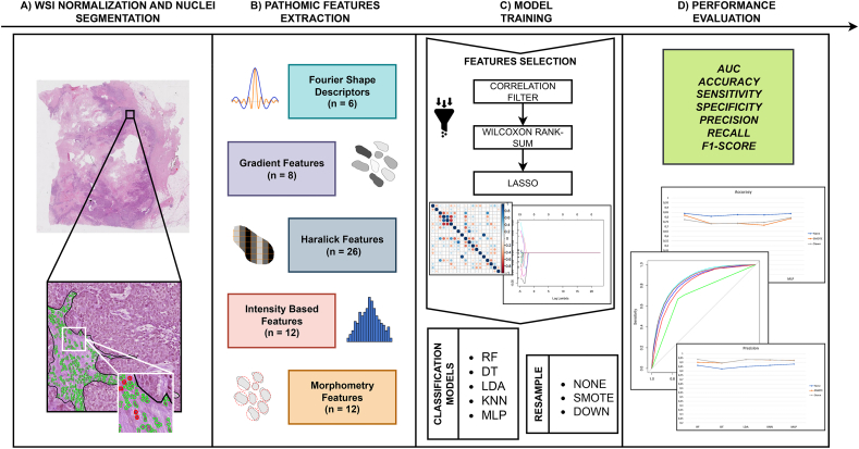
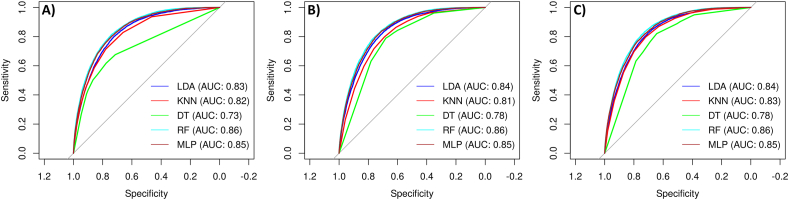
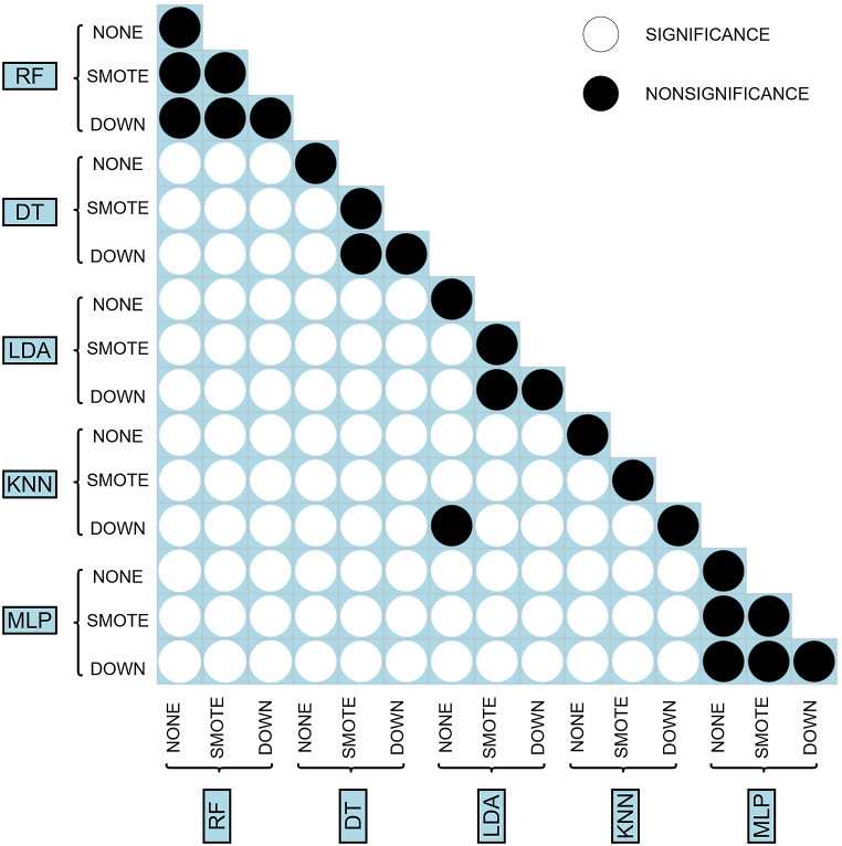
Methods: A dataset containing 1037 regions of interest with tissue compartments and TILs annotated on 195 TNBC and HER2+ BC hematoxylin and eosin (H&E)-stained WSI was used. After segmenting nuclei within tumor-associated stroma using a watershed-based approach, 71 pathomic features were extracted from each nucleus and reduced using a Spearman's correlation filter followed by a nonparametric Wilcoxon rank-sum test and least absolute shrinkage and selection operator. The relevant features were used to classify each candidate nucleus as either TILs or non-TILs using 5 multivariable machine learning classification models trained using 5-fold cross-validation (1) without resampling, (2) with the synthetic minority over-sampling technique and (3) with downsampling. The prediction performance of the models was assessed using ROC curves.
Results: 21 features were selected, with most of them related to the well-known TILs properties of having regular shape, clearer margins, high peak intensity, more homogeneous enhancement and different textural pattern than other cells. The best performance was obtained by Random-Forest with ROC AUC of 0.86, regardless of resampling technique.
Conclusions: The presented approach holds promise for the classification of TILs in BC H&E-stained WSI and could provide support to pathologists for a reliable, rapid and interpretable clinical assessment of TILs in BC.
Keywords: Breast cancer; Digital pathology; Machine learning; Pathomics; Tumor infiltrating lymphocytes.
© 2023 The Authors.
Conflict of interest statement
The authors declare no competing interests.
Figures

References
-
- Salemme V., Centonze G., Cavallo F., Defilippi P., Conti L. The crosstalk between tumor cells and the immune microenvironment in breast cancer: implications for immunotherapy. Front. Oncol. 2021:11. https://www.frontiersin.org/articles/10.3389/fonc.2021.610303 accessed. - DOI - PMC - PubMed
-
- El Bairi K., Haynes H.R., Blackley E., Fineberg S., Shear J., Turner S., de Freitas J.R., Sur D., Amendola L.C., Gharib M., Kallala A., Arun I., Azmoudeh-Ardalan F., Fujimoto L., Sua L.F., Liu S.-W., Lien H.-C., Kirtani P., Balancin M., El Attar H., Guleria P., Yang W., Shash E., Chen I.-C., Bautista V., Do Prado Moura J.F., Rapoport B.L., Castaneda C., Spengler E., Acosta-Haab G., Frahm I., Sanchez J., Castillo M., Bouchmaa N., Md Zin R.R., Shui R., Onyuma T., Yang W., Husain Z., Willard-Gallo K., Coosemans A., Perez E.A., Provenzano E., Ericsson P.G., Richardet E., Mehrotra R., Sarancone S., Ehinger A., Rimm D.L., Bartlett J.M.S., Viale G., Denkert C., Hida A.I., Sotiriou C., Loibl S., Hewitt S.M., Badve S., Symmans W.F., Kim R.S., Pruneri G., Goel S., Francis P.A., Inurrigarro G., Yamaguchi R., Garcia-Rivello H., Horlings H., Afqir S., Salgado R., Adams S., Kok M., Dieci M.V., Michiels S., Demaria S., Loi S. The tale of TILs in breast cancer: a report from the international immuno-oncology biomarker working group. Npj Breast Cancer. 2021;7:1–17. doi: 10.1038/s41523-021-00346-1. - DOI - PMC - PubMed
-
- Kurozumi S., Inoue K., Matsumoto H., Fujii T., Horiguchi J., Oyama T., Kurosumi M., Shirabe K. Prognostic utility of tumor-infiltrating lymphocytes in residual tumor after neoadjuvant chemotherapy with trastuzumab for HER2-positive breast cancer. Sci. Rep. 2019;9:1583. doi: 10.1038/s41598-018-38272-1. - DOI - PMC - PubMed
-
- Salgado R., Denkert C., Demaria S., Sirtaine N., Klauschen F., Pruneri G., Wienert S., Van den Eynden G., Baehner F.L., Penault-Llorca F., Perez E.A., Thompson E.A., Symmans W.F., Richardson A.L., Brock J., Criscitiello C., Bailey H., Ignatiadis M., Floris G., Sparano J., Kos Z., Nielsen T., Rimm D.L., Allison K.H., Reis-Filho J.S., Loibl S., Sotiriou C., Viale G., Badve S., Adams S., Willard-Gallo K., Loi S., International TILs Working Group The evaluation of tumor-infiltrating lymphocytes (TILs) in breast cancer: recommendations by an International TILs Working Group 2014. Ann. Oncol. 2014;26(2015):259–271. doi: 10.1093/annonc/mdu450. - DOI - PMC - PubMed
LinkOut - more resources
Full Text Sources
Research Materials
Miscellaneous
