. 2023 Apr 15;136(8):jcs261018.
doi: 10.1242/jcs.261018. Epub 2023 Apr 20.

Patch repair protects cells from the small pore-forming toxin aerolysin

Affiliations

Patch repair protects cells from the small pore-forming toxin aerolysin

Roshan Thapa et al. J Cell Sci. .

Abstract

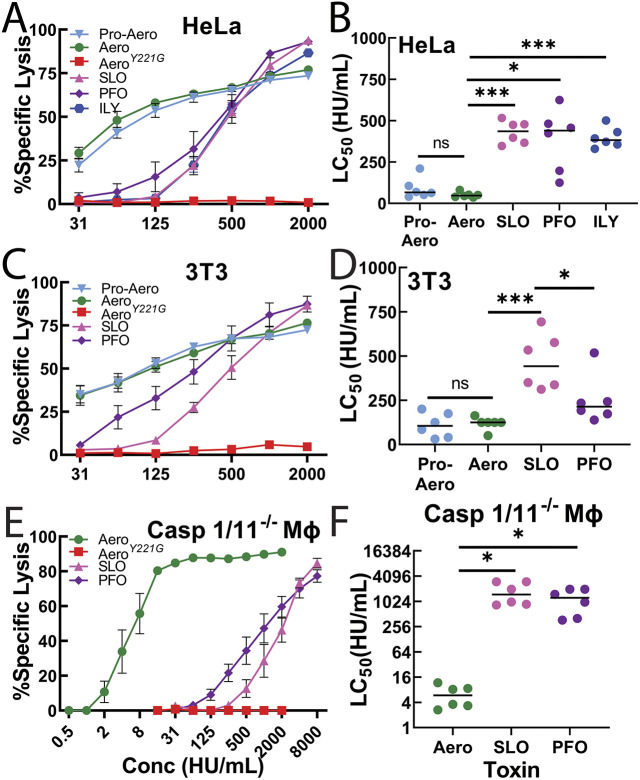
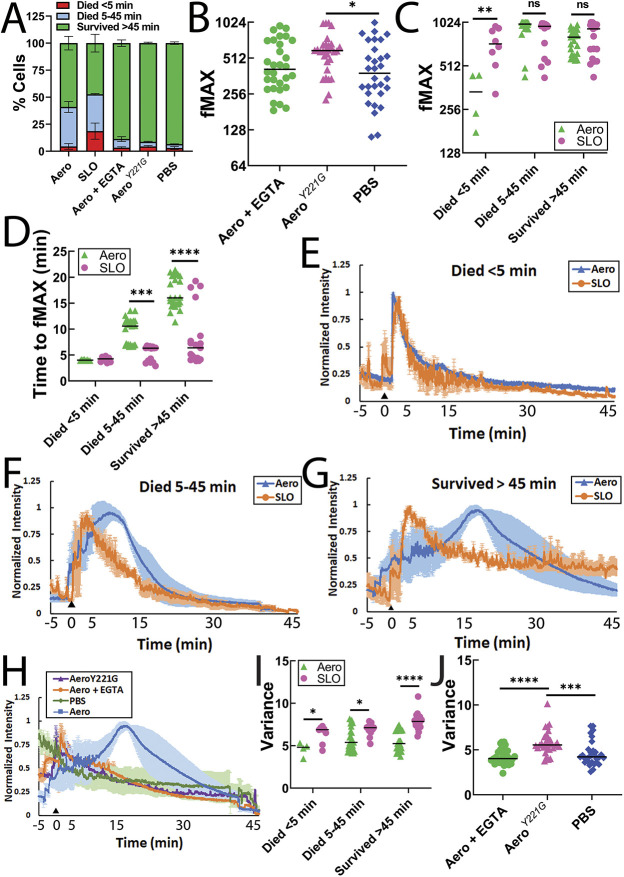
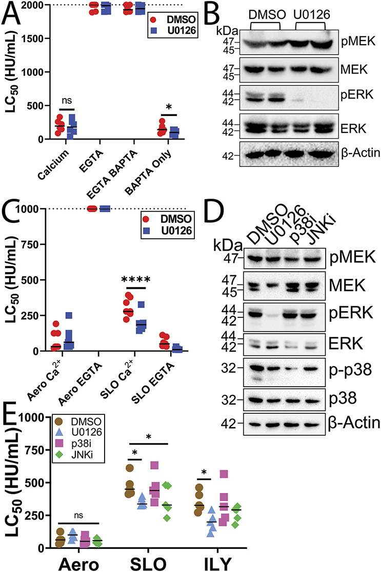
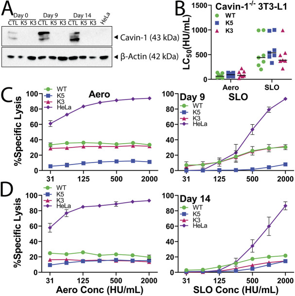
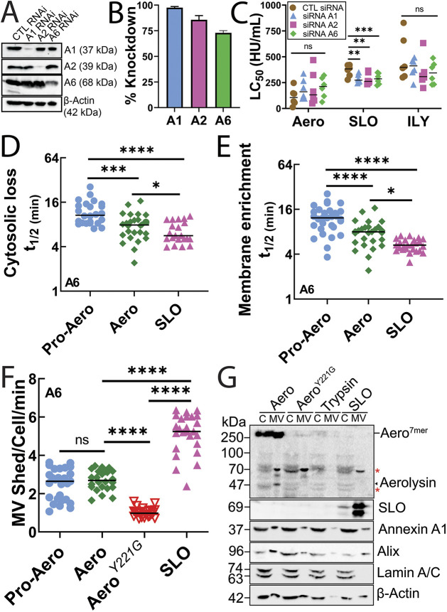
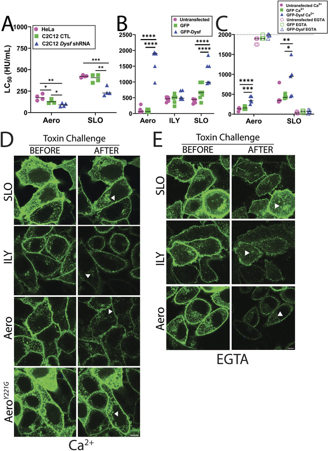
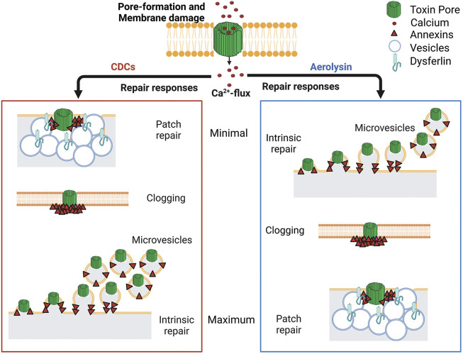
Aerolysin family pore-forming toxins damage the membrane, but membrane repair responses used to resist them, if any, remain controversial. Four proposed membrane repair mechanisms include toxin removal by caveolar endocytosis, clogging by annexins, microvesicle shedding catalyzed by MEK, and patch repair. Which repair mechanism aerolysin triggers is unknown. Membrane repair requires Ca2+, but it is controversial if Ca2+ flux is triggered by aerolysin. Here, we determined Ca2+ influx and repair mechanisms activated by aerolysin. In contrast to what is seen with cholesterol-dependent cytolysins (CDCs), removal of extracellular Ca2+ protected cells from aerolysin. Aerolysin triggered sustained Ca2+ influx. Intracellular Ca2+ chelation increased cell death, indicating that Ca2+-dependent repair pathways were triggered. Caveolar endocytosis failed to protect cells from aerolysin or CDCs. MEK-dependent repair did not protect against aerolysin. Aerolysin triggered slower annexin A6 membrane recruitment compared to CDCs. In contrast to what is seen with CDCs, expression of the patch repair protein dysferlin protected cells from aerolysin. We propose aerolysin triggers a Ca2+-dependent death mechanism that obscures repair, and the primary repair mechanism used to resist aerolysin is patch repair. We conclude that different classes of bacterial toxins trigger distinct repair mechanisms.

Keywords: Aeromonas; Annexin; Membrane repair; Patch repair; Pore-forming toxin; Streptolysin O.


Conflict of interest statement

Competing interests The authors declare no competing or financial interests.

Figures

Fig. 1.
Fig. 1.
Nucleated cells are more sensitive to aerolysin than CDCs. (A,B) HeLa or (C,D) 3T3 cells, or (E,F) Casp 1/11−/− bone-marrow derived macrophages (MΦ) were challenged with (A–D) 31–2000 HU/ml pro-aerolysin (Pro-Aero), aerolysin (Aero), streptolysin O (SLO), perfringolysin O (PFO), or (A,B) intermedilysin (ILY), or (E,F) 0.5–2000 HU/ml aerolysin, or 15–8000 HU/ml SLO or PFO, or (A,C,E) a mass equivalent to wild-type aerolysin of aerolysinY221G (AeroY221G) in 2 mM CaCl2 supplemented RPMI (RC) with 20 μg/ml propidium iodide (PI) for 30 min at 37°C. PI uptake was analyzed by flow cytometry. Specific lysis was determined and plotted against concentrations used (A,C,E). The LC50 was calculated as described in the Materials and Methods. Graphs display the mean (±s.e.m. for A,C,E) of eight (A,B) or six (C–F) independent experiments. Data points represent individual experiments. *P<0.05; ***P<0.001; ns, not significant (one-way ANOVA with Tukey post test).
Fig. 2.
Fig. 2.
Ca2+ influx drives aerolysin cytotoxicity. (A,B) HeLa cells were challenged with 31–2000 HU/ml pro-aerolysin (Pro-Aero), aerolysin (Aero) or SLO for 30 min at 37°C in either RPMI (A) with or without 150 mM KCl, (B) with 2 mM CaCl2, or 2 mM EGTA supplemented RPMI. The dotted line indicates the limit of detection. (C) HeLa cells were challenged with 31–2000 HU/ml aerolysin, SLO, or ILY for 30 min at 37°C in RC or Tyrode's buffer. (D) HeLa cells were challenged with 125 HU/ml pro-aerolysin, aerolysin, 250 HU/ml SLO, PFO or ILY, or mass equivalent to aerolysin of mutant aerolysinY221G (AeroY221G) for 30 min at 37°C in Tyrode's buffer supplemented with the indicated concentrations of CaCl2. PI uptake was analyzed by flow cytometry. The LC50 was calculated as described in the Materials and Methods. (A–C) Data points represent individual experiments. Graphs show the mean (±s.e.m. for D) of five (A,B) or six (C,D) independent experiments. **P<0.01; ****P<0.0001; ns, not significant (repeated-measures ANOVA with Tukey post test).
Fig. 3.
Fig. 3.
Aerolysin triggers delayed Ca2+ influx compared to CDCs. HeLa cells were labeled with Fluo-4 for 30 min in DMEM. The medium was replaced with 2 μg/ml TO-PRO3, 25 mM HEPES pH 7.4, and 2 mM CaCl2 supplemented RPMI (imaging buffer). 2 mM EGTA was substituted for the CaCl2 where indicated. Sublytic toxin [62 HU/ml aerolysin, 250 HU/ml SLO, mass equivalent of aerolysinY221G (AeroY221G)] or PBS was added 5 min after confocal imaging started. Cells were imaged for ∼45 min at 37°C. Ca2+ flux and cell death were recorded. Cells were categorized into three subsets by time of cell death: died <5 min, died between 5 and 45 min, and survived >45 min. (A) The percentage of each subset is shown. (B,C) Maximal Fluo-4 fluorescence intensities (fMAX) are shown for individual cells. (D) The time to reach fMAX is shown. (E–H) Fluo-4 fluorescence intensity was normalized to fMAX for individual cells and averaged. Ca2+ traces for (E) cells that died<5 min, (F) cells that died between 5 and 45 min, or (G) cells that survived >45 min after toxin challenge with are shown. (H) Ca2+ traces for cells challenged with aerolysin with 2 mM EGTA, AeroY221G or PBS alone are shown along with the wild-type aerolysin trace from (G). Arrowhead indicates toxin addition. (I,J) Ca2+ oscillations were quantified by determining the volatility of the Ca2+ flux for each cell. Data points represent individual cells from three independent experiments. Graphs show the (B,C) geometric mean or (A,D–J) mean (±s.e.m. for E–H), for of at least three independent experiments. *P<0.05; **P<0.01; ***P<0.001; ****P<0.0001; ns, not significant (one-way ANOVA with Tukey post test).
Fig. 4.
Fig. 4.
MEK-dependent repair does not contribute to aerolysin resistance. HeLa cells were serum starved for 30 min and pretreated with (A–E) vehicle (DMSO), (A) 100 μM BAPTA-AM, (A–E) 20 μM U0126 (MEK inhibitor), (D, E) 20 μM SB203580 (p38 inhibitor) or 20 μM SP600125 (JNK inhibitor) for 30 min, then either (A,C,E) challenged with toxin or (B,D) lysed for western blot analysis. HeLa cells were challenged in either (A,C) 2 mM EGTA or (A,C,E) 2 mM CaCl2 supplemented RPMI with 31–2000 HU/ml of the indicated toxins for 30 min at 37°C. PI uptake was analyzed by flow cytometry. The LC50 was calculated as described in the Materials and Methods. (B,D) Blots were probed with the indicated antibodies and HRP-conjugated secondary antibodies. Graphs show the mean of (A) six, (C) seven or (E) five independent experiments. The blots show one representative experiment from (B) seven or (D) five independent experiments. The dotted lines indicate the limit of detection. Points on this line had LC50 >2000 HU/ml. *P<0.05; ****P<0.0001; ns, not significant by (A,C) two-way or (E) one-way ANOVA with Tukey post test between groups.
Fig. 5.
Fig. 5.
Caveolar endocytosis does not mediate repair responses to aerolysin or CDCs. (A) Wild-type CRISPR control (CTL, WT) or two different cavin-1 (PTRF) CRISPR knockout 3T3-L1 cells (K5 and K3) were undifferentiated or differentiated into adipocytes and collected 0, 9 or 14 days after differentiation. HeLa cells and day 0, 9 or 14 3T3-L1 adipocytes were analyzed by western blotting. Portions of the blot were probed with anti-PTRF (anti-Cavin-1) or anti-β-actin antibodies, and HRP-conjugated secondary antibodies. (B–D) WT, K5 or K3 3T3-L1 cells were (B) undifferentiated or differentiated into adipocytes for (C) 9 or (D) 14 days and challenged with 31–2000 HU/ml aerolysin or SLO. PI uptake was analyzed by flow cytometry. The LC50 was calculated as described in the Materials and Methods. The blots show one representative experiment from three independent experiments. Graphs show the mean (±s.e.m. for C,D) of (B) seven, or (C,D) three independent experiments.
Fig. 6.
Fig. 6.
Annexins minimally resist aerolysin with limited shedding. HeLa cells were transfected with control (CTL), A1, A2 or A6 siRNA for 3 days and then (A,B) lysed for western blot analysis, or (C) challenged with 31–2000 HU/ml aerolysin (Aero), SLO or ILY for 30 min at 37°C. PI uptake was analyzed by flow cytometry. (A) Portions of the blot were probed with antibodies against A1, A2, A6 or β-actin followed by HRP-conjugated secondary antibodies. (B) Quantification of the knockdown efficiency compared to control siRNA is shown. (D,E) HeLa cells were transfected with A6–YFP and challenged with sublytic toxin concentrations (62 HU/ml pro-aerolysin or aerolysin, or 250 HU/ml SLO) in imaging buffer. The cells were imaged by confocal microscopy for ∼45 min at 37°C and then lysed with 1% Triton X-100. The time to half maximal (t1/2) (D) cytosolic depletion or (E) membrane accumulation of A6 is shown. (F) Shedding of annexin A6+ microvesicles was manually counted and expressed as microvesicles shed/cell/min. (G) HeLa cells were challenged for 15 min at 37°C with sublytic doses of aerolysin, SLO, mass equivalent of AeroY221G or sufficient trypsin to activate pro-aerolysin. Cells were pelleted at 2000 g for 5 min to yield cell pellets (denoted C). Cell supernatants were spun at 100,000 g for 40 min at 4°C and the microvesicle pellet (MV) collected. Fractions were analyzed by western blotting using anti-aerolysin, 6D11 anti-SLO, CPTC-A1-3 anti-annexin A1, anti-Alix, MANLAC-4A7 anti-lamin A/C or AC-15 anti-β-actin. Graphs show the mean (C,F) or geometric mean (D,E). Data points represent (C) individual experiments or (D,E) individual cells from three independent experiments. *P<0.05; **P<0.01; ***P<0.001; ****P<0.0001; ns, not significant (one-way ANOVA with Tukey post test). Asterisks in G indicates non-specific bands.
Fig. 7.
Fig. 7.
Patch repair protects cells from aerolysin. (A) HeLa, C2C12 control and Dysf shRNA cells were challenged with 15–1000 HU/ml aerolysin (Aero) or 31–2000 HU/ml SLO for 30 min at 37°C. PI uptake was analyzed by flow cytometry. HeLa cells were either untransfected or transfected with GFP or GFP–Dysferlin (GFP–Dysf) for 48 h and challenged with aerolysin (Aero), SLO or ILY at 31-2000 HU/ml for 30 min at 37°C either in (B,C) 2 mM CaCl2- or (C) 2 mM EGTA-supplemented RPMI. PI uptake was analyzed by flow cytometry. The LC50 was calculated as described in the Materials and Methods. (D,E) GFP–Dysf (green) transfected HeLa cells were challenged with sublytic toxin doses (250 HU/ml SLO or ILY, 62 HU/ml aerolysin or mass equivalent to aerolysin of mutant AeroY221G) in imaging buffer either supplemented with (D) 2 mM CaCl2 or (E) 2 mM EGTA, imaged for ∼45 min at 37°C, and then lysed with 1% Triton X-100. Depletion of GFP–Dysf-containing vesicles from cytosol are shown by arrowheads. Graphs show the mean±s.e.m. of (A) four, (B) seven or (C) five independent experiments. The dotted line indicates the limit of detection. Points on this line had LC50>2000 HU/ml. Micrographs show representative images from three independent experiments. **P<0.01, ***P<0.001, ****P<0.0001 (one-way ANOVA with Tukey post test). Scale bars: 5 μm.
Fig. 8.
Fig. 8.
Toxins trigger distinct repair responses. After pore formation and membrane damage, multiple repair pathways are triggered downstream of Ca2+ flux. CDCs, such as SLO and PFO, are predominantly resisted by MEK-dependent microvesicle shedding and by annexins that might clog pore access to a lesser extent. Patch repair is triggered as a last resort. In contrast, aerolysin does not stimulate MEK-dependent microvesicle shedding. Aerolysin triggers minimal annexin translocation. Instead, patch repair serves as the primary resistance mechanism to aerolysin.

Similar articles

Cited by

References

    1. Abrami, L., Fivaz, M., Decroly, E., Seidah, N. G., Jean, F., Thomas, G., Leppla, S. H., Buckley, J. T. and van der Goot, F. G. (1998a). The pore-forming toxin proaerolysin is activated by furin. J. Biol. Chem. 273, 32656-32661. 10.1074/jbc.273.49.32656 - DOI - PubMed
    1. Abrami, L., Fivaz, M., Glauser, P. E., Parton, R. G. and van der Goot, F. G. (1998b). A pore-forming toxin interacts with a GPI-anchored protein and causes vacuolation of the endoplasmic reticulum. J. Cell Biol. 140, 525-540. 10.1083/jcb.140.3.525 - DOI - PMC - PubMed
    1. Babiychuk, E. B., Monastyrskaya, K., Potez, S. and Draeger, A. (2009). Intracellular Ca(2+) operates a switch between repair and lysis of streptolysin O-perforated cells. Cell Death Differ. 16, 1126-1134. 10.1038/cdd.2009.30 - DOI - PubMed
    1. Bhattacharjee, P. and Keyel, P. A. (2018). Cholesterol-dependent cytolysins impair pro-inflammatory macrophage responses. Sci. Rep. 8, 6458. 10.1038/s41598-018-24955-2 - DOI - PMC - PubMed
    1. Bittel, D. C., Chandra, G., Tirunagri, L. M. S., Deora, A. B., Medikayala, S., Scheffer, L., Defour, A. and Jaiswal, J. K. (2020). Annexin A2 mediates dysferlin accumulation and muscle cell membrane repair. Cells 9, 1919. 10.3390/cells9091919 - DOI - PMC - PubMed

Publication types

MeSH terms