. 2023 Mar 13;164(5):bqad050.
doi: 10.1210/endocr/bqad050.

Activating Transcription Factor 3 Stimulates Follicle-Stimulating Hormone-β Expression In Vitro But Is Dispensable for Follicle-Stimulating Hormone Production in Murine Gonadotropes In Vivo

Affiliations

Activating Transcription Factor 3 Stimulates Follicle-Stimulating Hormone-β Expression In Vitro But Is Dispensable for Follicle-Stimulating Hormone Production in Murine Gonadotropes In Vivo

Carlos A I Alonso et al. Endocrinology. .

Abstract

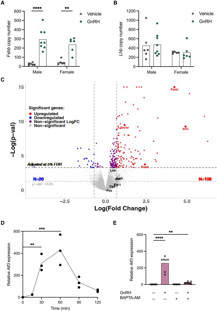
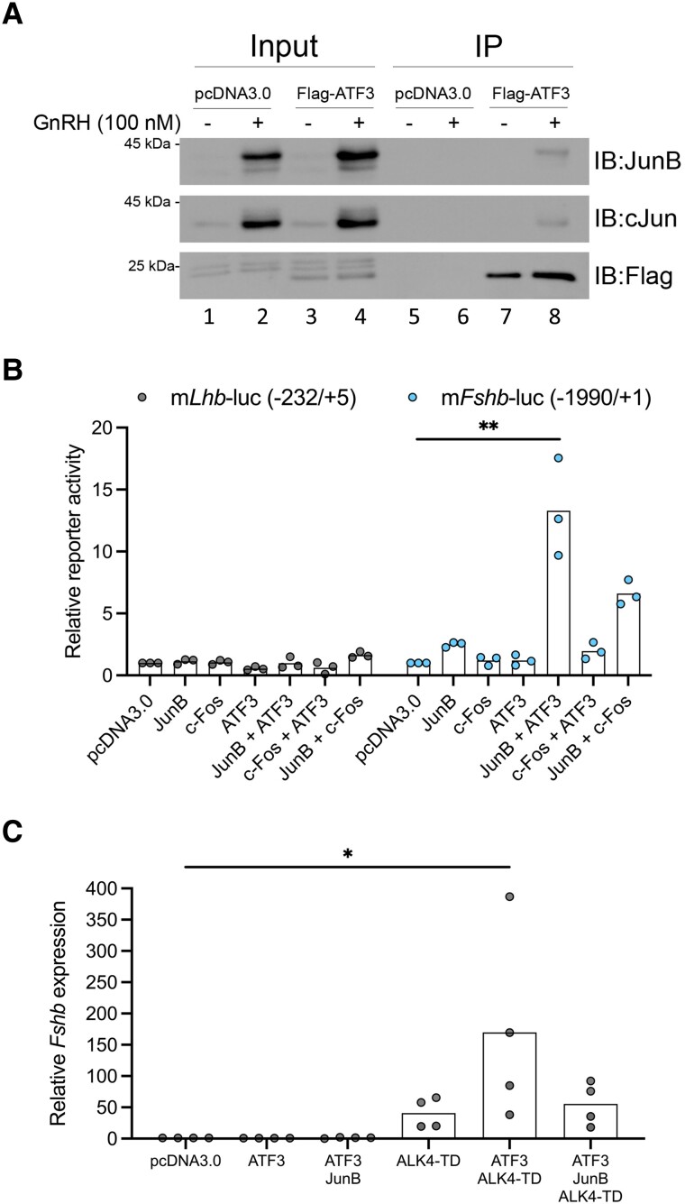
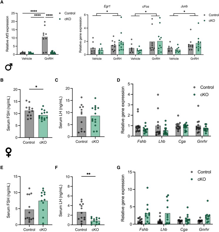
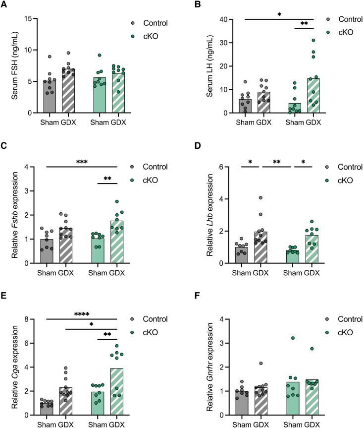
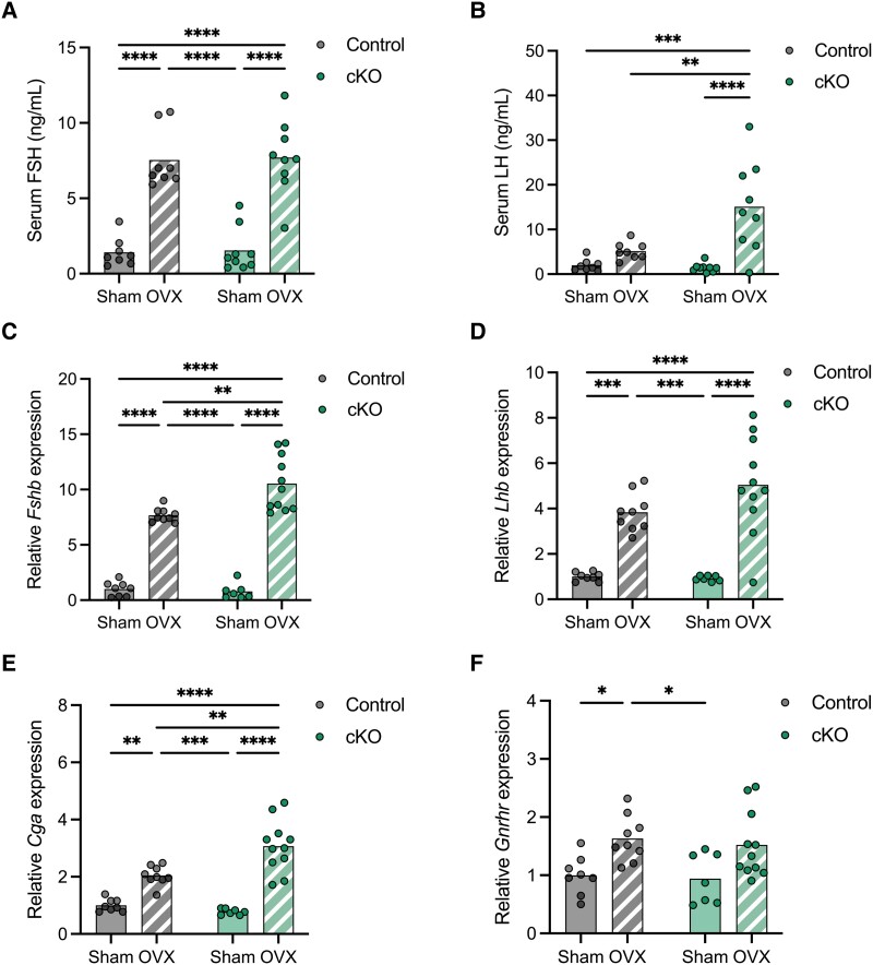
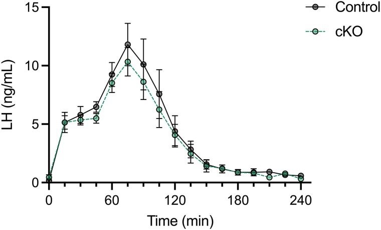
Follicle-stimulating hormone (FSH), a dimeric glycoprotein produced by pituitary gonadotrope cells, regulates spermatogenesis in males and ovarian follicle growth in females. Hypothalamic gonadotropin-releasing hormone (GnRH) stimulates FSHβ subunit gene (Fshb) transcription, though the underlying mechanisms are poorly understood. To address this gap in knowledge, we examined changes in pituitary gene expression in GnRH-deficient mice (hpg) treated with a regimen of exogenous GnRH that increases pituitary Fshb but not luteinizing hormone β (Lhb) messenger RNA levels. Activating transcription factor 3 (Atf3) was among the most upregulated genes. Activating transcription factor 3 (ATF3) can heterodimerize with members of the activator protein 1 family to regulate gene transcription. Co-expression of ATF3 with JunB stimulated murine Fshb, but not Lhb, promoter-reporter activity in homologous LβT2b cells. ATF3 also synergized with a constitutively active activin type I receptor to increase endogenous Fshb expression in these cells. Nevertheless, FSH production was intact in gonadotrope-specific Atf3 knockout [conditional knockout (cKO)] mice. Ovarian follicle development, ovulation, and litter sizes were equivalent between cKOs and controls. Testis weights and sperm counts did not differ between genotypes. Following gonadectomy, increases in LH secretion were enhanced in cKO animals. Though FSH levels did not differ between genotypes, post-gonadectomy increases in pituitary Fshb and gonadotropin α subunit expression were more pronounced in cKO than control mice. These data indicate that ATF3 can selectively stimulate Fshb expression in vitro but is not required for FSH production in vivo.

Keywords: GnRH; cell line; knockout mouse; pituitary; transcription.


Figures

Figure 1.
Figure 1.
GnRH induces Atf3 expression. (A-C) Hpg mice were treated once daily with vehicle or 1 µg GnRH i.p. for 10 consecutive days. GnRH-induced changes in pituitary (A) Fshb and (B) Lhb mRNA levels were assessed by qPCR from whole pituitaries. Shown are the relative copy number for each transcript for one representative experiment out of 2. Twenty-five animals were assessed, including 11 females (5 vehicle and 6 GnRH-treated) and 14 males (6 vehicle and 8 GnRH-treated). Data were analyzed by two-way ANOVA. (C) Pituitary RNA from male mice treated as in panels A and B was subjected to bulk RNA-seq. Volcano plot showing up (red) and down (blue) regulated genes. Select genes are labeled. (D) LβT2b cells were treated with 10 nM GnRH for the indicated lengths of time. RNA was collected at each time point and Atf3 expression was assessed by RT-qPCR. Each point represents an experimental replicate, n = 3. Groups were compared using one-way ANOVA followed by Tukey's multiple comparisons test. (E) LβT2b cells were pre-incubated with vehicle or 50 µM BAPTA-AM for 1 hour and then treated with 10 nM GnRH for 1 hour. Atf3 expression was assessed by RT-qPCR. Each point represents an experimental replicate. Bars represent the mean of the experimental replicates, n = 7. Groups were compared using the Kruskal-Wallis test followed by Dunn's correction for multiple comparisons. Asterisks indicate significant differences: ** P < 0.01; *** P < 0.001; **** P < 0.0001. Abbreviations: ANOVA, analysis of variance; GnRH, gonadotropin-releasing hormone; mRNA, messenger RNA; qPCR, quantitative polymerase chain reaction; RNA-seq, RNA sequencing; RT-qPCR, real time PCR.
Figure 2.
Figure 2.
ATF3 physically and functionally interacts with JunB. (A) LβT2b cells were transfected with 500 ng Flag-ATF3 or empty vector (pcDNA3.0). The next day, cells were treated with vehicle or 100 nM GnRH for 2 hours. Flag-ATF3 was immunoprecipitated with anti-Flag conjugated agarose beads. Input and eluates were subjected to sodium dodecyl sulfate-PAGE and immunoblotting with JunB, cJun, and Flag antibodies. Images shown are representative of 3 independent experiments. (B) LβT2b cells in 48-well plates were transfected with murine Lhb or Fshb promoter-reporters (225 ng/well) and with the indicated combinations of expression vectors for JunB (50 ng/well), c-Fos (50 ng/well), and/or ATF3 (50 ng/well). All conditions were balanced with pcDNA3.0. Cells were starved overnight, and reporter activity was measured the following day. Each point represents the average of technical triplicates in independent experiments, n = 3. (C) LβT2b cells in 6-well plates were transfected with the indicated expression vectors for ATF3, JunB, and ALK4-TD (500 ng/well each). The amount of transfected DNA was balanced with empty vector (pcDNA3.0). Cells were starved overnight, and RNA was extracted the following day. Fshb mRNA levels were measured by RT-qPCR using the pcDNA3.0 condition as control and ribosomal protein L19 (Rpl19) as the housekeeping gene. Each point represents the average of technical triplicates from independent experiments, n = 4. Bars represent the mean of the experimental replicates. Groups were compared using the Kruskal-Wallis test followed by Dunn's correction for multiple comparisons. Asterisks indicate significant differences: * P < 0.05, ** P < 0.01. Abbreviations: GnRH, gonadotropin-releasing hormone; mRNA, messenger RNA: RT-qPCR, real time quantitative polymerase chain reaction.
Figure 3.
Figure 3.
Gonadotrope-specific deletion of Atf3 does not impair gonadotropin production in male or female mice. (A) Adult gonadotrope-specific Atf3 cKO (green) or control mice (gray) of both sexes were treated once intraperitoneally with GnRH or vehicle. After 1 hour, pituitaries were harvested for analysis of Atf3, Egr1, Fos, and Junb mRNA levels by RT-qPCR. Data were normalized to the vehicle treated controls, and ribosomal protein L19 (Rpl19) was the housekeeping gene. Serum FSH (B) and LH (C) levels in 8- to 10-week-old control and cKO males were measured by ELISA; n = 12, Control; n = 14, cKO. Serum FSH (E) and LH (F) levels in 9- to 12-week-old control and cKO females on estrus morning were measured by ELISA. n = 13, Control; n = 12, cKO. Gene expression in pituitary glands from 8- to 10-week-old control and cKO males (D) or 9- to 12-week-old females (G). Fshb, Lhb, Cga, and Gnrhr mRNA levels were assessed by RT-qPCR. Results were normalized to control animals. Males: n = 12, Control; n = 12, cKO. Females: n = 12, Control; n = 11, cKO. In all panels bars represent group means. Each point represents an individual animal. In (A), groups were compared using two-way ANOVA followed by Bonferroni correction for multiple comparisons. In (B)-(G), all pairwise comparisons were done using t-tests. Asterisks indicate significant differences: * P < 0.05; ** P < 0.01; **** P < 0.0001. Abbreviations: ANOVA, analysis of variance; cKO, conditional knockout; ELISA, enzyme-linked immunosorbent assay; FSH, follicle-stimulating hormone; GnRH, gonadotropin-releasing hormone; LH, luteinizing hormone; mRNA, messenger RNA; RT-qPCR, real time quantitative polymerase chain reaction.
Figure 4.
Figure 4.
Gonadotrope-specific Atf3 knockout mice are normogonadal and fertile. Seminal vesicle (A) and testes (B) weights in control and cKO males. Organ weights were normalized to body weight. n = 13, Control; n = 13, cKO. (C) Epididymal sperm counts in control and cKO animals. n = 9 Control; n = 9, cKO. Uterine (D) and ovarian (E) weights in control or cKO females. Organ weights were normalized to body weight. n = 13, Control; n = 12, cKO. (F) Number of antral (preovulatory) follicles and corpora lutea in one ovary from control and cKO females. n = 4, Control; n = 4, cKO. (G) Control and cKO females were mated at 9 weeks of age with wild-type C57BL/6 males and cumulus-oocyte complexes counted on the morning of vaginal plugging. n = 5, Control; n = 6, cKO. (H) Nine-week-old females from both genotypes were mated with wild-type C57BL/6 males for 6 months and the number of pups per litter was monitored. The average per female is shown. n = 6, Control; n = 5, cKO. Each point represents one animal. Bars represent group means. In all cases groups were compared using t-tests. An asterisk (*) indicates significant differences: P < 0.05. Abbreviations: cKO, conditional knockout.
Figure 5.
Figure 5.
LH but not FSH secretion is enhanced in castrated Atf3 cKO males. Ten- to 12-week-old control or cKO males were sham-operated (Sham, solid color) or GDX (hatched). Serum FSH (A) and LH (B) levels were measured by ELISA. n = 9, Sham-Control; n = 9, GDX-Control; n = 9, Sham-cKO; n = 9, GDX-cKO. Pituitary RNA was isolated and Fshb (C), Lhb (D), Cga (E), and Gnrhr (F) mRNA levels were assessed by RT-qPCR. n = 8, Sham-Control; n = 10, GDX-Control; n = 8, Sham-cKO; n = 8, GDX-cKO. Points and bars represent individual animals and group means, respectively. Groups were compared using two-way ANOVA followed by the Bonferroni correction for multiple comparisons. Asterisks indicate significant differences: *: P < 0.05, ** P < 0.01, *** P < 0.001, **** P < 0.0001. Abbreviations: ANOVA, analysis of variance; cKO, conditional knockout; FSH, follicle-stimulating hormone; GDX, gonadectomized; LH, luteinizing hormone; mRNA, messenger RNA; RT-qPCR, real time quantitative polymerase chain reaction.
Figure 6.
Figure 6.
LH but not FSH secretion is enhanced in ovariectomized Atf3 cKO females. Ten- to 12-week-old control or cKO females were sham-operated (Sham, solid color) or OVX (hatched). Serum FSH (A) and LH (B) levels were measured by ELISA. n = 8, Sham-Control; n = 8, OVX-Control; n = 9, Sham-cKO; n = 9, OVX-cKO. Pituitary RNA was isolated and Fshb (C), Lhb (D), Cga (E), and Gnrhr (F) mRNA levels were assessed by RT-qPCR. n = 8, Sham-Control; n = 9, OVX-Control; n = 7, Sham-cKO; n = 11, OVX-cKO. Points and bars represent individual animals and group means, respectively. Groups were compared using two-way ANOVAs and the Bonferroni correction for multiple comparisons was used. Asterisks indicate significant differences: * P < 0.05, ** P < 0.01, *** P < 0.001, **** P < 0.0001. Abbreviations: ANOVA, analysis of variance; cKO, conditional knockout; FSH, follicle-stimulating hormone; LH, luteinizing hormone; mRNA, messenger RNA; OVX, ovariectomized; RT-qPCR, real time quantitative polymerase chain reaction.
Figure 7.
Figure 7.
Kisspeptin-induced LH release is not altered in Atf3 cKO females. Secretion of LH in response to kisspeptin-54 in juvenile control (grey, continuous line) and cKO (green, dashed line) females. Twenty-eight- to 30-day old females from each genotype were injected with 1 nmol of Kisspeptin-54 at time 0. Then blood was collected at 15-minute intervals and assayed for LH by ELISA. Each point represents the group mean ± SEM. n = 5 per genotype. Groups were compared using a mixed effects model using restricted maximum likelihood estimation. Abbreviations: cKO, conditional knockout; LH, luteinizing hormone.

Similar articles

Cited by

References

    1. Tapanainen JS, Aittomäki K, Min J, Vaskivuo T, Huhtaniemi IT. Men homozygous for an inactivating mutation of the follicle-stimulating hormone (FSH) receptor gene present variable suppression of spermatogenesis and fertility. Nat Genet. 1997;15(2):205‐206. - PubMed
    1. Kumar TR, Wang Y, Lu N, Matzuk MM. Follicle stimulating hormone is required for ovarian follicle maturation but not male fertility. Nat Genet. 1997;15(2):201‐204. - PubMed
    1. Ma X, Dong Y, Matzuk MM, Kumar TR. Targeted disruption of luteinizing hormone beta-subunit leads to hypogonadism, defects in gonadal steroidogenesis, and infertility. Proc Natl Acad Sci USA. 2004;101(49):17294‐17299. - PMC - PubMed
    1. Weiss J, Axelrod L, Whitcomb RW, Harris PE, Crowley WF, Jameson JL. Hypogonadism caused by a single amino acid substitution in the beta subunit of luteinizing hormone. N Engl J Med. 1992;326(3):179‐183. - PubMed
    1. Wildt L, Häusler A, Marshall G, et al. . Frequency and amplitude of gonadotropin-releasing hormone stimulation and gonadotropin secretion in the rhesus monkey. Endocrinology. 1981;109(2):376‐385. - PubMed

Publication types

MeSH terms

Substances