. 2023 May;10(13):e2207216.
doi: 10.1002/advs.202207216. Epub 2023 Mar 23.

Polydopamine Nanoparticles Targeting Ferroptosis Mitigate Intervertebral Disc Degeneration Via Reactive Oxygen Species Depletion, Iron Ions Chelation, and GPX4 Ubiquitination Suppression

Affiliations

Polydopamine Nanoparticles Targeting Ferroptosis Mitigate Intervertebral Disc Degeneration Via Reactive Oxygen Species Depletion, Iron Ions Chelation, and GPX4 Ubiquitination Suppression

Xiao Yang et al. Adv Sci (Weinh). 2023 May.

Abstract

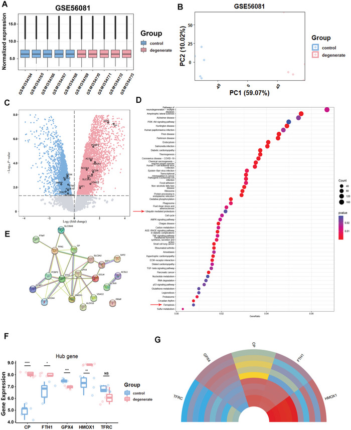
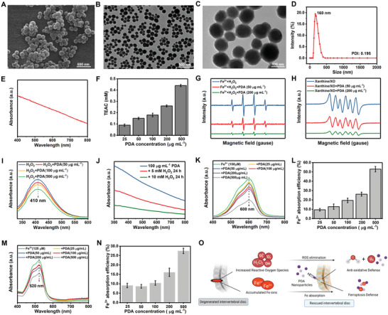
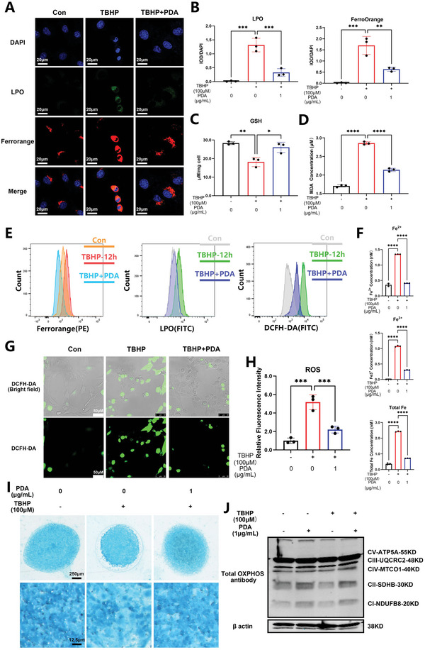
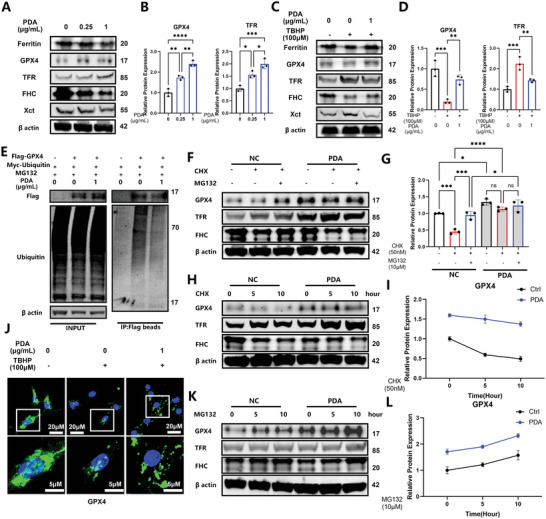
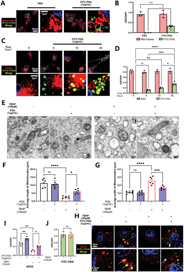
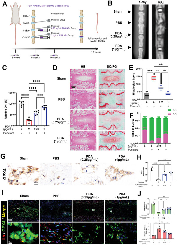
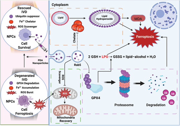
Intervertebral disc degeneration (IVDD)-induced lower back pain (LBP) is a common problem worldwide. The underlying mechanism is partially accredited to ferroptosis, based on sequencing analyses of IVDD patients from the gene expression omnibus (GEO) databases. In this study, it is shown that polydopamine nanoparticles (PDA NPs) inhibit oxidative stress-induced ferroptosis in nucleus pulposus (NP) cells in vitro. PDA NPs scavenge reactive oxygen species (ROS), chelate Fe2+ to mitigate iron overload, and regulate the expression of iron storage proteins such as ferritin heavy chain (FHC), ferritin, and transferrin receptor (TFR). More importantly, PDA NPs co-localize with glutathione peroxidase 4 (GPX4) around the mitochondria and suppress ubiquitin-mediated degradation, which in turn exerts a protective function via the transformation and clearance of phospholipid hydroperoxides. PDA NPs further down-regulate malondialdehyde (MDA) and lipid peroxide (LPO) production; thus, antagonizing ferroptosis in NP cells. Moreover, PDA NPs effectively rescue puncture-induced degeneration in vivo by targeting ferroptosis and inhibiting GPX4 ubiquitination, resulting in the upregulation of antioxidant pathways. The findings offer a new tool to explore the underlying mechanisms and a novel treatment strategy for IVDD-induced LBP.

Keywords: GPX4 ubiquitination; ferroptosis; intervertebral disc degeneration; nucleus pulposus; polydopamine nanoparticles.


Conflict of interest statement

The authors declare no conflict of interest.

Figures

Figure 1
Figure 1
Ferroptosis is involved in the progressive degeneration of intervertebral discs in humans, as determined by the gene expression omnibus (GEO) database. A,B) Normalized expression matrix and principal component analysis (PCA) diagram of GSE56081. C) The volcano plot of GSE56081. D,E) KEGG pathway enrichment analysis and genes associated with ferroptosis enriched by differentially expressed genes (DEGs). F) Validation of hub genes in GSE56081. G) Circos plot for the expression levels of hub genes among different samples. *P < 0.05, **P < 0.01, ***P < 0.001, and ****P < 0.0001.
Figure 2
Figure 2
Characterization of PDA NPs. A) Scanning electron microscopy (SEM) image of PDA NPs. B,C) Transmission electron microscopy (TEM) images of PDA NPs. D) The average hydrodynamic particle diameter of PDA NPs. E) Ultraviolet–visible (UV–vis) absorption spectroscopy of PDA NPs. F) Antioxidant capacity of PDA NPs measured by the 2,2″‐azino‐bis (3‐ethylbenzothiazoline‐6‐sulfonic acid) (ABTS) method. G,H) Electron spin resonance (ESR) spectra of DMPO/•OH adducts (G) and DMPO/O2•− (H), indicating the depletion of •OH and O2•− upon incubation with PDA NPs. I) UV–vis absorption spectra of peroxide–titanium after the addition of PDA NPs, indicating the H2O2 depletion of PDA NPs (titanium sulfate method). J) UV–vis absorption spectra and PDA NPs after reaction with H2O2, indicating the degradation of PDA NPs. K) UV–vis absorption spectra of blue tripyridyltriazine‐Fe2+ complex after the addition of PDA NPs, indicating the Fe2+‐chelating capacity of PDA NPs. L) Fe2+ ions chelation efficiency of PDA NPs. M) UV–vis absorption spectra of pink 2,2″‐bipyridine‐Fe3+ complex after the addition of PDA NPs, indicating the Fe3+‐chelating capacity of PDA NPs. N) Fe3+ ions chelation efficiency of PDA NPs. O) Illustration of the reactive oxygen species (ROS) depletion and Fe ions chelation activities of PDA NPs, which are expected to facilitate the anti‐oxidative and ferroptosis defense microenvironment and rescue the degenerated intervertebral disc.
Figure 3
Figure 3
PDA NPs scavenge reactive oxygen species (ROS) and chelate Fe2+ ions to mitigate the ferroptosis of nucleus pulposus (NP) cells in vitro. A) Immunofluorescence analysis of lipid peroxide (LPO) and FerroOrange of NP cells stimulated with tert‐butyl hydroperoxide (TBHP) (100 µm) for 12 h and/or pretreated with PDA NPs (1 µg mL−1) for 24 h. B) Quantification of integrated optical density (IOD)/4′,6‐diamidino‐2‐phenylindole (DAPI) of the LPO and FerroOrange. C) GSH assay of NP cells stimulated with TBHP (100 µm) for 12 h and/or pretreated with PDA NPs (1 µg mL−1) for 24 h. D) Malondialdehyde (MDA) quantification of NP cells stimulated with TBHP (100 µm) for 12 h and/or pretreated with PDA NPs (1 µg mL−1) for 24 h. E) Flowcytometry analysis of PE‐FerroOrange, FITC‐LPO, and FITC‐ROS in NP cells stimulated with TBHP (100 µm) for 12 h and/or pretreated with PDA NPs (1 µg mL−1) for 24 h. F) Quantification of the iron amount (Fe2+, Fe3+) in NP cells stimulated with TBHP (100 µm) for 12 h and/or pretreated with PDA NPs (1 µg mL−1) for 24 h. G) Immunofluorescence analysis of ROS (DCFH‐DA) in NP cells stimulated with TBHP (100 µm) for 12 h and/or pretreated with PDA NPs (1 µg mL−1) for 24 h. H) Quantification of relative fluorescence intensity of the NP cells. I) Alcian blue stain of the high‐density culture of NP cells stimulated with TBHP (100 µm) for 12 h and/or pretreated with PDA NPs (1 µg mL−1) for 24 h. J) Western blot analysis of total oxidative phosphorylation (OXPHOS) in NP cells stimulated with PDA NPs alone for 24 h, TBHP (100 µm) for 12 h, and/or pretreated with PDA NPs (1 µg mL−1) for 24 h. All data are presented as mean ± SD from three replicates. *P < 0.05, **P < 0.01, ***P < 0.001, and ****P < 0.0001.
Figure 4
Figure 4
PDA NPs inhibit the ubiquitination of GPX4 to suppress the ferroptosis of nucleus pulposus (NP) cells in vitro. A) Western blot analysis of ferritin, GPX4, TFR, FHC, and Xct in NP cells treated with different concentrations of PDA NPs (0.25, 1 µg mL−1) for 24 h. B) Relative protein quantification of grey scale value for GPX4 and TFR. C) Western blot analysis of ferritin, GPX4, TFR, FHC, and Xct in NP cells stimulated with tert‐butyl hydroperoxide (TBHP) (100 µm) for 12 h and/or pretreated with PDA NPs (1 µg mL−1) for 24 h. D) Relative protein quantification of grey scale value for GPX4 and TFR. E) Ubiquitylation analysis of GPX4 in 293T cells treated with MG132 (10 µm) and/or PDA NPs (1 µg mL−1) using Flag‐GPX4 and Myc‐ubiquitin plasmids with Flag‐tagged beads. F) Western blot analysis of GPX4, TFR, and FHC using β‐actin as the loading control in NP cells (pretreated with or without PDA NPs) treated with cycloheximide (50 nm) and MG132 (10 µm). G) Relative protein quantification of grey scale value for GPX4. H) Western blot analysis of GPX4, TFR, and FHC using β‐actin as the loading control in NP cells (pretreated with or without PDA NPs) treated with cycloheximide (50 nm) for 0, 5, and 10 h. I) Relative protein quantification of grey scale value for GPX4. J) Immunofluorescence analysis of GPX4 in NP cells stimulated with TBHP (100 µm) for 12 h and/or pretreated with PDA NPs (1 µg mL−1) for 24 h. K) Western blot analysis of GPX4, TFR, and FHC using β‐actin as the loading control in NP cells (pretreated with or without PDA NPs) treated with MG132 (10 µm) for 0, 5, and 10 h. L) Relative protein quantification of grey scale value for GPX4. All data are presented as mean ± SD from three replicates. *P < 0.05, **P < 0.01, ***P < 0.001, and ****P < 0.0001.
Figure 5
Figure 5
PDA NPs were absorbed via endocytosis and co‐localized with the mitochondria and GPX4 in NP cells in vitro. A) Immunofluorescence analysis of Mito‐tracker and FITC‐PDA NPs in NP cells treated with or without PDA NPs (1 µg mL−1) for 24 h. B) Quantification of integrated optical density (IOD)/4′,6‐diamidino‐2‐phenylindole (DAPI) for the Mito‐tracker and FITC‐PDA NPs. C) Immunofluorescence analysis of Rab5 and FITC‐PDA NPs in NP cells treated with PDA NPs (1 µg mL−1) for 0, 6, 12, and 24 h. D) Quantification of IOD/DAPI for the Rab5 and FITC‐PDA NPs. E) Transmission electron microscopy (TEM) analysis of NP cells stimulated with PDA NPs alone for 24 h, TBHP (100 µm) for 12 h, and/or pretreated with PDA NPs (1 µg mL−1) for 24 h. F,G) Quantification of average length and width of mitochondria. H) Immunofluorescence analysis of GPX4 and FITC‐PDA NPs in NP cells stimulated with FITC‐PDA NPs alone for 24 h, TBHP (100 µm) for 12 h, and/or pretreated with FITC‐PDA NPs (1 µg mL−1) for 24 h. I) Quantification of IOD/DAPI for the GPX4. J) Quantification of IOD/DAPI for the FITC‐PDA NPs. All data are presented as mean ± SD from three or six replicates. *P < 0.05, **P < 0.01, ***P < 0.001, and ****P < 0.0001.
Figure 6
Figure 6
PDA NPs reduce ferroptosis and mitigate IVDD in rats by targeting GPX4 ubiquitination in vivo. All rats (n = 6) were punctured at Co7/8, Co8/9, and Co9/10; then, Co8/9 and Co9/10 were rescued by PDA NPs in a concentration of 0.25 and 1 µg mL−1 (Sham group: Co6/7, PBS group: Co7/8, PDA Low concentration group: Co8/9, PDA high concentration group: Co9/10). A) Puncture induced IVDD model in rats and relative PDA NPs treatment methods. B) X‐ray and MRI of rat tails focused on the area of operation. C) Quantification of relative DHI in the sections. D) Safranin O‐Fast Green and hematoxylin and eosin (H&E) staining of paraffin sections of the rat tails. E) Quantification of histological score in the sections. F) Quantification of IOD ratio of SO/FG stain of the sections. G) Immunohistochemistry analysis of paraffin sections of the rat tails. H) Quantification of the relative IOD of GPX4. I) Immunofluorescence analysis of paraffin sections of the rat tails. J) Quantification of relative IOD/DAPI of GPX4 and ubiquitin in the sections. All data are presented as mean ± SD from six replicates. *P < 0.05, **P < 0.01, ***P < 0.001, and ****P < 0.0001.
Figure 7
Figure 7
PDA NPs targeting ferroptosis mitigate intervertebral disc degeneration via three main pathways: 1) suppression of the ubiquitylation of GPX4 to maintain its expression and function; 2) chelation of Fe2+ to inhibit lipid peroxidation and malondialdehyde (MDA) formation; and 3) recovering mitochondrial function as a reactive oxygen species (ROS) scavenger. Created with BioRender.com.

Similar articles

Cited by

References

    1. GBD 2019 Diseases and Injuries Collaborators , Lancet 2020, 396, 1204. - PMC - PubMed
    1. Hartvigsen J., Hancock M. J., Kongsted A., Louw Q., Ferreira M. L., Genevay S., Hoy D., Karppinen J., Pransky G., Sieper J., Smeets R. J., Underwood M., Lancet 2018, 391, 2356. - PubMed
    1. DePalma M. J., Ketchum J. M., Saullo T., Pain Medicine 2011, 12, 224. - PubMed
    1. Chou D., Samartzis D., Bellabarba C., Patel A., Luk K. D. K., Kisser J. M. S., Skelly A. C., Spine 2011, 36, S43. - PubMed
    1. Takatalo J., Karppinen J., Niinimäki J., Taimela S., Näyhä S., Mutanen P., Sequeiros R. B., Kyllönen E., Tervonen O., Spine 2011, 36, 2180. - PubMed

Publication types

MeSH terms

LinkOut - more resources