. 2023 Apr 26;89(4):e0191922.
doi: 10.1128/aem.01919-22. Epub 2023 Mar 23.

Identification of lthB, a Gene Encoding a Putative Glycosyltransferase Family 8 Protein Required for Leptothrix Sheath Formation

Affiliations

Identification of lthB, a Gene Encoding a Putative Glycosyltransferase Family 8 Protein Required for Leptothrix Sheath Formation

Tatsuki Kunoh et al. Appl Environ Microbiol. .

Abstract

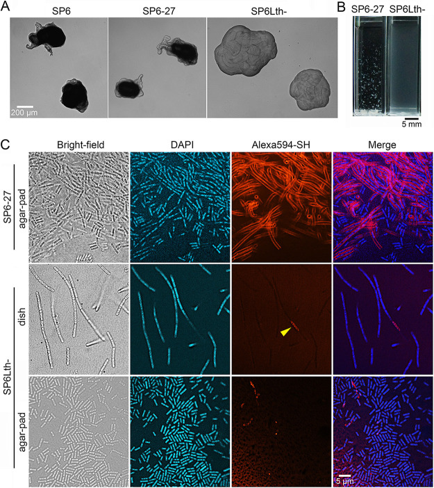
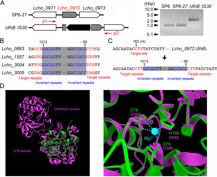
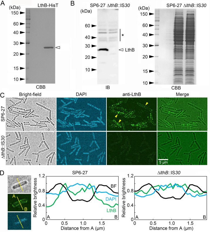
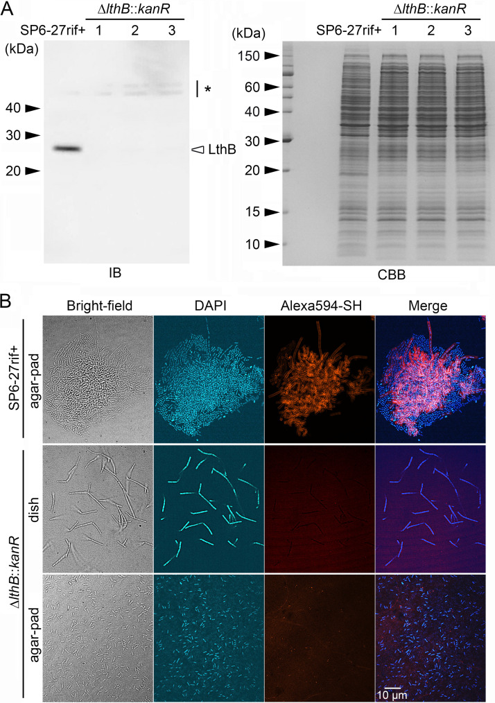
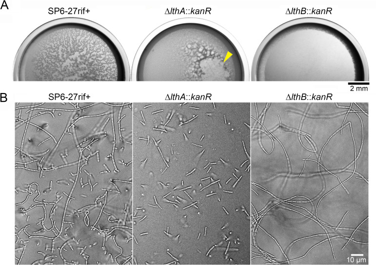
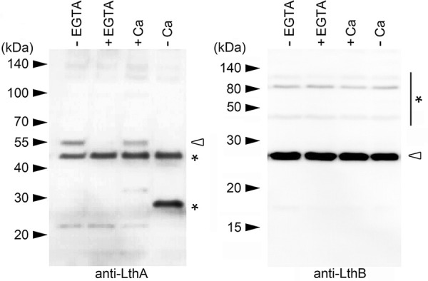
The bacterium Leptothrix cholodnii generates cell chains encased in sheaths that are composed of woven nanofibrils. The nanofibrils are mainly composed of glycoconjugate repeats, and several glycosyltransferases (GTs) are required for its biosynthesis. However, only one GT (LthA) has been identified to date. In this study, we screened spontaneous variants of L. cholodnii SP6 to find those that form smooth colonies, which is one of the characteristics of sheathless variants. Genomic DNA sequencing of an isolated variant revealed an insertion in the locus Lcho_0972, which encodes a putative GT family 8 protein. We thus designated this protein LthB and characterized it using deletion mutants and antibodies. LthB localized adjacent to the cell envelope. ΔlthB cell chains were nanofibril free and thus sheathless, indicating that LthB is involved in nanofibril biosynthesis. Unlike the ΔlthA mutant and the wild-type strain, which often generate planktonic cells, most ΔlthB organisms presented as long cell chains under static conditions, resulting in deficient pellicle formation, which requires motile planktonic cells. These results imply that sheaths are not required for elongation of cell chains. Finally, calcium depletion, which induces cell chain breakage due to sheath loss, abrogated the expression of LthA, but not LthB, suggesting that these GTs cooperatively participate in glycoconjugate biosynthesis under different signaling controls. IMPORTANCE In recent years, the regulation of cell chain elongation of filamentous bacteria via extracellular signals has attracted attention as a potential strategy to prevent clogging of water distribution systems and filamentous bulking of activated sludge in industrial settings. However, a fundamental understanding of the ecology of filamentous bacteria remains elusive. Since sheath formation is associated with cell chain elongation in most of these bacteria, the molecular mechanisms underlying nanofibril sheath formation, including the intracellular signaling cascade in response to extracellular stimuli, must be elucidated. Here, we isolated a sheathless variant of L. cholodnii SP6 and thus identified a novel glycosyltransferase, LthB. Although mutants with deletions of lthA, encoding another GT, and lthB were both defective for nanofibril formation, they exhibited different phenotypes of cell chain elongation and pellicle formation. Moreover, LthA expression, but not LthB expression, was influenced by extracellular calcium, which is known to affect nanofibril formation, indicating the functional diversities of LthA and LthB. Such molecular insights are critical for a better understanding of ecology of filamentous bacteria, which, in turn, can be used to improve strategies to control filamentous bacteria in industrial facilities.

Keywords: Leptothrix; filamentous bacterium; insertion sequence; next-generation sequencing; sheath formation; sheathless variant.


Conflict of interest statement

The authors declare no conflict of interest.

Figures

FIG 1
FIG 1
Isolation of a spontaneous mutant, SP6Lth. (A) Colony morphology of SP6 (left), SP6-27 (middle), and SP6Lth (right) cells on MSVP plates incubated for 3 days. (B) Images of SP6-27 and SP6Lth cells from 2-day-old shaking cultures. (C) Fluorescent staining of nanofibril sheaths and DNA with Alexa Fluor 594-SH and DAPI, respectively, in SP6-27 and SP6Lth cells cultured under shaking conditions. Stained cells were imaged in liquid culture in glass-bottom dishes (middle) or after being spotted and dried on agar pads (top and bottom). The arrowhead indicates intracellular fluorescence.
FIG 2
FIG 2
Determination of an insertion sequence within the Lcho_0972 gene, which encodes a putative glycosyltransferase. (A) (Left) Schematic of an insertion sequence within the Lcho_0972 (lthB) gene. A primer set is indicated by red arrows. (Right) PCR confirmation of an insertion into the Lcho_0972 gene using gDNA isolated from SP6, SP6-27, and SP6Lth cells. Size markers are indicated on the left. (B) Loci containing identical insertion sequences in the reference SP6 genome. Numbers indicate the nucleotide position relative to the start codon. (C) Detailed sequence of the Lcho_0972 gene before (top) and after (bottom) the insertion. Numbers indicate nucleotide position relative to the start codon of Lcho_0972 and the transposase, respectively. (D) Superposition of the Lcho_0972 protein (green) and WbbM (magenta). Structural alignments show that the conserved aspartic acid and histidine residues in the Lcho_0972 protein are predicted to be located around the Mg2+ ion, as in WbbM. The right image is focused on the active center of GT8 with the cofactor Mg2+.
FIG 3
FIG 3
Immunoblotting and immunostaining of SP6-27 and ΔlthB::IS30 (SP6Lth) cells using an anti-LthB antibody. (A) Purified LthB-HisT protein that was used to acquire rabbit anti-LthB antibodies. (B) Immunoblotting of LthB in cell extracts from SP6-27 and ΔlthB::IS30 cells using anti-LthB antibodies (left). CBB staining of an SDS-PAGE gel loaded with SP6-27 and ΔlthB::IS30 cell extracts, showing that almost the same amount was loaded (right). Size markers are indicated on the left of each gel. The asterisk indicates nonspecific bands. (C) Immunostaining of SP6-27 and ΔlthB::IS30 cells using anti-LthB antibodies. DNA was stained with DAPI. Fluorescence detected around septa between two connected cells is indicated by arrowheads. (D) Relative signal intensities of bright-field (black), DAPI (blue), and anti-LthB (green) at a cell cross section of SP6-27 (left) and ΔlthB::IS30 (right) cells. Average intensities from >15 cells were plotted.
FIG 4
FIG 4
Replacement of the Lcho_0972 (lthB) and Lcho_0973 genes with kanR in SP6-27 cells resulted in nanofibril-free cells that were similar to ΔlthB::IS30 cells. (A) Immunoblotting of LthB in cell extracts from SP6-27rif+ cells and their ΔlthB::kanR disruptants using an anti-LthB antibody (left). The asterisk on the right indicates nonspecific bands. An SDS-PAGE gel loaded with cell extracts from SP6-27rif+ cells and their ΔlthB::kanR disruptants was stained with CBB (right). Size markers are indicated on the left. (B) Fluorescent staining of nanofibril sheaths and DNA with Alexa Fluor 594-SH and DAPI, respectively, from SP6-27rif+ cells and their ΔlthB::kanR disruptant mutants under shaking conditions. Stained cells were imaged in liquid culture in glass-bottom dishes (middle) or after they were spotted and dried on agar pads (top and bottom).
FIG 5
FIG 5
Different cell properties in ΔlthA and ΔlthB cells. (A) Pellicle formation in static dish cultures showing that ΔlthB::kanR cells failed to form pellicles. The arrowhead indicates the collapse of the ΔlthA::kanR pellicle. (B) Chain formation of SP6-27rif+, ΔlthA::kanR, and ΔlthB::kanR cells at the bottom surface of the glass.
FIG 6
FIG 6
Abrogation of LthA expression by Ca2+ depletion. Immunoblots of cell extracts from SP6rif+ cells using anti-LthA (left) and anti-LthB (right) antibodies, respectively. SP6rif+ cells were cultured in EGTA-free (lane 1) and EGTA-containing (lane 2) MSVP for 6 h. SP6rif+ cells were also cultured in MSVP (lane 3) and Ca2+-free MSVP (−Ca) (lane 4) for 12 h. Size markers are indicated on the left. Asterisks indicate nonspecific bands.

Similar articles

Cited by

References

    1. Sawayama M, Suzuki T, Hashimoto H, Kasai T, Furutani M, Miyata N, Kunoh H, Takada J. 2011. Isolation of a Leptothrix strain, OUMS1, from ocherous deposits in groundwater. Curr Microbiol 63:173–180. 10.1007/s00284-011-9957-6. - DOI - PubMed
    1. Kvartenko O, Sabliy L, Kovalchuk N, Lysytsya A. 2018. The use of the biological method for treating iron containing underground waters. J Water Land Dev 39:77–82. 10.2478/jwld-2018-0061. - DOI
    1. Kunoh T, Matsumoto S, Nagaoka N, Kanashima S, Hino K, Uchida T, Tamura K, Kunoh H, Takada J. 2017. Amino group in Leptothrix sheath skeleton is responsible for direct deposition of Fe(III) minerals onto the sheaths. Sci Rep 7:6498. 10.1038/s41598-017-06644-8. - DOI - PMC - PubMed
    1. Kunoh T, Kunoh H, Takada J. 2015. Perspectives on the biogenesis of iron oxide complexes produced by Leptothrix, an iron-oxidizing bacterium and promising industrial applications for their functions. J Microb Biochem Technol 7::419–426. 10.4172/1948-5948.1000249. - DOI
    1. Spring S. 2006. The genera Leptothrix and Sphaerotilus, p 758–777. In Prokaryotes: a handbook on the biology of bacteria, vol 5, 3rd ed. Springer, Berlin, Germany.

Publication types

MeSH terms

LinkOut - more resources