. 2023 May 1;133(9):e166028.
doi: 10.1172/JCI166028.

IL-18-secreting CAR T cells targeting DLL3 are highly effective in small cell lung cancer models

Affiliations

IL-18-secreting CAR T cells targeting DLL3 are highly effective in small cell lung cancer models

Janneke E Jaspers et al. J Clin Invest. .

Abstract

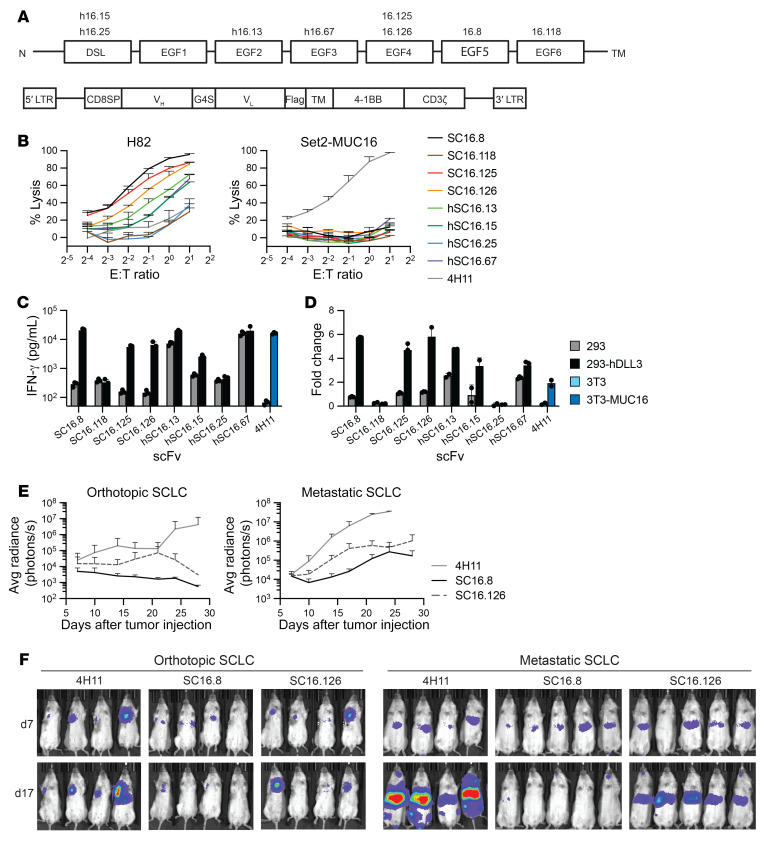
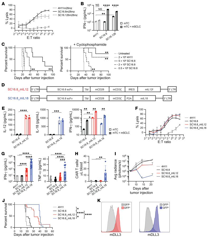
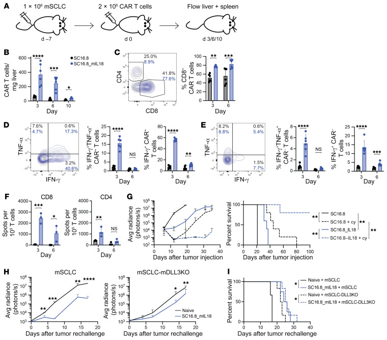
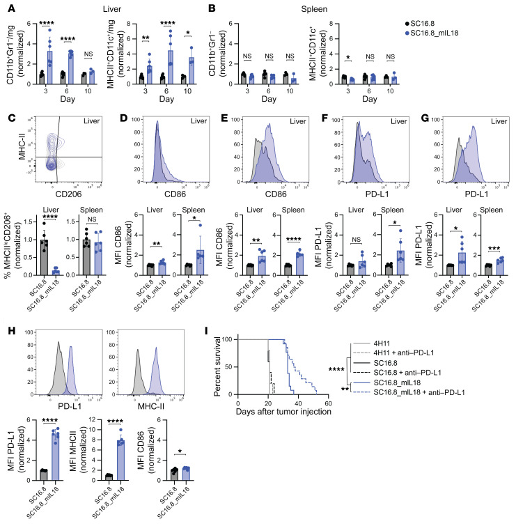
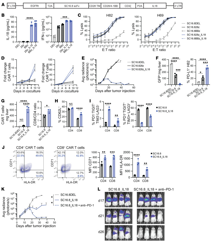
Patients with small cell lung cancer (SCLC) generally have a poor prognosis and a median overall survival of only about 13 months, indicating the urgent need for novel therapies. Delta-like protein 3 (DLL3) has been identified as a tumor-specific cell surface marker on neuroendocrine cancers, including SCLC. In this study, we developed a chimeric antigen receptor (CAR) against DLL3 that displays antitumor efficacy in xenograft and murine SCLC models. CAR T cell expression of the proinflammatory cytokine IL-18 greatly enhanced the potency of DLL3-targeting CAR T cell therapy. In a murine metastatic SCLC model, IL-18 production increased the activation of both CAR T cells and endogenous tumor-infiltrating lymphocytes. We also observed an increased infiltration, repolarization, and activation of antigen-presenting cells. Additionally, human IL-18-secreting anti-DLL3 CAR T cells showed an increased memory phenotype, less exhaustion, and induced durable responses in multiple SCLC models, an effect that could be further enhanced with anti-PD-1 blockade. All together, these results define DLL3-targeting CAR T cells that produce IL-18 as a potentially promising novel strategy against DLL3-expressing solid tumors.

Keywords: Cancer immunotherapy; Cellular immune response; Immunology; Lung cancer; Oncology.


Conflict of interest statement

Conflict of interest: CMR has consulted with AbbVie, Amgen, AstraZeneca, D2G, Daiichi Sankyo, Epizyme, Genentech/Roche, Ipsen, Jazz, Kowa, Lilly, Merck, and Syros on oncology drug development. He serves on the scientific advisory boards of Bridge Medicines, Earli, and Harpoon Therapeutics. RJB has licensed intellectual property to and collect royalties (US-7446190-B2 and US20190099447A1) from Bristol-Myers Squibb, Caribou, and Sanofi and has received research funding from Bristol-Myers Squibb. RJB is a consultant for Bristol-Myers Squibb, Atara Biotherapeutics Inc., CoImmune, and Triumvira and was previously a consultant for Gracell Biotechnologies Inc. (with employment ending within the past 30 months. RJB is a member of the CoImmune and Triumvira scientific advisory boards and has ownership interest (including patent PCT/US2017/045550) in IL-18.

Figures

Figure 1
Figure 1. Selection of scFv for effective CAR T cells against DLL3 on small cell lung cancer.
(A) Schematic of extracellular DLL3 domains with binding location of anti-DLL3 SC16 antibody clones (top), and schematic of CAR design for initial selection of single-chain variable fragments (bottom). LTR, long terminal repeats; CD8SP, CD8 signal peptide; VH, heavy chain; VL, light chain; TM, transmembrane domain; h, humanized. (B) Luciferase killing assay with CAR T cells cocultured with DLL3-expressing H82-SCLC or DLL3 Set2 cells. The Set2 cells overexpress MUC16 as positive control for the MUC16-targeting 4H11 CAR (n = 3). (C and D) DLL3-specific activation of the SC16.8, SC16.125, and SC16.126 CARs in cocultures with DLL3+ 293 cells, (C) as measured by IFN-γ production (see also Supplemental Figure 1 for IL-2, GM-CSF, and TNF-α levels) and (D) 7-day proliferation (n = 3). 3T3-MUC16 cells were used as positive control for the 4H11 CAR. (E and F) Orthotopic or metastatic H82-SCLC tumor growth following administration of the indicated CAR T cells (n = 4–5).
Figure 2
Figure 2. CAR T cell–secreted IL-12 and, especially, IL-18 prolong survival in murine SCLC model.
(A) Luciferase killing assay with murine CAR T cells and mSCLC cells that endogenously express murine DLL3 (n = 3). The CAR consists of the SC16.8 single-chain variable fragment (scFv) and murine CD28 and CD3ζ domains. (B) IFN-γ levels in supernatant of 24-hour coculture of murine CAR T cells with or without mSCLC cells. **P < 0.01, ****P < 0.0001 (2-way ANOVA, n = 2). (C) Survival of mice with metastatic mSCLC that were treated with the indicated doses of SC16.8m28mz CAR T cells (day 7) with or without 50 mg/kg cyclophosphamide pretreatment (day 6). **P < 0.01 (log-rank test, n = 4–6). (D) Schematic of CAR design with SC16.8 scFv, murine CD28 and CD3ζ signaling domains, and the murine Il12 fusion transgene or murine Il18 transgene. IRES, internal ribosome entry site. (E) IFN-γ production by the respective CAR T cells in the absence and presence of mSCLC cells. *P < 0.05, **P < 0.01, ***P < 0.001, ****P < 0.0001 (2-way ANOVA and Student’s t test, n = 2–3). (F) Luciferase killing assay with SC16.8 CAR T cells that express IL-12, IL-18, or no cytokine and 4H11 CAR T cells as negative control (n = 3). (G and H) Serum IFN-γ and TNF-α levels (n = 12–15) and circulating CAR T cells (n = 5) in the blood of mice collected 3 days after treatment with the indicated CAR T cells. **P < 0.01, ***P < 0.001, ****P < 0.0001 (1-way ANOVA). (I) Tumor growth in mice that were treated with 2 × 10⁶ CAR T cells intravenously 7 days after systemic mSCLC administration (n = 12–15). (J) Survival of mice in I. *P < 0.05, ****P < 0.0001 (log-rank test). (K) DLL3 surface expression on end-stage tumors from mice treated with SC16.8m28mz_mIL12 (red) or SC16.8m28mz_mIL18 (blue) CAR T cells.
Figure 3
Figure 3. IL-18 increases activation of both genetically engineered and endogenous T cells.
(A) Experimental setup for BE and Figure 4, A–H. See also Supplemental Figure 2, A–C. 2 × 10⁶ murine mCherry-expressing SC16.8m28mz or SC16.8m28mz_mIL18 CAR T cells were administered 7 days after systemic mSCLC injection. 3, 6, or 10 days later livers and spleens were harvested for flow cytometry analysis (day 3 and day 6, n = 6 from 2 independent experiments; day 10, n = 3). (B) Levels of CAR T cells detected in the liver over time. (C) Example (day 3) and quantification of the CD8+ CAR T cell population. (D and E) Example (day 3) and quantification of (D) intracellular IFN-γ and TNF-α in CAR+ T cells and (E) CAR endogenous T cells in the liver. (F) CD4+ or CD8+ mCherry endogenous T cells were sorted on day 3 and 6 after CAR T cell treatment and cocultured with mSCLC, and IFN-γ release was measured with an ELISpot assay (n = 3). (BF) *P < 0.05, **P < 0.01, ***P < 0.001, ****P < 0.0001 (2-way ANOVA). (G) Systemic mSCLC growth curve (left) and survival curve (right). Tumor-bearing mice were treated with low-dose 50 mg/kg cyclophosphamide (day 6) followed by low-dose 0.5 × 10⁶ CAR T cells (day 7). **P < 0.01 (log-rank test, n = 5). (H) Tumor growth of wild-type (n = 3–4) or DLL3KO mSCLC (n = 6, see Supplemental Figure 2D) in long-term surviving mice after cyclophosphamide plus SC16.8m28mz_mIL18 treatment with no evidence of tumor on day 42 (see G) compared with age-matched naive control mice. *P < 0.05, **P < 0.01, ***P < 0.001, ****P < 0.0001 (Student’s t test). (I) Survival of mice in H. *P < 0.05 (log-rank test, n = 6).
Figure 4
Figure 4. IL-18 recruits and reprograms myeloid cells in the tumor microenvironment.
(A and B) Levels of CD11b+Gr1 macrophages and MHC-II+CD11c+ dendritic cells detected (A) in the liver TME and (B) in the spleen 3, 6, and 10 days after CAR T cell treatment. *P < 0.05, **P < 0.01, ****P < 0.0001 (2-way ANOVA). (CH) Representative flow plots and quantification on day 6 after CAR T cell administration. See Supplemental Figure 3 for quantification on day 3, 6, and 10. *P < 0.05, **P < 0.01, ***P < 0.001, ****P < 0.0001 (Student’s t test). (C) Levels of CD206+MHC-IIlo M2-like macrophages. (D and E) Expression of activation marker CD86 on (D) macrophages and (E) dendritic cells in liver TME and spleen. (F and G) Expression of checkpoint molecule PD-L1 on (F) macrophages and (G) dendritic cells in liver TME and spleen. (H) Expression of PD-L1, MHC-II, and CD86 on F4/80+ macrophages in the liver TME. (I) Survival of mice with metastatic mSCLC that received 2 × 10⁶ CAR T cells on day 7 and 250 μg anti–PD-L1 antibody on days 14, 17, and 20. **P < 0.01, ****P < 0.0001 (log-rank test, n = 5 for 4H11 and SC16.8, n = 14 for SC16.8_mIL18).
Figure 5
Figure 5. Human DLL3-targeting CAR T cells that secrete IL-18 are effective against SCLC and can be further potentiated by late-onset anti–PD-1 therapy.
(A) Schematic overview of the CAR design. EGFRt, truncated EGFR; scFv, single-chain variant fragment. (B) Levels of IL-18 and IFN-γ in cell culture supernatant of cocultures of the indicated CAR T cells and DLL3-expressing target cells. ***P < 0.001, ****P < 0.0001 (Student’s t test, n = 4). (C) Luciferase killing assay with H82 (DLL3+) and H69 (DLL3lo) SCLC cells. (D) CAR T cell proliferation in cocultures with H82 or H69 SCLC cells (E/T ratio of 1:5, n = 3, representative result from 3 independent experiments). (E) Growth curve of metastatic H82-SCLC tumors in mice that received 1 × 10⁶ SC16.8 CAR T cells that did or did not secrete IL-18 (n = 4–5). (FJ) Livers of mice were subjected to flow cytometry analysis 9 days after CAR T cell treatment of mice with metastatic H82-SCLC tumors. *P < 0.05, **P < 0.01, ***P < 0.001, ****P < 0.0001 (2-way ANOVA, n = 3). (F) Cell count of GFP+ H82 tumor cells (left) and PD-L1 expression on these GFP+ cells (right). (G) Cell count of EGFRt+ CAR T cells in the liver (left) and CD8/CD4 ratio of the CAR T cells (right). (H) Quantification of CD62L+ central memory CAR T cells. (I) Quantification of CD4+ and CD8+ CAR T cells that are quadruple negative (left) or quadruple positive (right) for the exhaustion markers PD-1, TIGIT, TIM3, and LAG3. (J) Representative flow plots and mean fluorescence intensity (MFI) of T cell activation markers CD71 and HLA-DR on CAR T cells. (K) Growth curve of metastatic H82-SCLC tumors in mice that received 0.3 × 10⁶ CAR T cells alone or in combination with twice weekly 250 μg anti-human anti–PD-1 antibody, starting on day 18 (n = 4). (L) Bioluminescence imaging of same mice as in K, on days 17, 21, and 26 after tumor cell injection.

Comment in

References

    1. Rudin CM, et al. Small-cell lung cancer. Nat Rev Dis Primers. 2021;7(1):3. doi: 10.1038/s41572-020-00235-0. - DOI - PMC - PubMed
    1. Horn L, et al. First-line atezolizumab plus chemotherapy in extensive-stage small-cell lung cancer. N Engl J Med. 2018;379(23):2220–2229. doi: 10.1056/NEJMoa1809064. - DOI - PubMed
    1. Alexandrov LB, et al. Signatures of mutational processes in human cancer. Nature. 2013;500(7463):415–421. doi: 10.1038/nature12477. - DOI - PMC - PubMed
    1. Doyle A, et al. Markedly decreased expression of class I histocompatibility antigens, protein, and mRNA in human small-cell lung cancer. J Exp Med. 1985;161(5):1135–1151. doi: 10.1084/jem.161.5.1135. - DOI - PMC - PubMed
    1. Mahadevan NR, et al. Intrinsic immunogenicity of small cell lung carcinoma revealed by its cellular plasticity. Cancer Discov. 2021;11(8):1952–1969. doi: 10.1158/2159-8290.CD-20-0913. - DOI - PMC - PubMed

Publication types