. 2023 May 15;133(10):e162800.
doi: 10.1172/JCI162800.

Memory T cells possess an innate-like function in local protection from mucosal infection

Affiliations

Memory T cells possess an innate-like function in local protection from mucosal infection

Tanvi Arkatkar et al. J Clin Invest. .

Abstract

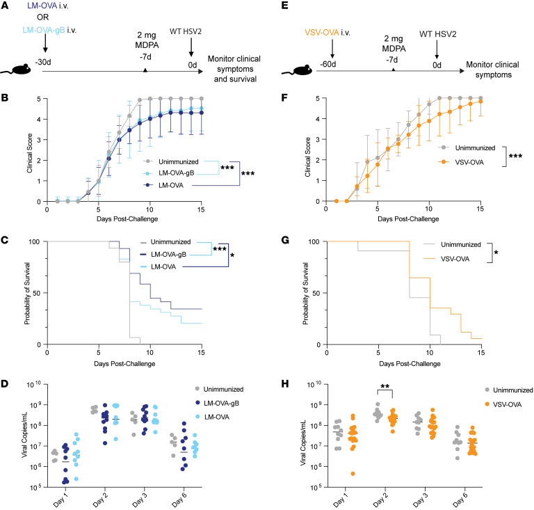
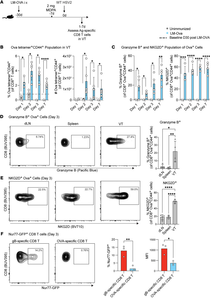
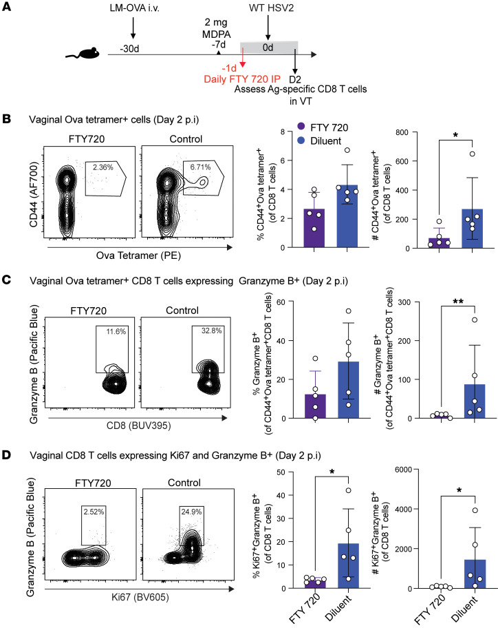
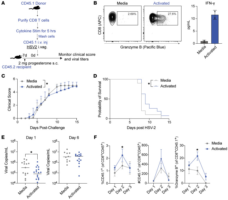
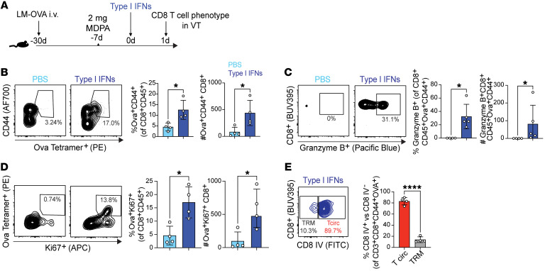
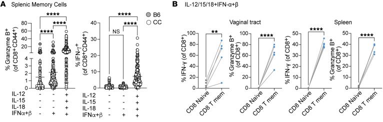
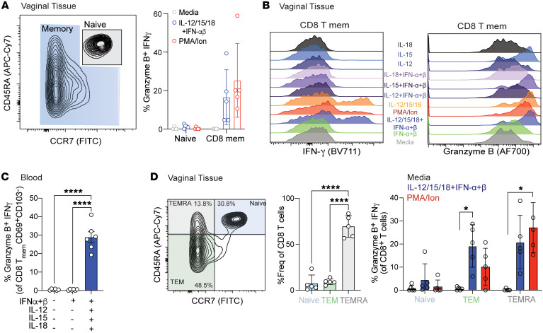
Mucosal infections pose a significant global health burden. Antigen-specific tissue-resident T cells are critical to maintaining barrier immunity. Previous studies in the context of systemic infection suggest that memory CD8+ T cells may also provide innate-like protection against antigenically unrelated pathogens independent of T cell receptor engagement. Whether bystander T cell activation is also an important defense mechanism in the mucosa is poorly understood. Here, we investigated whether innate-like memory CD8+ T cells could protect against a model mucosal virus infection, herpes simplex virus 2 (HSV-2). We found that immunization with an irrelevant antigen delayed disease progression from lethal HSV-2 challenge, suggesting that memory CD8+ T cells may mediate protection despite the lack of antigen specificity. Upon HSV-2 infection, we observed an early infiltration, rather than substantial local proliferation, of antigen-nonspecific CD8+ T cells, which became bystander-activated only within the infected mucosal tissue. Critically, we show that bystander-activated CD8+ T cells are sufficient to reduce early viral burden after HSV-2 infection. Finally, local cytokine cues within the tissue microenvironment after infection were sufficient for bystander activation of mucosal tissue memory CD8+ T cells from mice and humans. Altogether, our findings suggest that local bystander activation of CD8+ memory T cells contributes a fast and effective innate-like response to infection in mucosal tissue.

Keywords: Immunology; Infectious disease; T cells.


Conflict of interest statement

Conflict of interest: The authors have declared that no conflict of interest exists.

Figures

Figure 1
Figure 1. Antigen-nonspecific CD8+ T cells provide partial protection from genital HSV-2 infection.
(A) Experimental schematic to compare protective efficacy of LM-OVA-gB (HSV-specific) immunization and LM-OVA (HSV-nonspecific) immunization. (B and C) Mice were monitored for 2 weeks for clinical score (B) and survival (C) after lethal HSV-2 challenge. (D) Vaginal swabs were collected on days 1, 2, 3, and 6 from HSV-2–infected mice that were either unimmunized, LM-OVA–immunized, or immunized with LM-OVA-gB. Viral titers were measured by RT-PCR. (E) Experimental schematic to assess protective efficacy of VSV-OVA immunization. (F and G) Mice were monitored for 2 weeks for clinical score (F) and survival (G) after lethal HSV-2 challenge. (H) Vaginal swabs were collected on days 1, 2, 3, and 6 from HSV-2–infected mice that were either unimmunized or immunized with VSV-OVA. Viral titers were measured by real time PCR (RT-PCR). For viral titers in D and H, each dot represents an individual mouse and data are pooled from 2 or 3 experiments with 10–12 mice per group. Error bars represent mean ± SD. Statistical significance was determined by 2-way ANOVA with Tukey’s multiple comparisons for B and F, by log-rank test for C and G, and by unpaired t test for D and H. *P < 0.05, **P < 0.01, ***P < 0.001.
Figure 2
Figure 2. OVA-specific CD8+ T cells are present in the vagina after HSV-2 infection and display a bystander-activated phenotype.
(A) Experimental schematic to generate an OVA-specific memory compartment followed by intravaginal challenge with wild-type (WT) HSV-2. Mice were euthanized on days 1–7 after WT HSV-2 challenge to assess OVA-specific CD8+ T cells in the vaginal tract (VT). (B) Frequency and counts of CD44+ OVA tetramer–specific cells on days 1–7 after HSV-2 challenge in LM-OVA–immunized mice. Dashed lines indicate mean of OVA-specific cells in the VT of LM-OVA–immunized mice prior to HSV-2 challenge. (C) Frequency of granzyme B+ and NKG2D+ population gated on OVA tetramer–positive population. (D and E) Frequency of granzyme B+ and NKG2D+ population gated on OVA tetramer–positive population within draining lymph nodes (dLN; iliac and inguinal), spleen, and VT. Each dot represents an individual mouse, and data are representative of 1–3 experiments with 4–5 mice per group. (F) HSV gB-specific and OVA-specific memory compartments were generated by immunization of Nur77-GFP mice with LM-OVA-gB or LM-OVA followed by intravaginal challenge with WT HSV-2. Mice were euthanized on day 3 after WT HSV-2 challenge to assess Nur77-expressing populations within gB- and OVA-specific CD8+ T cells in the VT. Red bars indicate HSV gB-specific population within LM-OVA-gB–immunized mice, and blue bars indicate OVA-specific population within LM-OVA–immunized mice. Data are representative of 3 experiments, each with 3 mice per group. Error bars represent mean ± SD. Statistical significance was determined by unpaired t test for B, C, and F, and 1-way ANOVA for D and E. *P < 0.05, **P < 0.01, ***P < 0.001, ****P < 0.0001.
Figure 3
Figure 3. FTY720 treatment prevents accumulation of bystander-activated OVA-specific CD8+ T cells in the infected mucosa.
(A) Experimental schematic to generate OVA-specific memory compartment followed by intravaginal challenge with WT HSV-2. FTY720 (1 mg/kg) or 2% cyclodextrin was administered by an intraperitoneal route on days –1, 0, 1, and 2 relative to HSV-2 infection. (B) Representative flow plots and graphs to assess OVA tetramer–positive subset in the VT in LM-OVA–immunized mice infected with WT HSV-2 that received either FTY720 or diluent (2% cyclodextrin). (C) Representative flow plots and graphs of granzyme B–expressing population within OVA+CD44+ subset. (D) Representative flow plots and graphs of Ki67+granzyme B+ population plotted on total CD8 gate. Each dot represents an individual mouse, and data are representative of 2 experiments with 4–5 mice per group. Error bars represent mean ± SD. Statistical significance was determined by unpaired t test. *P < 0.05, **P < 0.01.
Figure 4
Figure 4. Adoptively transferred bystander-activated CD8+ T cells delay the progression of lethal HSV-2 infection and lower the viral burden.
(A) Experimental outline to compare the viral burden of mice receiving CD8+ T cells stimulated with medium or cytokines (IL-12/15/18 plus IFN-α/β) after WT HSV-2 challenge. CD8+ T cells were purified from splenocytes and draining lymph nodes and were derived from CD45.1 donor mice. The cells were stimulated with medium (RP10) or cytokines (10 ng/mL IL-12/15/18 plus 1,000 U IFN-α/β; activated) for 5 hours and washed before intravenous injection into CD45.2 recipient mice. (B) Representative flow plots showing intracellular granzyme B staining in medium- and cytokine-treated memory CD8+ T cells at 5 hours. IFN-γ levels were measured by ELISA from supernatants obtained after 5 hours of incubation. (C and D) Mice were monitored for clinical score and survival after the lethal HSV-2 challenge. (E) Vaginal washes were obtained after HSV-2 infection, and viral titers were measured by RT-PCR on days 1 and 6 after HSV-2 infection. (F) Mice were euthanized on days 1–3 after WT HSV-2 challenge and adoptive transfer to assess frequency and counts of donor CD45.1+ CD8+ T cells in the VT. Percent frequency of granzyme B+ population was gated on CD45.1+ CD8+ T cells. Data are pooled from at least 3 independent experiments with n = 5–10 for the control and experimental groups. Error bars represent mean ± SEM. Statistical significance was determined by 2-way ANOVA with Tukey’s multiple comparisons and by unpaired t test (E and F). *P < 0.05.
Figure 5
Figure 5. Vaginal memory CD8+ T cells increase in numbers and function in vivo upon type I IFN treatment.
(A) Experimental schematic to assess CD8+ T cell phenotype after intravaginal treatment with type I IFNs in LM-OVA–immunized mice. (B) Representative flow plots and graphs to assess OVA tetramer–positive subset in the VT in LM-OVA–immunized mice that received either PBS alone or IFN-α/β. (C) Representative flow plots and graphs of granzyme B–expressing CD62L CD8+ T cells within the OVA tetramer–positive population. (D) Representative flow plots and graphs showing the OVA tetramer– and Ki67-positive population plotted on total CD8 gate. (E) Mice received intravenous injection with anti-CD8 antibody (CD8 IV) 5 minutes before euthanasia. OVA+CD44+ population was categorized as circulating T cells (CD8 IV+) and TRM (CD8 IV) by intravascular staining. Each dot represents an individual mouse, and data are representative of at least 2 experiments with 4–5 mice per group. Error bars represent mean ± SD. Statistical significance was determined by Mann-Whitney test. *P < 0.05, ****P < 0.0001.
Figure 6
Figure 6. Memory CD8+ T cells acquire a bystander phenotype upon cytokine exposure.
(A) Splenocytes from C57BL/6 (gray dots) and Collaborative Cross (CC-RIX) mice (white dots) were cultured in vitro for 24 hours with IFN-α/β (1,000 U) alone or with IL-12/15/18 (100 ng/mL). Granzyme B and IFN-γ expression was assessed within activated memory CD8+ T cells (CD44+) gated by flow cytometry. (B) Vaginal cells and splenic cells from LM-OVA memory mice were cultured in vitro for 24 hours with IFN-α/β (1,000 U) and IL-12/15/18 (100 ng/mL). IFN-γ and granzyme B expression within naive (CD44) and memory CD8+ T (CD44+) cell populations was measured. Each dot represents an individual mouse, and data are representative of at least 2 experiments with 4–10 mice per mouse strain. Error bars represent mean ± SD. Statistical significance was determined by 1-way ANOVA with Tukey’s multiple comparisons (A) or unpaired t test (B). **P < 0.01, ****P < 0.0001.
Figure 7
Figure 7. Human memory CD8+ T cells in the vaginal tissue acquire bystander phenotype upon cytokine treatment.
Cells from vaginal tissues obtained from prolapse repair surgeries were cultured in vitro for 24 hours with varying combinations of IFN-α/β (1,000 U) and IL-12/15/18 (100 ng/mL). (A) Left: Representative flow plot shows the distribution of CD8+ T cells based on CD45RA and CCR7 markers highlighting memory (blue) and naive (gray) compartments. Right: Graph plot shows coexpression of granzyme B and IFN-γ within each compartment after treatment with medium or cytokines. Each dot represents an individual condition, and the color code for each condition is the same as in B. (B) Staggered histogram separated per different cytokine treatments showing IFN-γ and granzyme B expression within the memory CD8+ T cell compartment. (C) Human PBMCs were cultured for 24 hours with IFN-α/β (1,000 U) alone or with IL-12/15/18 (100 ng/mL). Graph plots show memory CD8+ T cells expressing granzyme B and IFN-γ with varying cytokine combinations. (D) Flow plot shows the distribution of CD8+ T cells as naive (CCR7+CD45RA+), TEM (CCR7CD45RA), and TEMRA (CCR7CD45RA+). Graph plot represents each CD8+ T cell subset within an individual donor followed by expression of granzyme B and IFN-γ within each subset. Each dot represents an individual donor, and data are pooled from 5 separate donors. Error bars represent mean ± SD. Statistical significance was determined by 1-way ANOVA with Tukey’s multiple comparisons. *P < 0.05, ****P < 0.0001.

Similar articles

Cited by

References

    1. Ariotti S, et al. T cell memory. Skin-resident memory CD8+ T cells trigger a state of tissue-wide pathogen alert. Science. 2014;346(6205):101–105. doi: 10.1126/science.1254803. - DOI - PubMed
    1. Schenkel JM, et al. T cell memory. Resident memory CD8+ T cells trigger protective innate and adaptive immune responses. Science. 2014;346(6205):98–101. doi: 10.1126/science.1254536. - DOI - PMC - PubMed
    1. Berg RE, Forman J. The role of CD8 T cells in innate immunity and in antigen non-specific protection. Curr Opin Immunol. 2006;18(3):338–343. doi: 10.1016/j.coi.2006.03.010. - DOI - PubMed
    1. Maurice NJ, et al. The ugly duckling turned to swan: a change in perception of bystander-activated memory CD8 T cells. J Immunol. 2021;206(3):455–462. doi: 10.4049/jimmunol.2000937. - DOI - PMC - PubMed
    1. Raué HP, et al. Cytokine-mediated programmed proliferation of virus-specific CD8(+) memory T cells. Immunity. 2013;38(1):131–139. doi: 10.1016/j.immuni.2012.09.019. - DOI - PMC - PubMed

Publication types