. 2023 May 15;133(10):e164575.
doi: 10.1172/JCI164575.

Intrathecal AAV9/AP4M1 gene therapy for hereditary spastic paraplegia 50 shows safety and efficacy in preclinical studies

Affiliations

Intrathecal AAV9/AP4M1 gene therapy for hereditary spastic paraplegia 50 shows safety and efficacy in preclinical studies

Xin Chen et al. J Clin Invest. .

Abstract

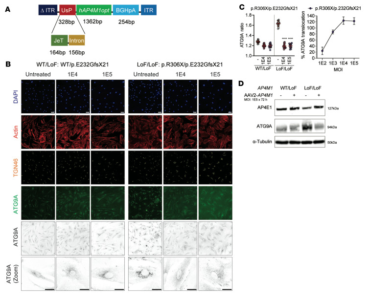
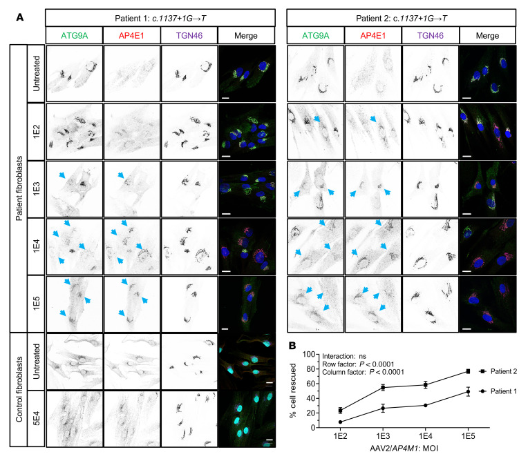
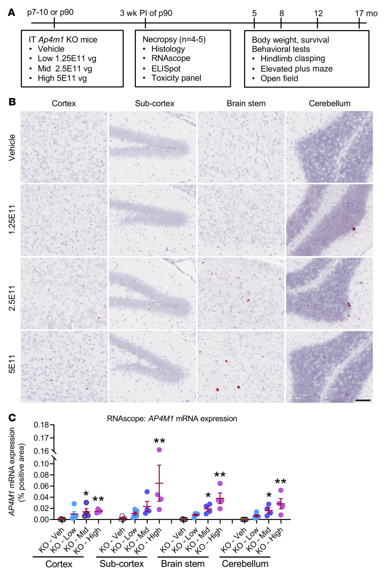
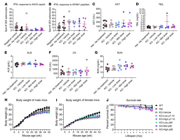
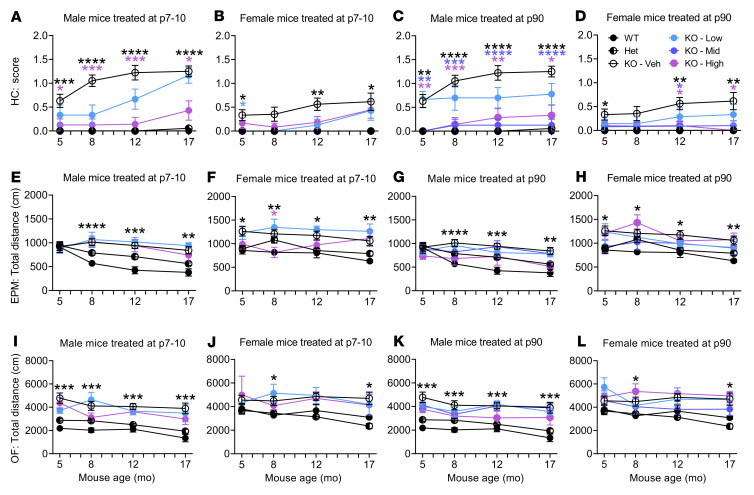
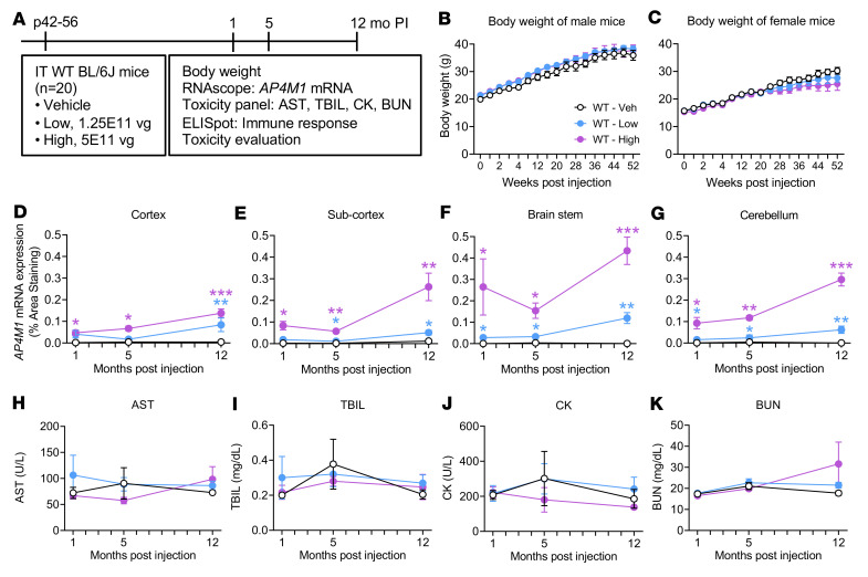
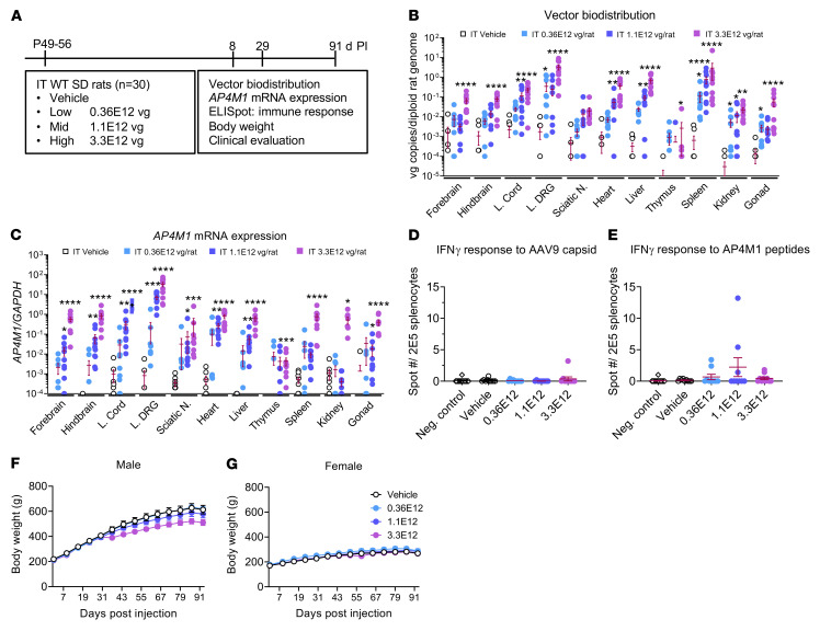
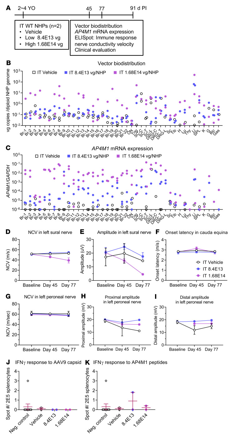
Spastic paraplegia 50 (SPG50) is an ultrarare childhood-onset neurological disorder caused by biallelic loss-of-function variants in the AP4M1 gene. SPG50 is characterized by progressive spastic paraplegia, global developmental delay, and subsequent intellectual disability, secondary microcephaly, and epilepsy. We preformed preclinical studies evaluating an adeno-associated virus (AAV)/AP4M1 gene therapy for SPG50 and describe in vitro studies that demonstrate transduction of patient-derived fibroblasts with AAV2/AP4M1, resulting in phenotypic rescue. To evaluate efficacy in vivo, Ap4m1-KO mice were intrathecally (i.t.) injected with 5 × 10¹¹, 2.5 × 10¹¹, or 1.25 × 10¹¹ vector genome (vg) doses of AAV9/AP4M1 at P7-P10 or P90. Age- and dose-dependent effects were observed, with early intervention and higher doses achieving the best therapeutic benefits. In parallel, three toxicology studies in WT mice, rats, and nonhuman primates (NHPs) demonstrated that AAV9/AP4M1 had an acceptable safety profile up to a target human dose of 1 × 10¹⁵ vg. Of note, similar degrees of minimal-to-mild dorsal root ganglia (DRG) toxicity were observed in both rats and NHPs, supporting the use of rats to monitor DRG toxicity in future i.t. AAV studies. These preclinical results identify an acceptably safe and efficacious dose of i.t.-administered AAV9/AP4M1, supporting an investigational gene transfer clinical trial to treat SPG50.

Keywords: Neurological disorders; Neuroscience.


Conflict of interest statement

Conflict of interest: SJG received patent royalties for intellectual property (patent US7465583B2) licensed to Asklepios Biophama, but this intellectual property was not used in this study. UT Southwestern Medical Center, SJG, and XC may benefit from the potential future commercialization of the technology described herein (US provisional patent application 63/177,559).

Figures

Figure 1
Figure 1. AAV2/AP4M1 vector construct expressing human AP4M1 and its restoration of ATG9A trafficking and AP4E1 level in primary fibroblasts from a patient with SPG50.
(A) Schematic of AAV2/AP4M1 construct comprising a mutant AAV2 ITR with the D element deleted (Δ ITR), UsP promoter (JeT + Intron), hAP4M1opt, synthetic BGHpA signal, and WT AAV2 ITR. (B–D) Fibroblasts from a clinically unaffected heterozygous carrier (same-sex parent, WT/pE232GfsX21) or patient with biallelic LoF variants in AP4M1 (p.R306X/pE232GfsX21) were treated with AAV2/AP4M1 vector at an MOI of 1 × 10⁴ or 1 × 10⁵ for 72 hours. The fibroblasts were then fixed for (B and C) immunocytochemistry and automated image analysis or harvested for (D) Western blotting. Scale bar: 20 μm. (C) Left: The mean ATG9A ratio for all conditions. Scatter plots show the mean ± SD for each well (n = 16 wells/group). Data sets were compared using 1-way ANOVA, with α set at 0.05, and Dunnett’s correction. Right: The percentage translocation of ATG9A. Translocation refers to the change (in %) relative to the difference between the positive (ATG9A ratio of fibroblasts with heterozygous AP4M1 variants) and negative controls (ATG9A ratio of fibroblasts with homozygous LoF variants in AP4M1) of the same assay plate. A dose-dependent effect becomes evident. ****P < 0.0001 compared with untreated fibroblasts. (D) Western blot of whole-cell lysates of fibroblasts from a clinically unaffected heterozygous carrier (parent) and patient with SPG50 treated with AAV2/AP4M1 at an MOI of 1 × 10⁵ for 72 hours. AAV2, adeno-associated virus 2; AP4E1, adaptor protein complex, subunit ɛ; AP4M1, adaptor protein complex, subunit μ4; ATG9A, autophagy-related protein 9A; BGHpA, bovine growth hormone polyadenylation; hAP4M1opt, human AP4M1 codon-optimized coding sequence; ITR, inverted terminal repeat; LoF, loss of function; TGN, trans-Golgi network.
Figure 2
Figure 2. AAV2/AP4M1 vector restored ATG9A trafficking and AP4E1 levels in primary fibroblasts from patients with SPG50.
(A) Fibroblasts from 2 sibling patients with a donor splice site pathogenic mutation in intron 14 of the AP4M1 gene (c.1137+1GT) or normal control fibroblasts were treated without or with AAV2/AP4M1 vector at the indicated MOI for 72 hours. Fibroblasts were then fixed for immunofluorescence analysis of ATG9A, AP4E1, and TGN46, as described in Methods. Nuclei were stained with DAPI (in blue, merge). Single channels are shown in inverted grayscale. Note that expression of AP4M1 by the viral vector caused dispersal of the ATG9A signal and increased AP4E1 staining at the TGN (i.e., phenotypic rescue; indicated by arrows). Scale bar: 20 μm. (B) The percentage of rescued cells was counted and is represented as the mean ± SEM from 2 independent experiments (see Supplemental Table 2 for details). Statistical analysis was done using 2-way ANOVA with repeated measures. AAV2, adeno-associated virus 2; AP4E1, adaptor protein complex, subunit ɛ; AP4M1, adaptor protein complex, subunit μ4; ATG9A, autophagy-related protein 9A; SPG50, spastic paraplegia 50; TGN, trans-Golgi network.
Figure 3
Figure 3. The experimental design for in vivo efficacy study and increased AP4M1 mRNA expression in the CNS of Ap4m1-KO mice following i.t. treatment with AAV9/AP4M1.
(A) Vehicle or low (1.25 × 10¹¹ vg/mouse), mid (2.5 × 10¹¹ vg/mouse), or high (5 × 10¹¹ vg/mouse) doses of AAV9/AP4M1 vector were administered intrathecally to balanced numbers of male and female Ap4m1-KO mice at P7–P10 (before manifesting) or P90 (early manifestation). Study readouts at each time point at specified ages are listed from left to right. (B) Brains from mice treated at P90 for 3 weeks were analyzed by RNAscope staining to detect hAP4M1opt mRNA. Histology images with 1 section per animal were digitized with a ScanScope slide scanner and analyzed using custom analysis settings in HALO Image Analysis Platform. Scale bars: 100 μm. (C) Results are presented as percentage area positively stained for hAP4M1opt mRNA in the indicated brain regions. Each data point represents measurement from an individual animal (n = 4–5), with lines representing the mean ± SEM. Data sets that passed tests for normality or homogeneity of variance were analyzed using 1-way ANOVA, with α set at 0.05, and Dunnett’s correction for relevant pairwise comparisons. Data sets that did not pass tests for normality or homogeneity of variance were analyzed using Kruskal-Wallis test, with α set at 0.05, and Dunn’s correction for relevant pairwise comparisons. *P < 0.05, **P < 0.01 compared with KO mice treated with vehicle (Veh). AAV9, adeno-associated virus 9; AP4M1, adaptor protein complex, subunit μ4; hAP4M1opt, human AP4M1 codon-optimized coding sequence; i.t., intrathecal; PI, postinjection; vg, vector genome.
Figure 4
Figure 4. i.t. AAV9/AP4M1 treatment generated minimal IFN-γ responses to AAV9 capsid or AP4M1 peptides, minimal effects on serum toxicity panels, male or female body weight, or survival rates.
(A–G) Vehicle or low (1.25 × 10¹¹ vg/mouse), mid (2.5 × 10¹¹ vg/mouse), or high (5 × 10¹¹ vg/mouse) doses of AAV9/AP4M1 vector were administered intrathecally to balanced numbers of male and female KO mice at P90, with WT and Het mice as normal controls. At 3 weeks after injection, (A and B) mouse splenocytes and serum were collected for ELISpot and (C–G) toxicity panel analyses. Each data point represents a measurement from an individual animal (n = 5–8), with lines representing the mean ± SEM. Data sets that passed tests for normality or homogeneity of variance were analyzed using 1-way ANOVA, with α set at 0.05, and Dunnett’s correction for relevant pairwise comparisons. Data sets that did not pass tests for normality or homogeneity of variance were analyzed using Kruskal-Wallis test, with α set at 0.05, and Dunn’s correction for relevant pairwise comparisons. No significant differences were observed. (H and I) Male (n = 7–26) and female (n = 5–24) mouse body weights were monitored up to 52 weeks of age. Two-way ANOVA with repeated measures was used for statistical analysis. (J) Mouse survival shown with Kaplan-Meier survival curves compared with log-rank (Mantel-Cox) test. No significant differences were observed. AAV9, adeno-associated virus 9; ALB, albumin; AP4M1, adaptor protein complex, subunit μ4; AST, aspartate transaminase; BUN, blood urea nitrogen; CK, creatine kinase; Het, heterozygotes; i.t., intrathecal; TBIL, total bilirubin; vg, vector genome.
Figure 5
Figure 5. i.t. AAV9/AP4M1 treatment partially improved abnormal behavioral phenotypes in Ap4m1-KO mice.
Vehicle or low (1.25 × 10¹¹ vg/mouse), mid (2.5 × 10¹¹ vg/mouse), or high (5 × 10¹¹ vg/mouse) doses of AAV9/AP4M1 vector were administered intrathecally to balanced numbers of male and female mice at P7–P10 or P90, with WT and Het mice as normal controls. The mice were subjected to (A–D) hind limb clasping, (E–H) elevated plus maze, and (I–L) open-field tests at 5, 8, 12, and 17 months of age. All data are presented as mean ± SEM (male, n = 7–26, and female, n = 5–24). Two-way ANOVA with repeated measures was used for statistical analysis. *P < 0.05, **P < 0.01, ***P < 0.001, ****P < 0.0001 compared with KO mice treated with vehicle (Veh). AAV9, adeno-associated virus 9; AP4M1, adaptor protein complex, subunit μ4; EPM, elevated plus maze test; HC, hind limb clasping test; Het, Heterozygotes; i.t., intrathecal; OF, open-field test; vg, vector genome.
Figure 6
Figure 6. Experimental design for in vivo safety study in WT mice and changes in body weight, hAP4M1opt mRNA expression in the CNS, and serum toxicity panels in i.t. AAV9/AP4M1-treated mice.
(A) Vehicle or low (1.25 × 10¹¹ vg/mouse), or high (5 × 10¹¹ vg/mouse) doses of AAV9/AP4M1 vector were administered intrathecally to male and female mice at P42–P56 (n = 10/group/sex). Study readouts at each time point and specified ages are listed from left to right. (B and C) Body weights were monitored regularly up to 52 weeks of age. At 1, 5, and 12 months after injection, mouse brain and serum were harvested for RNAscope staining (D–G) to detect hAP4M1opt mRNA expression and (H–K) for assessment of serum toxicity, respectively. Histology images (1 section per animal) were digitized with a ScanScope slide scanner and analyzed using custom analysis settings in HALO Image Analysis Platform. (D–G) Results are presented as percentage area staining positive for hAP4M1opt mRNA by tissue region. All data are presented as the mean ± SEM. Two-way ANOVA with repeated measures was used for statistical analysis. *P < 0.05, **P < 0.01, ***P < 0.001 compared with WT mice treated with vehicle. AAV9, adeno-associated virus 9; AP4M1, adaptor protein complex, subunit μ4; AST, aspartate transaminase; BUN, blood urea nitrogen; CK, creatine kinase; hAP4M1opt, human AP4M1 codon-optimized coding sequence; i.t., intrathecal; PI, postinjection; TBIL, total bilirubin; vg, vector genome.
Figure 7
Figure 7. Experimental design for in vivo safety study in WT rats and vector biodistribution, AP4M1 mRNA expression, effects on IFN-γ responses to AAV9 capsid or AP4M1 peptides, and body weight in i.t. AAV9/AP4M1-treated rats.
(A) Vehicle or low (0.36 × 10¹² vg/rat), mid (1.1 × 1012 × 1012 vg/rat), or high (3.3 × 10¹² vg/rat) doses of AAV9/AP4M1 vector were administered intrathecally to male and female rats at P49–P56 (n = 5/group/sex). Study readouts at each time point and specified ages are listed from left to right. At 29 days after injection, rat organs were harvested for (B) measurement of vector biodistribution and (C) AP4M1 mRNA expression by qPCR. Rat splenocytes were prepared for IFN-γ responses to (D) AAV9 capsid or (E) AP4M1 peptides by ELISpot. (F and G) Rat body weights were monitored regularly up to 91 days after injection. All data in B–G are presented as the mean ± SEM. (B–E) Data sets that passed tests for normality or homogeneity of variance were analyzed using 1-way ANOVA, with α set at 0.05, and Dunnett’s correction for relevant pairwise comparisons. Data sets that did not pass tests for normality or homogeneity of variance were analyzed using Kruskal-Wallis test, with α set at 0.05, and Dunn’s correction for relevant pairwise comparisons. *P < 0.05, **P < 0.01, ***P < 0.001, ****P < 0.0001 compared with i.t. vehicle. (F and G) Two-way ANOVA with repeated measures was used for statistical analysis of male and female rat body weight. AAV9, adeno-associated virus 9; AP4M1, adaptor protein complex, subunit μ4; i.t., intrathecal; L. Cord, lumbar spinal cord; L. DRG, lumbar dorsal root ganglion; Sciatic N., sciatic nerve; Neg, negative; PI, postinjection; SD, Sprague-Dawley; vg, vector genome.
Figure 8
Figure 8. Experimental design for in vivo safety study in WT NHPs and vector biodistribution, AP4M1 mRNA expression, effects on NCV or amplitude in peripheral nerves, and IFN-γ responses to AAV9 capsid or AP4M1 peptides in i.t. AAV9/AP4M1-treated NHPs.
(A) Vehicle or low (8.4 × 10¹³ vg/NHP), or high (1.68 × 10¹⁴ vg/NHP) doses of AAV9/AP4M1 vector were administered intrathecally to NHPs at 2–4 years of age (n = 2/group). Study readouts at each time point and specified ages are listed from left to right. At 91 days after injection, monkey organs were harvested for (B) measurement of vector biodistribution and (C) AP4M1 mRNA expression by qPCR. (D–I) NCV tests were performed at baseline and day 45 and day 77 after injection. Monkey splenocytes were prepared for IFN-γ responses to (J) AAV9 capsid or (K) AP4M1 peptides by ELISpot. Each dot in B and C represents an individual monkey. All data in D–K are presented as the mean measurement ± SEM. AAV9, adeno-associated virus 9; AP4M1, adaptor protein complex, subunit μ4; i.t., intrathecal; YO, years old; PI, postinjection; NHPs, nonhuman primates; vg, vector genome; Br, brain; ON, optic nerve; Trig, trigeminal ganglion; SC-C, cervical spinal cord; SC-T, thoracic spinal cord; SC-L, lumbar spinal cord; DRG-C, cervical dorsal root ganglion; DRG-T, thoracic dorsal root ganglion; DRG-L, lumbar dorsal root ganglion; Sc, sciatic nerve; Tib, tibia nerve; H, heart; Lu, lung; Thy, Thymus; Li, liver; SPL, spleen; K, kidney; G, gonad; Bic, biceps femoris; Gas, gastrocnemius; NCV, nerve conduction velocity; Neg, Negative.

Comment in

References

    1. Ebrahimi-Fakhari D, et al. Defining the clinical, molecular and imaging spectrum of adaptor protein complex 4-associated hereditary spastic paraplegia. Brain. 2020;143(10):2929–2944. - PMC - PubMed
    1. Ebrahimi-Fakhari D, et al. AP-4-Associated hereditary spastic paraplegia. In: Adam MP, et al., eds. GeneReviews. University of Washington; 2018:1993–2023. https://www.ncbi.nlm.nih.gov/books/NBK535153/ Accessed May 3, 2023.
    1. Verkerk AJ, et al. Mutation in the AP4M1 gene provides a model for neuroaxonal injury in cerebral palsy. Am J Hum Genet. 2009;85(1):40–52. doi: 10.1016/j.ajhg.2009.06.004. - DOI - PMC - PubMed
    1. Ebrahimi-Fakhari D, et al. Systematic analysis of brain MRI findings in adaptor protein complex 4-associated hereditary spastic paraplegia. Neurology. 2021;97(19):e1942–e1954. doi: 10.1212/WNL.0000000000012836. - DOI - PMC - PubMed
    1. Dell’Angelica EC, et al. AP-4, a novel protein complex related to clathrin adaptors. J Biol Chem. 1999;274(11):7278–7285. doi: 10.1074/jbc.274.11.7278. - DOI - PubMed

Publication types