. 2023 Aug;52(8):1557-1566.
doi: 10.1007/s00256-023-04323-6. Epub 2023 Mar 23.

Scaphoid kinematics in scapholunate instability: a dynamic CT study

Affiliations

Scaphoid kinematics in scapholunate instability: a dynamic CT study

Melanie Amarasooriya et al. Skeletal Radiol. 2023 Aug.

Abstract

Objective: The scaphoid is proposed to be driven by the distal carpal row in scapholunate instability (SLI) as it is dissociated from the proximal row. The aim of this study was to describe the 6 degrees of freedom kinematics of the scaphoid using dynamic CT in the normal and SLI wrists. We hypothesised that the SLI scaphoid would demonstrate kinematic evidence conforming to distal row motion.

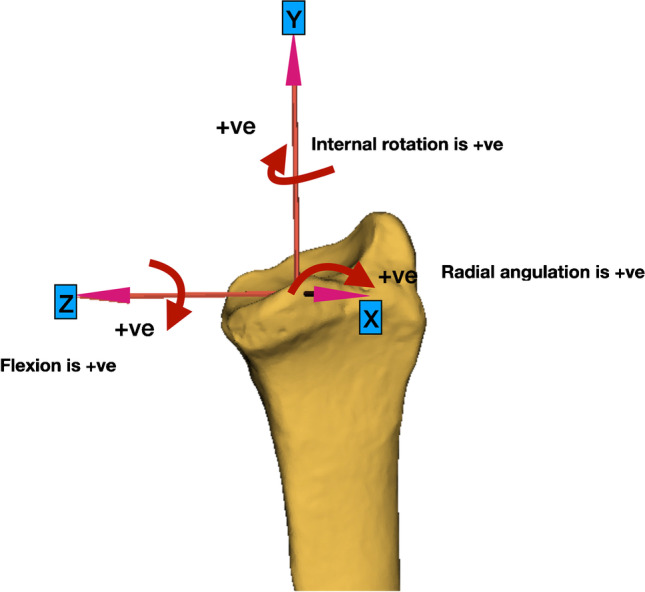
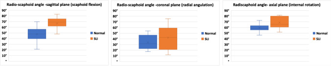
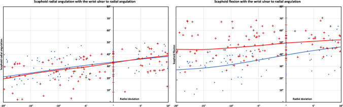
Materials and methods: We studied dynamic CT scans of 17 SLI and 17 normal wrists during ulnar to radial deviation and extension to flexion. The radio-scaphoid angles in three anatomic planes were calculated in the wrist neutral position and during wrist motion. The centroid position was also calculated in the wrist neutral position and during wrist motion. The scapho-capitate motion index (SCI) was calculated as a ratio between the scaphoid and the capitate motion.

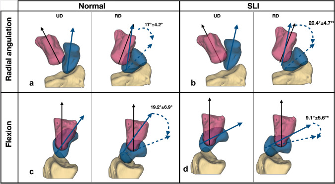
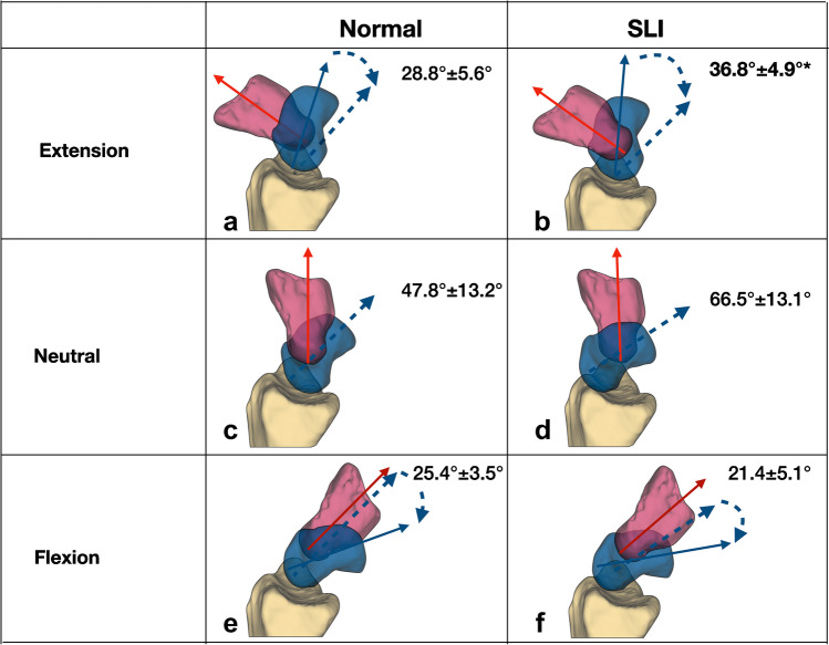
Results: In the neutral position of the wrist, the SLI scaphoid was flexed, internally rotated, and radially translated compared to the normal scaphoid. During wrist motion, the SLI scaphoid had more 'in-plane' motion and less 'out-of-plane' motion with a higher SCI during wrist neutral to radial deviation and extension to neutral.

Conclusion: We have described the malalignment of the SLI scaphoid in the neutral position of the wrist and 6 degrees of freedom kinematics during wrist motion of the SLI scaphoid compared to the normal. The SLI scaphoid conformed more to the distal row motion than the normal scaphoid. This information may help define the surgical reconstruction techniques for SLI.

Keywords: Carpal instability; Dynamic CT; Scaphoid kinematics; Scapholunate.


Conflict of interest statement

The authors declare no competing interests.

Figures

Fig. 1
Fig. 1
Scaphoid positions during wrist ulnar to radial deviation, with its relation to the radius. The scaphoid is segmented at each time point and saved as a 3-dimensional mesh in the stereolithography (STL) format. The image shows the relationship of the scaphoid to the radius. Every 4th consecutive frame/time point of a single representative normal wrist is depicted
Fig. 2
Fig. 2
Radius coordinate system and definitions of directions of motion. The iso-centre was at the midpoint of the ridge between the scaphoid and the lunate facets. The y-axis is the longitudinal axis of the radius. The z-axis is the line perpendicular to the y-axis, passing from the iso-centre to the radial styloid. The x-axis is orthogonal to z and y. Positive rotation is illustrated with arrows in flexion, radial, and distal directions
Fig. 3
Fig. 3
The radio-scaphoid angle of the normal and the SLI wrists with the wrist in the neutral position, in three anatomic planes, coronal, sagittal, and axial. Note that the SLI scaphoid is more flexed and internally rotated than the normal scaphoid
Fig. 4
Fig. 4
Scaphoid angulations during the wrist ulnar to radial deviation a. Scaphoid radial angulation. (in-plane motion) The SLI scaphoid had more radial angulation (in-plane motion) (p < 0.05) than the normal scaphoid. b. Scaphoid flexion (out-of-plane motion). The SLI scaphoid is more flexed than the normal scaphoid. The flexion arc of the SLI scaphoid is less than the flexion arc of the normal scaphoid (p < 0.01). The results presented are the radio-scaphoid angle, with the wrist moving from radial to ulnar deviation. The markers indicate the individual raw data points, and the lines are the mean radio-scaphoid angle following linear interpolation
Fig. 5
Fig. 5
Scaphoid angulations during wrist ulnar to radial deviation. Scaphoid radial angulation (in-plane motion), a normal 17° ± 4.2° (SCI 56.8%). b SLI 20.4° ±4.7°, (SCI 68%). *(p < 0.05). Flexion (out of plane motion). c Normal 19.2° ±6.9° d. SLI 9.1° ±5.6°* (p < 0.01). The wrist is moving from 20° ulnar to 10° radial deviation, indicated by the black arrow on the capitate. Images are from a single representative right wrist (volar a and b, ulnar c and d views) with mean and standard deviation for the arc of scaphoid motion mentioned
Fig. 6
Fig. 6
Scaphoid angulations during wrist extension to flexion. During wrist extension to neutral, there was more scaphoid flexion in the SLI wrists. SLI wrists had a greater scapho-capitate motion index than the normal wrist. a Normal 28.8° ± 5.6°, 92%, b SLI 36.8° ± 4.9, 71%. p < 0.01. The wrist in the neutral position, the SLI scaphoid (d) was more flexed than the normal scaphoid (c) (SLI 66.5°± 13.1° vs normal 47.8° ± 13.2° p < 0.01) During wrist flexion, the SLI wrist motion was not significantly different. e Normal 25.4° ± 3.5°, 82%, f SLI 21.4° ± 5.1°, 71%, p = 0.16. The results presented are the arc scaphoid motion, indicated by the curved blue arrow. Images are the ulnar views of a single representative right wrist with mean and standard deviation
Fig. 7
Fig. 7
Scaphoid flexion during wrist extension to flexion. During wrist extension to the neutral position, the ‘in-plane motion of the SLI scaphoid’ (SLI-red) almost resembles the capitate motion (capitate-purple). so that the scaphoid flexion closely follows the capitate motion. The SLI scaphoid has a higher scapho-capitate index (SCI) during wrist extension to the neutral position (p < 0.01)
Fig. 8
Fig. 8
Possible explanation for kinematic changes seen in the SLI. During wrist ulnar to radial deviation the normal scaphoid (a and c) naturally flexes in an “out of plane” motion. This is likely due to the influence of the SL ligament and DIC. With SLI, the proximal pole becomes unstable, as the SLL and DIC have been disrupted. As the distal restraints are likely to be intact, they provide a deforming force for the scaphoid, leading to less out-of-plane motion and more in-plane motion (b and d). This is reflected in the increased scaphoid capitate angulation index. Images are the volar views of a single normal wrist and a single SLI wrist

Similar articles

References

    1. Linscheid RL, Dobyns JH, Beabout JW, Bryan RS. Traumatic instability of the wrist. Diagnosis, classification, and pathomechanics. J Bone Joint Surg Am. 1972;54(8):1612–1632. doi: 10.2106/00004623-197254080-00003. - DOI - PubMed
    1. Mathoulin CL. From scapholunate interosseus ligament to scapholunate ligament complex. J Wrist Surg. 2013;2(2):98. doi: 10.1055/s-0033-1341961. - DOI - PMC - PubMed
    1. Chan K, Vutescu ES, Wolfe SW, Lee SK. Radiographs detect dorsal scaphoid translation in scapholunate dissociation. J Wrist Surg. 2019;8(3):186–191. doi: 10.1055/s-0038-1677536. - DOI - PMC - PubMed
    1. Omori S, Moritomo H, Omokawa S, Murase T, Sugamoto K, Yoshikawa H. In vivo 3-dimensional analysis of dorsal intercalated segment instability deformity secondary to scapholunate dissociation: a preliminary report. J Hand Surg Am. 2013;38(7):1346–1355. doi: 10.1016/j.jhsa.2013.04.004. - DOI - PubMed
    1. Garcia-Elias M, Lluch AL. Green’s operative hand surgery. 7. Philadelphia: Elsevier; 2017. Wrist instabilities, misalignments, and dislocations; pp. 418–478.