. 2023 Mar 23;80(4):106.
doi: 10.1007/s00018-023-04747-2.

Podocyte-specific deletion of ubiquitin carboxyl-terminal hydrolase L1 causes podocyte injury by inducing endoplasmic reticulum stress

Affiliations

Podocyte-specific deletion of ubiquitin carboxyl-terminal hydrolase L1 causes podocyte injury by inducing endoplasmic reticulum stress

Yuan Hu et al. Cell Mol Life Sci. .

Abstract

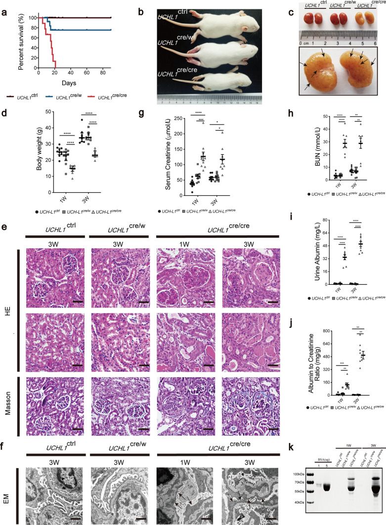
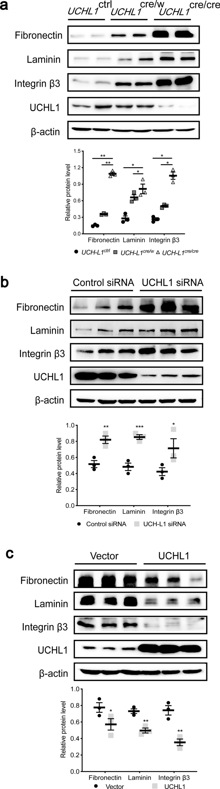
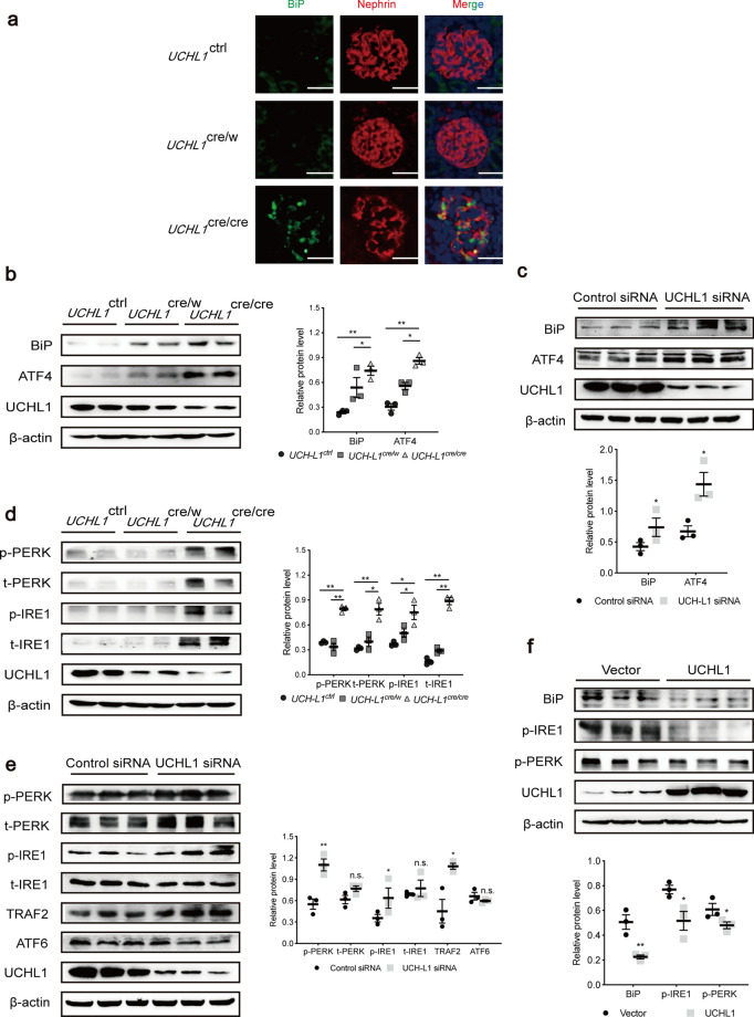
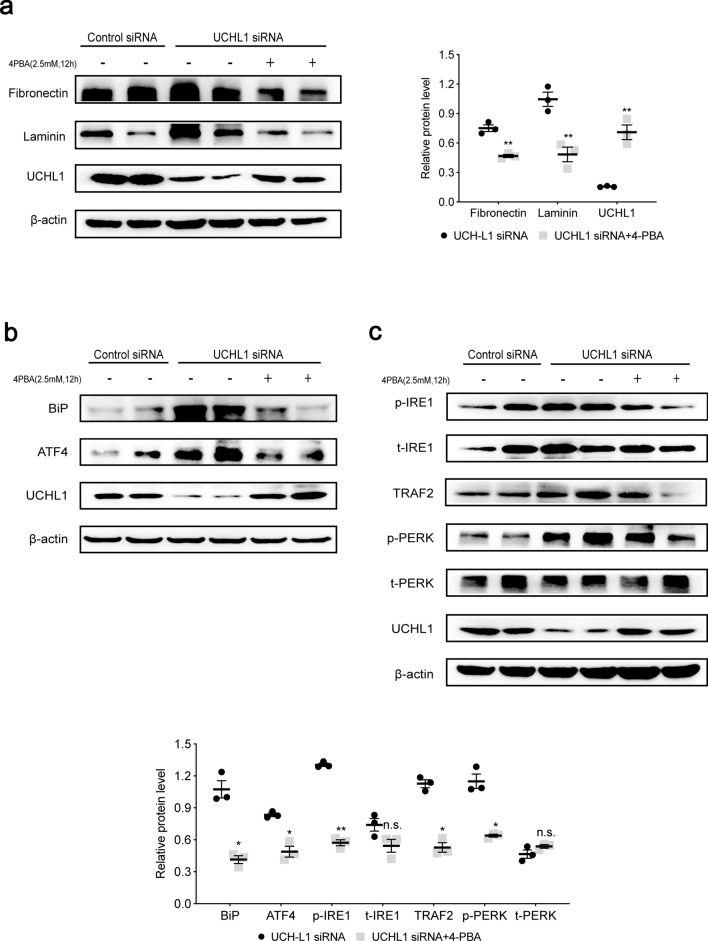
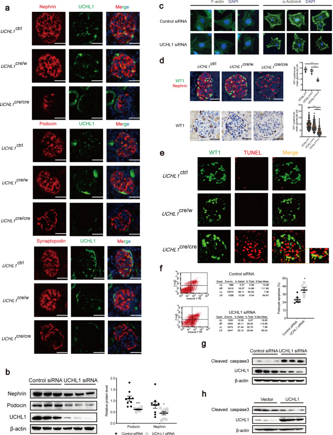
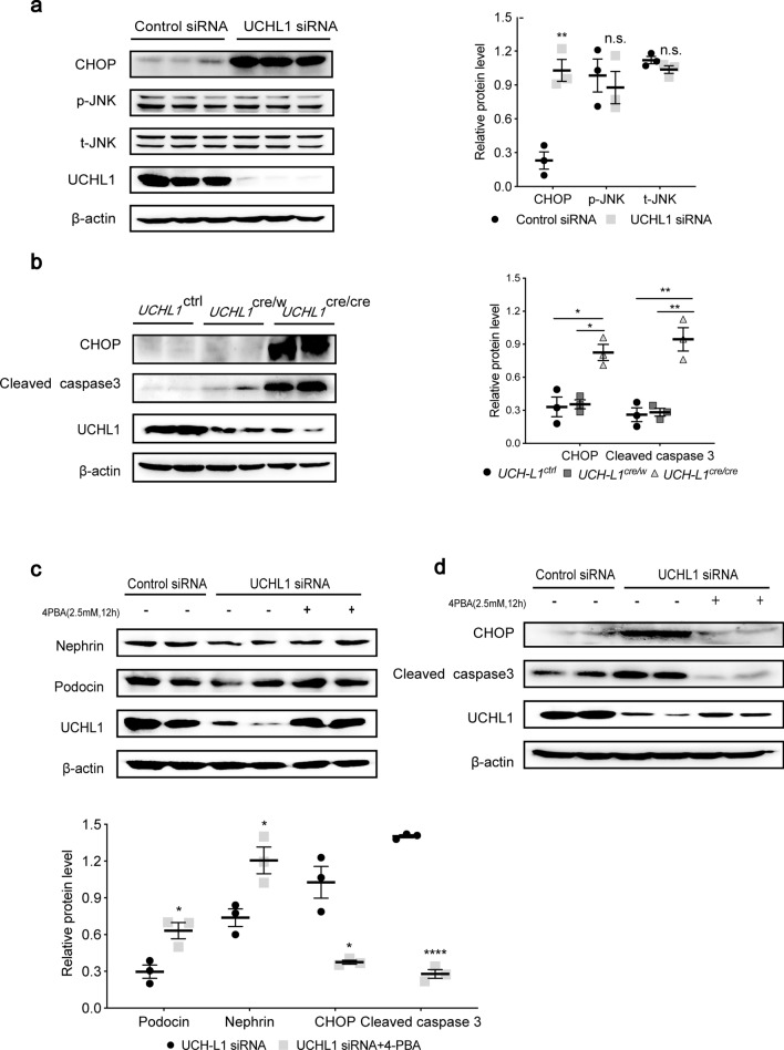
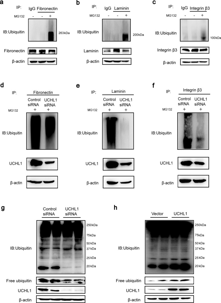
Ubiquitin carboxyl-terminal hydrolase L1 (UCHL1) is a unique component of the ubiquitin-proteasome system (UPS), which has multiple activities in maintaining intracellular ubiquitin levels. We previously reported the aberrant low expression of UCHL1 in podocytes of non-immune complex-mediated glomerulonephritis, and recent studies indicate that anti-UCHL1 antibody was responsible for the refractory minimal change disease (MCD), but the specific effect of UCHL1 to the podocytopathy has not been determined. Therefore, we generated podocyte-specific UCHL1 gene knockout (UCHL1cre/cre) rats model. Podocyte-specific UCHL1 knockout rats exhibited severe kidney damage, including segmental/global glomerulosclerosis, kidney function damage and severe proteinuria, compared with littermate control. Subsequently, by carrying out mass spectrometry analysis of isolated glomeruli of rats, abnormal protein accumulation of ECM-receptor Interaction was found in UCHL1cre/cre rats. Mechanistic studies in vivo and in vitro revealed that aberrant protein accumulation after UCHL1 deficiency induced endoplasmic reticulum (ER) stress, unfolded protein reaction (UPR) to reduce the protein level of podocyte skeleton proteins, and CHOP mediated apoptosis as well, which related to the dysfunction of the ubiquitin-proteasome system with decreased free monomeric ubiquitin level, thereby affecting protein ubiquitination and degradation. In addition, inhibition of ER stress by 4-PBA could attenuate the degree of ER stress and podocyte dysfunction. Our study indicates that UCHL1 is a potential target for preventing podocytes injury in some non-immune complex-mediated glomerulopathy.

Keywords: Endoplasmic reticulum stress; Podocyte apoptosis; Podocytopathy; Ubiquitin carboxyl-terminal hydrolase L1 (UCHL1); Ubiquitin–proteasome system (UPS).


Conflict of interest statement

The authors have no relevant financial or non-financial interests to disclose.

Figures

Fig. 1
Fig. 1
Characterization of UCHL1cre/cre and UCHL1cre/w rats. a Graph depicting the survival rate of UCHL1cre/cre rats, UCHL1cre/w rats and UCHL1w/w rats. n = 10–12 rats per group. Representative rats (b) and kidney (c) images at 3 weeks of age. (c) Pale kidneys and red miliary dots (arrows) in kidneys were observed in UCHL1cre/cre rats. d Body weight of all three genotypes of rats at the indicated age. Values were expressed as means ± SEM, n = 8–10 rats per group, **P < 0.01, ***P < 0.001, ****P < 0.0001. e H&E staining and Masson trichome staining showed mesangial proliferation by 1 week of age, and segmental or global glomerulosclerosis with tubular atrophy and interstitial fibrosis by 3 weeks of age in UCHL1cre/cre rats, compared with UCHL1cre/w and UCHL1ctrl rats. Scale bar: 50 μm. f Electron microscopy showed extensive foot process effacement (arrows) in UCHL1cre/cre rats at 1 and 3 weeks of age, compared with UCHL1cre/w and UCHL1ctrl littermates. Scale bar:1 μm. Serum creatinine (g), BUN (h), urine albumin levels (i) and urine albumin/creatinine ratios (ACR) (j) were significantly elevated in UCHL1cre/cre rats by 1 and 3 weeks of age. Values were expressed as means ± SEM, n = 8–10 rats per group, *P < 0.05, **P < 0.01, ***P < 0.001, ****P < 0.0001. (k) Coomassie-Brilliant Blue staining of urine samples processed by SDS-PAGE indicated that the urine albumin levels (~ 67 kDa bands) were elevated in UCHL1cre/cre rats at 1 week, which were increased obviously by 3 weeks. Standard BSA concentrations are shown in lanes 2 and 3. n = 6 rats per group. Shown are representative immunoblots from at least three independent experiments with similar results
Fig. 2
Fig. 2
UCHL1 deletion induced abnormal protein accumulation including fibronectin, laminin and integrin β3 in podocytes. a Western blot examined the protein expression of fibronectin, laminin and integrin β3 in isolated glomeruli of UCHL1cre/cre, UCHL1cre/w and UCHL1ctrl rats. n = 6 rats per group. b, c Western blot examined the protein level of fibronectin, laminin and integrin β3 in podocytes transfected with control siRNA or UCHL1 siRNA (b), or in podocytes expressing vector control (Vector) or UCHL1 plasmids (c) in vitro. Values were expressed as means ± SEM, *P < 0.05, **P < 0.01, ***P < 0.001. β-Actin was as a loading control for all western blots. Shown are representative immunoblots from at least three independent experiments with similar results
Fig. 3
Fig. 3
Endoplasmic reticulum (ER) stress of podocytes was activated by UCHL1 deletion. a Double immunofluorescence staining of BiP, an ER stress marker, and nephrin in podocytes of UCHL1cre/cre, UCHL1cre/w and UCHL1ctrl rats. Scale bar: 50 μm. n = 3 rats per group. b, c Western blot examined the protein level of BiP and ATF4 in isolated glomeruli of UCHL1cre/cre, UCHL1cre/w and UCHL1ctrl rats (b), or podocytes transfected with control siRNA or UCHL1 siRNA (c) in vitro. n = 6 rats per group. d Western blot examined the protein level of UPR containing PERK, IRE1 and ATF6 in isolated glomeruli of UCHL1cre/cre, UCHL1cre/w and UCHL1ctrl rats. n = 6 rats per group. e, f Western blot examined the three downstream of UPR (PERK, IRE1 and ATF6) in podocytes transfected with control siRNA or UCHL1 siRNA (e), or in podocytes expressing vector control (Vector) or UCHL1 plasmids (f) in vitro. Values were expressed as means ± SEM, *P < 0.05, **P < 0.01, n.s. (no significance). β-Actin was a loading control for all western blots. Shown are representative immunoblots from at least three independent experiments with similar results
Fig. 4
Fig. 4
4-PBA inhibited abnormal protein accumulation and ER stress induced by UCHL1 deficiency in podocytes. a Western blot showed that the increase of fibronectin and laminin after UCHL1 siRNA transfection in podocytes was significantly reduced with treatment of 2.5 mM 4-PBA for 12 h. b, c Western blot examined the protein level of BiP, ATF4 and UPR in podocytes transfected with UCHL1 siRNA and treated with or without 2.5 mM 4-PBA for 12 h. Values were expressed as means ± SEM, *P < 0.05, **P < 0.01, n.s. (no significance). β-Actin was a loading control for all western blots. Shown are representative immunoblots from at least three independent experiments with similar results
Fig. 5
Fig. 5
UCHL1 deficiency reduced the expression of podocyte-specific marker and induced apoptosis in podocytes. a Representative double immunofluorescence images showed the expression and distribution of synaptopodin, podocin and nephrin in glomeruli of UCHL1cre/cre, UCHL1cre/w or UCHL1ctrl rats. Scale bar: 50 μm. n = 3 rats per group. b Western blot examined the protein level of nephrin and podocin in podocytes transfected with control siRNA or UCHL1 siRNA in vitro. c Phalloidin and α-actinin 4 staining of podocyte after UCHL1 siRNA or control siRNA transfection. d Quantification of podocytes per glomerular. Double immunofluorescence staining with WT1 to label podocyte nuclei and nephrin to label podocyte cytoplasm, and Immunohistochemistry staining of WT1 in a thick and thin section method were used to count the podocytes per glomerular. The mean number of WT1/nephrin co-positive cells per glomerulus of each rat was calculated by counting 20 randomly selected glomeruli of each rat (n = 6 rats per group) and is shown as the upper right graphic. The mean number of WT1 positive cells per glomerulus was calculated from 20 randomly selected glomeruli of each rat (n = 10 rats per group) in two thickness slices and is shown as the lower right graphic. Number of podocytes per glomerular tuft was obviously decreased in UCHL1cre/cre rats, compared with UCHL1cre/w rats and UCHL1ctrl rats. Scale bar: 50 μm. e Representative double immunofluorescence images of WT1 and TUNEL of all three genotypes of rats showing the TUNEL-positive nuclei in podocytes (yellow arrows are podocytes). Scale bar: 50 μm. n = 3 rats per group. f Flow cytometry analysis showing the apoptosis rate of podocytes with control siRNA or UCHL1 siRNA transfection. The cells in the upper right and lower right were considered as apoptotic cells. The level of apoptosis rate was higher in podocytes with UCHL1 siRNA transfection. g, h Western blot examined the protein level of cleaved caspase3 in podocytes transfected with control siRNA or UCHL1 siRNA (g), or in podocytes expressing vector control (Vector) or UCHL1 plasmids (h) in vitro. Values were expressed as means ± SEM, *P < 0.05, ***P < 0.001, ***P < 0.0001. β-Actin was a loading control for all western blots. Shown are representative immunoblots from at least three independent experiments with similar results
Fig. 6
Fig. 6
UCHL1 deficiency induced ER stress-associated apoptosis and injury of podocytes, which was ameliorated by 4-PBA. a Western blot examined the protein level of CHOP and JNK signaling in podocytes with control siRNA or UCHL1 siRNA transfection, showing UCHL1 siRNA transfection upregulated CHOP rather than p-JNK and t-JNK. b Western blot examined the protein level of CHOP and cleaved caspase3 in isolated glomeruli of all three genotypes of the rat. n = 6 rats per group. c Western blot showed that the decreased protein level of nephrin and podocin after UCHL1 siRNA transfection in podocytes was increased with treatment of 2.5 mM 4-PBA for 12 h. d Western blot showed the increased protein level of CHOP and cleaved caspase 3 after UCHL1 siRNA transfection in podocytes were significantly reduced with treatment of 2.5 mM 4-PBA for 12 h. Values were expressed as means ± SEM, *P < 0.05, **P < 0.01, ****P < 0.0001. β-Actin was a loading control for all western blots. Shown are representative immunoblots from at least three independent experiments with similar results
Fig. 7
Fig. 7
UCHL1 deficiency reduced the ubiquitinated fibronectin, laminin or integrin β3 and the free monomeric ubiquitin level. ac Podocytes were cultured with MG132 for 12 h before being collected. Immunoprecipitation and western blot analysis showed the ubiquitination level of fibronectin, laminin and integrin β3 were significantly elevated, which indicated all of them were degraded by the ubiquitin–proteasome pathway in podocytes. df Immunoprecipitation and western blot analysis demonstrated that compared to the control group, the ubiquitination level of fibronectin, laminin and integrin β3 were significantly decreased in podocytes transfected with UCHL1 siRNA. g, h Immunoprecipitation and western blot analysis examined the levels of poly-ubiquitin and free monomeric ubiquitin in podocytes transfected with control siRNA or UCHL1 siRNA (g), or podocytes expressing vector or exogenous UCHL1 (h) in vitro. β-Actin was a loading control for all western blots. Shown are representative immunoblots from at least three independent experiments with similar results

Similar articles

Cited by

References

    1. Shankland SJ. The podocyte’s response to injury: role in proteinuria and glomerulosclerosis. Kidney Int. 2006;69(12):2131–2147. doi: 10.1038/sj.ki.5000410. - DOI - PubMed
    1. Nagata M. Podocyte injury and its consequences. Kidney Int. 2016;89(6):1221–1230. doi: 10.1016/j.kint.2016.01.012. - DOI - PubMed
    1. Bishop P, Rocca D, Henley JM. Ubiquitin C-terminal hydrolase L1 (UCH-L1): structure, distribution and roles in brain function and dysfunction. Biochem J. 2016;473(16):2453–2462. doi: 10.1042/BCJ20160082. - DOI - PMC - PubMed
    1. Day IN, Thompson RJ. UCHL1 (PGP 9.5): neuronal biomarker and ubiquitin system protein. Prog Neurobiol. 2010;90(3):327–362. doi: 10.1016/j.pneurobio.2009.10.020. - DOI - PubMed
    1. Bett JS, Ritorto MS, Ewan R, et al. Ubiquitin C-terminal hydrolases cleave isopeptide- and peptide-linked ubiquitin from structured proteins but do not edit ubiquitin homopolymers. Biochem J. 2015;466(3):489–498. doi: 10.1042/BJ20141349. - DOI - PMC - PubMed

Supplementary concepts