. 2023 May;299(5):104632.
doi: 10.1016/j.jbc.2023.104632. Epub 2023 Mar 22.

The intramembrane COOH-terminal domain of PRRT2 regulates voltage-dependent Na+ channels

Affiliations

The intramembrane COOH-terminal domain of PRRT2 regulates voltage-dependent Na+ channels

Francesca Franchi et al. J Biol Chem. 2023 May.

Abstract

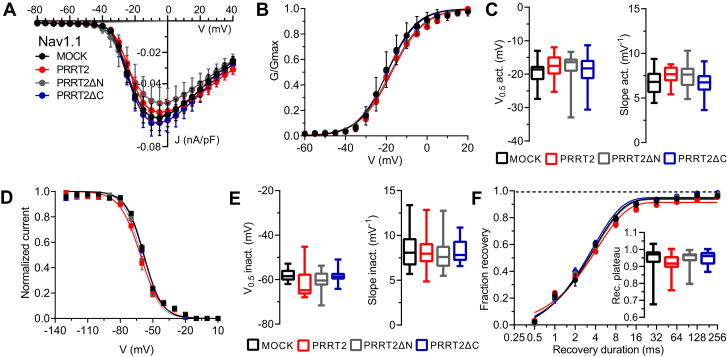
Proline-rich transmembrane protein 2 (PRRT2) is the single causative gene for pleiotropic paroxysmal syndromes, including epilepsy, kinesigenic dyskinesia, episodic ataxia, and migraine. PRRT2 is a neuron-specific type-2 membrane protein with a COOH-terminal intramembrane domain and a long proline-rich NH2-terminal cytoplasmic region. A large array of experimental data indicates that PRRT2 is a neuron stability gene that negatively controls intrinsic excitability by regulating surface membrane localization and biophysical properties of voltage-dependent Na+ channels Nav1.2 and Nav1.6, but not Nav1.1. To further investigate the regulatory role of PRRT2, we studied the structural features of this membrane protein with molecular dynamics simulations, and its structure-function relationships with Nav1.2 channels by biochemical and electrophysiological techniques. We found that the intramembrane COOH-terminal region maintains a stable conformation over time, with the first transmembrane domain forming a helix-loop-helix motif within the bilayer. The unstructured NH2-terminal cytoplasmic region bound to the Nav1.2 better than the isolated COOH-terminal intramembrane domain, mimicking full-length PRRT2, while the COOH-terminal intramembrane domain was able to modulate Na+ current and channel biophysical properties, still maintaining the striking specificity for Nav1.2 versus Nav1.1. channels. The results identify PRRT2 as a dual-domain protein in which the NH2-terminal cytoplasmic region acts as a binding antenna for Na+ channels, while the COOH-terminal membrane domain regulates channel exposure on the membrane and its biophysical properties.

Keywords: PRRT2; intrinsic excitability; molecular dynamics; structure-function relationships; voltage-dependent sodium channels.


Conflict of interest statement

Conflict of interest The authors declare that they have no conflicts of interest with the contents of this article.

Figures

Figure 1
Figure 1
Generation and characterization of the PRRT2 deletion mutants. A, schematics of the PRRT2 domain constructs. PRRT2 WT-HA is the entire protein (violet). PRRT2 ΔC-HA is a chimeric protein composed of the cytoplasmic PRRT2 (violet) domain anchored to the membrane by the transmembrane domain of IFITM1 (green). PRRT2 ΔN-HA is composed of the transmembrane domain of PRRT2. B, Hek- Nav1.2 cells transfected with (from left to right) PRRT2-HA, PRRT2ΔC-HA, and PRRT2ΔN-HA were permeabilized and subsequently labeled for anti-HA and pan-Nav antibodies, respectively, with nuclei marked with DAPI. The individual HA and pan-Nav staining are shown together with the respective merge images (top row). C, Hek-Nav cells transfected with PRRT2-HA, PRRT2ΔC-HA, and PRRT2ΔN-HA were surface-labeled with anti-HA to stain the membrane-exposed domains of the proteins. Scale bar, 10 μm. IFITM1, interferon-induced transmembrane protein 1; PRRT2, proline-rich transmembrane protein 2.
Figure 2
Figure 2
Molecular modeling ofthePRRT2 transmembrane segment.A, snapshot of the simulated system extracted from a trajectory; PRRT2 is represented in blue cartoon, phospholipids heads and tails as gray spheres and sticks, respectively, water molecules as red and white sticks, and sodium and chloride ions as orange and purple spheres, respectively. B, alpha-carbon RMSD of PRRT2 calculated along the two simulated trajectories at different timesteps (Δt); average values and S.D. are indicated. C, cartoon representation of the PRRT2 transmembrane domain; residues involved in the calculated cross-distances are represented as sticks; the values reported are those at the beginning of the simulations. D, cross-distances evolution for PRRT2 trajectories. E, lateral and extracellular views of Robetta (blue) AlphaFold2 (green), and ESMFold (orange) PRRT2 models, superimposed via PyMOL. PRRT2, proline-rich transmembrane protein 2.
Figure 3
Figure 3
Binding of PRRT2 deletion mutants to Nav1.2 and Nav1.1 channels. A, representative immunoblot of co-immunoprecipitation of PRRT2 variants. HA-tagged bacterial alkaline phosphatase (BAP), HA-tagged full-length PRRT2 (PRRT2-FL), or its deletion mutants PRRT2ΔN and PRRT2ΔC were expressed in naive Hek293 cells and purified by HA-immunoprecipitation. The extract of Hek293 stable clones expressing either human Nav1.2 or human Nav1.1 was added to the HA-immunoprecipitated BAP, PRRT2, or PRRT2 deletion mutants. Cell lysates (INPUT, 10 μg protein) and samples immunoprecipitated by anti-HA beads were analyzed by Western blotting with pan-Nav and HA antibodies. Molecular mass standards are reported on the right. The representative blots were cut from the same gel. B, quantification of the immunoreactive signals in PRRT2-HA immunoprecipitates. Box plots of n = 6 and 4 independent experiments for Hek-Nav1.2 and Hek-Nav1.1, respectively. ∗p < 0.05, ∗∗∗p < 0.001 versus full-length PRRT2; °p < 0.05, °°°p < 0.001 Nav1.1 versus Nav1.2 for each PRRT2 variant. Two-way ANOVA/Fisher’s tests. PRRT2, proline-rich transmembrane protein 2.
Figure 4
Figure 4
Effects of PRRT2 deletion mutants on the transient Nav1.2 current. A, representative whole-cell Na+ currents recorded in Hek293 cells stably expressing Nav1.2 α-subunits and transiently transfected with empty vector (MOCK, black), full-length PRRT2 (red), PRRT2ΔN (gray), and PRRT2ΔC (blue). Currents were elicited by a protocol (inset) consisting of 5-mV depolarization steps from −80 to 40 mV from a holding potential of −100 mV. For clarity, the first 20 ms of the 100-ms steps for eight representative traces per condition are plotted. B, current density (J) versus voltage (V) relationship for the four experimental conditions. C, box plots of J values at three representative voltages (−20/-10/0 mV). MOCK, n = 24; full-length PRRT2, n = 15; PRRT2ΔN, n = 19; PRRT2ΔC, n = 21. ∗∗p < 0.01, ∗∗∗p < 0.001 versus MOCK; °p < 0.05 versus full-length PRRT2. One-way ANOVA/uncorrected Fisher's LSD test. PRRT2, proline-rich transmembrane protein 2.
Figure 5
Figure 5
Effects of PRRT2 deletion mutants on the activation and inactivation kinetics of Nav1.2 channels. Hek293 cells stably expressing Nav1.2 α-subunits were transiently transfected with empty vector (MOCK, black), full-length PRRT2 (red), PRRT2ΔN (gray), and PRRT2ΔC (blue). A, Left: Voltage-dependence of activation. The lines are the best-fitted Boltzmann curves. Right: Box plots of the half-maximal voltage of activation (V0.5) and slope (MOCK, n = 24; full-length PRRT2, n = 15; PRRT2ΔN, n = 19; PRRT2ΔC, n = 21). B, Left: Steady-state inactivation curves. The lines are the best-fitted Boltzmann curves. Right: Means (±SEM) values of the half-maximal voltages for inactivation (V0.5 inact.) and slopes (MOCK, n = 20; full-length PRRT2, n = 18; PRRT2ΔN, n = 15; PRRT2ΔC, n = 21). ∗∗p < 0.01, ∗∗∗p < 0.001 versus MOCK; °°°p < 0.001 versus full-length PRRT2. One-way ANOVA/Dunnett’s test. PRRT2, proline-rich transmembrane protein 2.
Figure 6
Figure 6
Effects of PRRT2 deletion mutants on the recovery from inactivation of Nav1.2 channels. A, representative traces showing current recovery from inactivation for all the experimental conditions. Recordings were obtained prepulsing cells to −20 mV for 20 ms to inactivate Na+ currents and then coming back to a recovery potential of −100 mV for increasing durations before the repetition of test pulse to −20 mV. For clarity, 6 of the 9 time-intervals are shown. B, left: the time courses of the recovery from inactivation of peak currents at −20 mV are plotted on a semi-logarithmic scale for the four experimental conditions. Right: Box plots of τ and plateau of recovery estimated from one-phase decay fit to the data (MOCK, n = 24; full-length PRRT2, n =16; PRRT2ΔN, n = 18; PRRT2ΔC, n = 21). ∗∗∗p < 0.001 versus MOCK; °°°p < 0.001 versus full-length PRRT2. Kruskal–Wallis/Dunn’s tests. PRRT2, proline-rich transmembrane protein 2.
Figure 7
Figure 7
PRRT2 deletion mutants are ineffective on the transient current and biophysical properties of Nav1.1 channels. Hek293 cells stably expressing Nav1.1 α-subunits were transiently transfected with empty vector (MOCK, black), full-length PRRT2 (red), PRRT2ΔN (gray), and PRRT2ΔC (blue). A, current density (J) versus voltage (V) relationship for the four experimental conditions, B, voltage-dependence of activation. The lines are the best-fitted Boltzmann curves. C, box plots of the half-maximal voltage of activation (V0.5) and slope (MOCK, n = 21; full-length PRRT2, n = 19; PRRT2ΔN, n = 18; PRRT2ΔC, n = 23). D, steady-state inactivation curves. The lines are the best-fitted Boltzmann curves. E, box plots of the half-maximal voltages for inactivation (V0.5 inact.) and slopes (MOCK, n = 18; full-length PRRT2, n = 18; PRRT2ΔN, n = 16; PRRT2ΔC, n = 18). F, the time courses of the recovery from inactivation of peak currents at −20 mV are plotted on a semi-logarithmic scale for the four experimental conditions. Inset: Box plots of the plateau of recovery estimated from one-phase decay fit to the data (MOCK, n = 19; full-length PRRT2, n = 18; PRRT2ΔN, n = 17; PRRT2ΔC, n = 18). p > 0.05 versus MOCK. One-way ANOVA/Dunnett’s test. PRRT2, proline-rich transmembrane protein 2.
Figure 8
Figure 8
The tandem domain hypothesis of PRRT2-Nav1.2 specific interactions. A, structure of the transmembrane domains of the human Nav1.2 channel (PDB code: 6J8E; (64)), lateral (left) and extracellular (right) view. Residues that differ with Nav1.1 are represented as blue spheres. B, schematic representation of the interactions between Nav1.2 and PRRT2. The NH2-terminal intracellular domain may act as a Nav docking module favoring the action of the intramembrane COOH-terminal domain on the plasma membrane exposure and biophysical properties that are specific for Nav1.2. PRRT2, proline-rich transmembrane protein 2.

References

    1. Chen W.-J., Lin Y., Xiong Z.-Q., Wei W., Ni W., Tan G.-H., et al. Exome sequencing identifies truncating mutations in PRRT2 that cause paroxysmal kinesigenic dyskinesia. Nat. Genet. 2011;43:1252–1255. - PubMed
    1. Lee H.Y., Huang Y., Bruneau N., Roll P., Roberson E.D., Hermann M., et al. Mutations in the gene PRRT2 cause paroxysmal kinesigenic dyskinesia with infantile convulsions. Cell Rep. 2012;1:2–12. - PMC - PubMed
    1. Ebrahimi-Fakhari D., Saffari A., Westenberger A., Klein C. The evolving spectrum of PRRT2-associated paroxysmal diseases. Brain. 2015;138:3476–3495. - PubMed
    1. Gardiner A.R., Jaffer F., Dale R.C., Labrum R., Erro R., Meyer E., et al. The clinical and genetic heterogeneity of paroxysmal dyskinesias. Brain. 2015;138:3567–3580. - PMC - PubMed
    1. Heron S.E., Dibbens L.M. Role of PRRT2 in common paroxysmal neurological disorders: a gene with remarkable pleiotropy. J. Med. Genet. 2013;50:133–139. - PubMed

Publication types

LinkOut - more resources