

Investigation into the genetics of fetal congenital lymphatic anomalies
- PMID: 36959127
- PMCID: PMC10330091
- DOI: 10.1002/pd.6345
Investigation into the genetics of fetal congenital lymphatic anomalies
Abstract
Objective: Congenital lymphatic anomalies (LAs) arise due to defects in lymphatic development and often present in utero as pleural effusion, chylothorax, nuchal and soft tissue edema, ascites, or hydrops. Many LAs are caused by single nucleotide variants, which are not detected on routine prenatal testing.
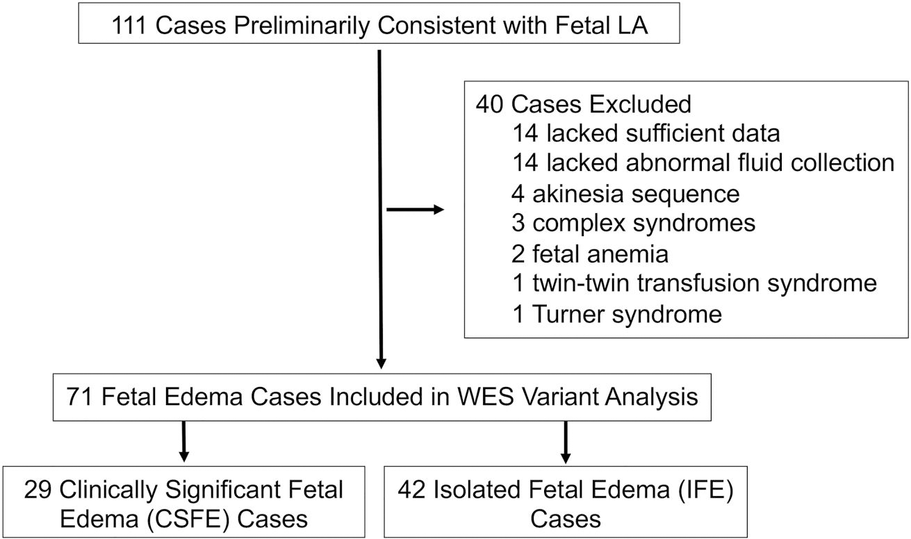
Methods: Demographic data were compared between two subcohorts, those with clinically significant fetal edema (CSFE) and isolated fetal edema. A targeted variant analysis of LA genes was performed using American College of Medical Genetics criteria on whole exome sequencing (WES) data generated for 71 fetal edema cases who remained undiagnosed after standard workup.
Results: CSFE cases had poor outcomes, including preterm delivery, demise, and maternal preeclampsia. Pathogenic and likely pathogenic variants were identified in 7% (5/71) of cases, including variants in RASopathy genes, RASA1, SOS1, PTPN11, and a novel PIEZO1 variant. Variants of uncertain significance (VOUS) were identified in 45% (32/71) of cases. In CSFEs, VOUS were found in CELSR1, EPHB4, TIE1, PIEZO1, ITGA9, RASopathy genes, SOS1, SOS2, and RAF1.
Conclusions: WES identified pathogenic and likely pathogenic variants and VOUS in LA genes in 51% of fetal edema cases, supporting WES and expanded hydrops panels in cases of idiopathic fetal hydrops and fluid collections.
© 2023 The Authors. Prenatal Diagnosis published by John Wiley & Sons Ltd.
Conflict of interest statement
CONFLICT OF INTEREST STATEMENT
None of the authors have conflicts of interest to disclose.
Figures
References
-
- Anomalies ISftSoV. ISSVA Classification for Vascular Anomalies; 2014.
MeSH terms
Substances
Grants and funding
LinkOut - more resources
Full Text Sources
Research Materials
Miscellaneous
