. 2023 Mar 23;14(1):1606.
doi: 10.1038/s41467-023-37225-1.

Modular-designed engineered bacteria for precision tumor immunotherapy via spatiotemporal manipulation by magnetic field

Affiliations

Modular-designed engineered bacteria for precision tumor immunotherapy via spatiotemporal manipulation by magnetic field

Xiaotu Ma et al. Nat Commun. .

Erratum in

Abstract

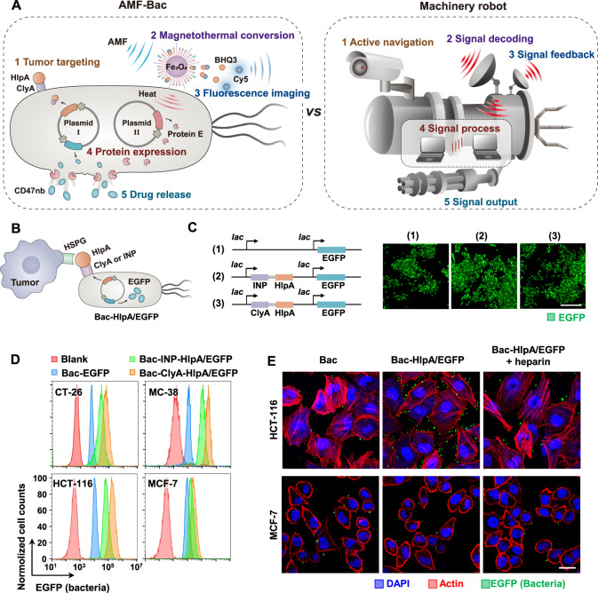
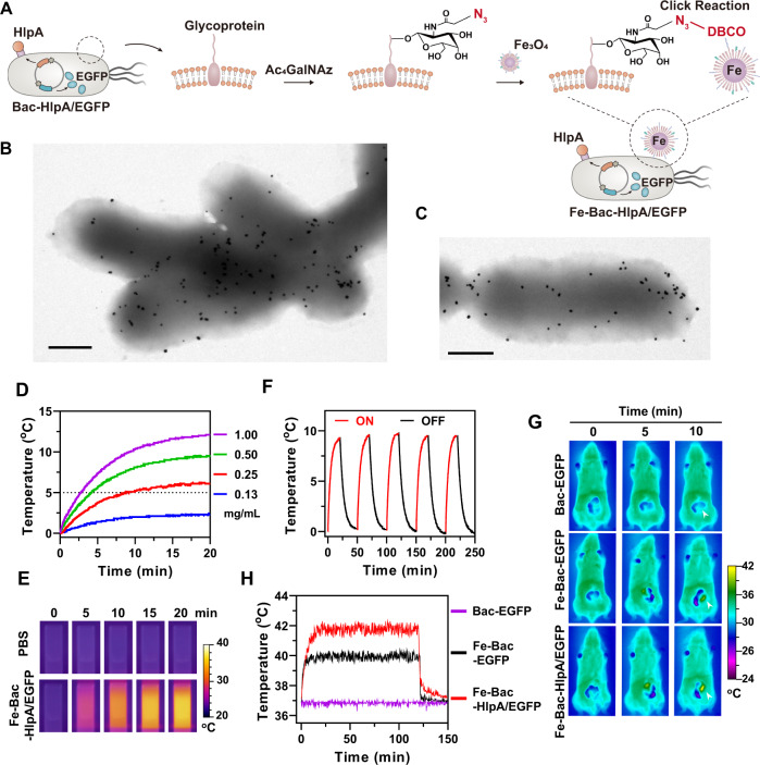
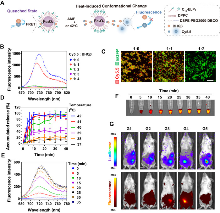
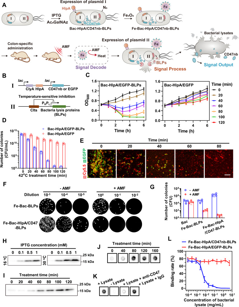
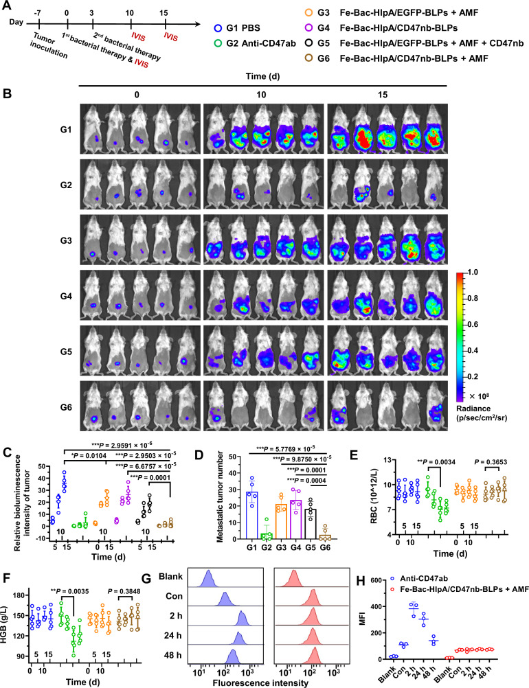
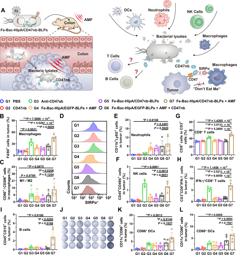
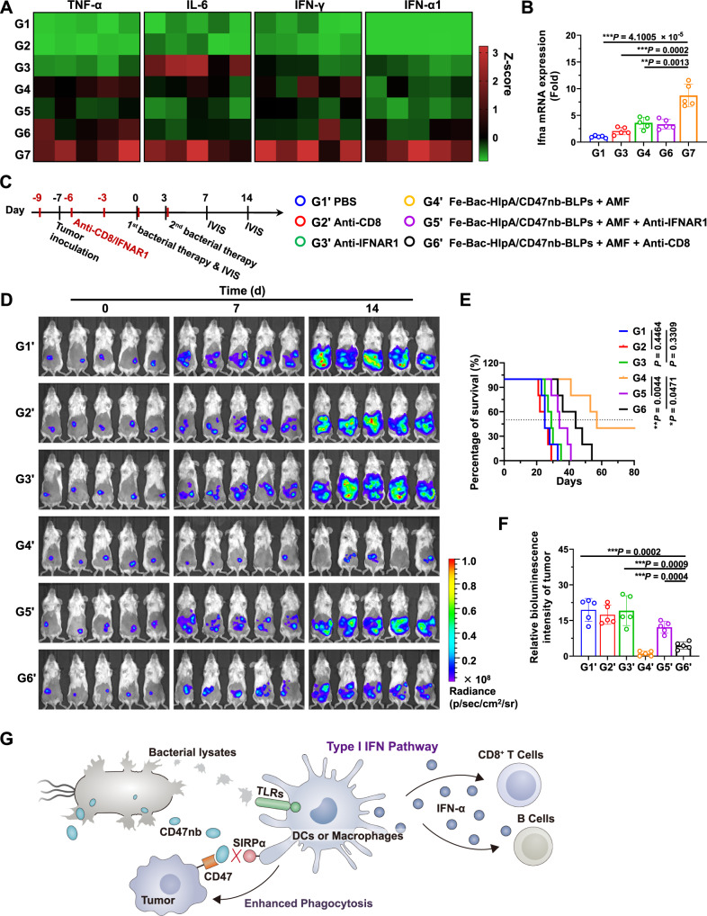
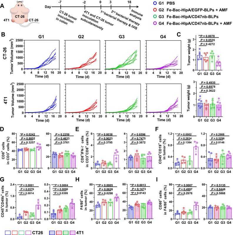
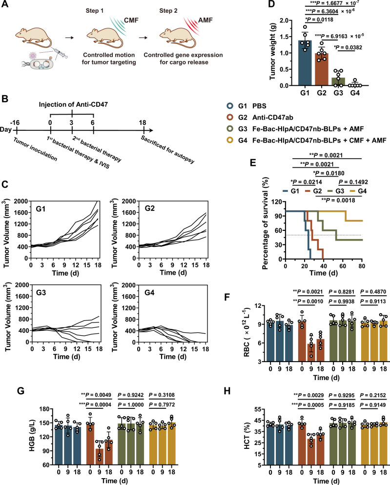
Micro-nano biorobots based on bacteria have demonstrated great potential for tumor diagnosis and treatment. The bacterial gene expression and drug release should be spatiotemporally controlled to avoid drug release in healthy tissues and undesired toxicity. Herein, we describe an alternating magnetic field-manipulated tumor-homing bacteria developed by genetically modifying engineered Escherichia coli with Fe3O4@lipid nanocomposites. After accumulating in orthotopic colon tumors in female mice, the paramagnetic Fe3O4 nanoparticles enable the engineered bacteria to receive and convert magnetic signals into heat, thereby initiating expression of lysis proteins under the control of a heat-sensitive promoter. The engineered bacteria then lyse, releasing its anti-CD47 nanobody cargo, that is pre-expressed and within the bacteria. The robust immunogenicity of bacterial lysate cooperates with anti-CD47 nanobody to activate both innate and adaptive immune responses, generating robust antitumor effects against not only orthotopic colon tumors but also distal tumors in female mice. The magnetically engineered bacteria also enable the constant magnetic field-controlled motion for enhanced tumor targeting and increased therapeutic efficacy. Thus, the gene expression and drug release behavior of tumor-homing bacteria can be spatiotemporally manipulated in vivo by a magnetic field, achieving tumor-specific CD47 blockage and precision tumor immunotherapy.


Conflict of interest statement

The authors declare no competing interests.

Figures

Fig. 1
Fig. 1. Design of AMF-Bac and its analogy with a machinery robot, and the assembly of the active navigation module.
A Illustration of AMF-Bac, comprising five modules: active navigation, signal decoding, signal feedback, signal process, and signal output. The five modules respectively carry out the physiological functions of tumor targeting, magnetothermal conversion, fluorescence imaging, protein expression, and drug release. B Diagram illustrating the tumor-targeting ability of Bac-HlpA/EGFP. HlpA is displayed on the surface of Bac-HlpA/EGFP for targeting to the HSPG on colon tumor cells, through two different surface display scaffolds, ClyA and INP. C Expression plasmid design and EGFP expression verification. Bacteria were transformed with the indicated plasmids, including pACYC-EGFP (1), pACYC-INP-HlpA-EGFP (2), and pACYC-ClyA-HlpA-EGFP (3), and the corresponding engineered bacteria were termed Bac-EGFP, Bac-INP-HlpA/EGFP and Bac-ClyA-HlpA/EGFP, respectively. lac represents the IPTG-inducible promotor. Successful expression of EGFP was verified by observation under a confocal laser scanning microscope (CLSM). Scale bar, 10 μm. D, E The tumor-targeting ability of Bac-HlpA/EGFP. Engineered bacteria were incubated with the indicated cell lines for 2 h at 4 °C, and the bacterial fluorescence intensities of the cells were examined using flow cytometry (D). The binding of bacteria onto cells was also directly visualized using CLSM (E). Bacteria were tracked by their expression of EGFP (green), while tumor cell actin was stained with phalloidine (red). Cell nuclei were stained with DAPI (blue). Scale bar, 40 μm. These experiments (C-E) were repeated three times independently with similar results. Source data are provided as a Source Data file.
Fig. 2
Fig. 2. Assembly of the signal decoding module.
A Diagram of site-specific coupling of DBCO-Fe3O4 onto the tumor-homing bacteria. The reactive functional groups (-N3) were introduced onto the bacterial surface via metabolic oligosaccharide engineering, through culturing bacteria with a non-natural form of galactosamine (Ac4GalNAc). DBCO-Fe3O4 were conjugated onto the N3-modified bacteria through the click chemistry reaction between DBCO and -N3 moieties. B, C TEM images of bacteria modified with Fe3O4 NPs. Site-specific (B) and non-site-specific (C) labeling methods were employed to conjugate Fe3O4 NPs onto bacteria. Scale bar, 0.5 μm. D Temperature elevation of aqueous solutions of Fe-Bac-HlpA/EGFP at different Fe3O4 concentrations (0.13–1.00 mg/mL) during treatment with AMF at a fixed frequency of 310 kHz and intensity of 14.6 kA/m. E Infrared imaging of Fe-Bac-HlpA/EGFP solutions (1.00 mg/mL Fe3O4) upon AMF treatment (310 kHz and 14.6 kA/m) for the indicated time intervals (0–20 min). F Temperature changes of Fe-Bac-HlpA/EGFP solutions (0.5 mg/mL Fe3O4) during five cycles of AMF treatment (ON; 310 kHz and 14.6 kA/m) and natural cooling-down (OFF). G Infrared imaging of BALB/c mice with CT-26 colonic orthotopic xenografts upon AMF treatment (310 kHz and 23.8 kA/m) for the indicated time intervals (0–10 min). The mice were treated with the indicated engineered bacteria (1 × 108 CFU) by colon-specific administration at 24 h prior to AMF treatment, and laparotomized before infrared imaging. The white arrows indicate the tumor. H Intratumoral temperature of CT-26 colonic orthotopic xenografts upon AMF treatment (310 kHz and 23.8 kA/m) for 120 min. The temperature in tumors was monitored by inserting an electronic needle thermometer into the tumor. These experiments (B-H) were repeated three times independently with similar results. Source data are provided as a Source Data file.
Fig. 3
Fig. 3. Assembly of signal feedback module.
A Illustration of signal feedback module for AMF-responsive fluorescence imaging. Fe3O4 NPs was coated with a monolayer of LTSL (LTSL@Fe), which was composed of DSPE-PEG2000-DBCO and temperature-sensitive lipids, including DPPC and C18-ELP3. The fluorescent dye, Cy5.5, and its black-hole quencher, BHQ3, were co-loaded into the LTSL monolayer (LTSL@Fe/C/B). In the LTSL@Fe/C/B, the fluorescence of Cy5.5 was quenched by BHQ3, which is recovered by the heat-induced conformational change and Cy5.5 release. B Fluorescence emission spectra of LTSL@Fe/C/B at different molar ratios of Cy5.5 and BHQ3. C Observation of LTSL@Fe/C/B-Bac-HlpA/EGFP at different molar ratios of Cy5.5 and BHQ3 using CLSM. Bacteria were tracked by EGFP expression. Scale bar, 20 μm. D The percentage of accumulated release of Cy5.5 from LTSL@Fe/C/B-Bac-HlpA/EGFP upon water-bath treatment for 40 min at the indicated temperatures (37–42 °C). The data are shown as the mean ± SD (n = 3 independent experiments). E, F Fluorescence emission spectra (E) and fluorescence imaging (F) of LTSL@Fe/C/B-Bac-HlpA/EGFP after AMF treatment (310 kHz and 23.8 kA/m) for the indicated time intervals. G Fluorescence imaging of mice with CT-26-luc colonic orthotopic xenografts after the indicated treatments: G1, Fe/C/B-Bac-HlpA/EGFP + AMF; G2, LTSL@Fe/C/B-Bac-EGFP; G3, LTSL@Fe/C/B-Bac-EGFP + AMF; G4, LTSL@Fe/C/B-Bac-HlpA/EGFP + AMF; G5, PBS + AMF. The mice were treated with the indicated engineered bacteria (1 × 108 CFU) via colon-specific administration at 24 h before AMF treatment. The AMF treatment (310 kHz and 23.8 kA/m) lasted for 30 min prior to fluorescence imaging. The tumor sites were tracked by bioluminescence imaging of luciferase expression in the tumor cells. For Fe/C/B-Bac-HlpA/EGFP, Fe3O4 NPs were coated with the temperature-insensitive lipid DSPC (distearoyl phosphatidylcholine), rather than LTSL. These experiments (B, C, EG) were repeated three times independently with similar results. Source data are provided as a Source Data file.
Fig. 4
Fig. 4. Assembly of signal process and signal output modules.
A Illustration of the assembly process and working principle of AMF-Bac. B The design of plasmids I and II. C The growth of Bac-HlpA/EGFP-BLPs and Bac-HlpA/EGFP after 42 °C treatment for the indicated times (0–120 min), as monitored by OD600. The data are shown as the mean ± SD (n = 3 independent experiments). D Bacteria from the different groups at 6 h in C were seeded on agar plates, and the number of colonies formed was counted. CFU, colony forming unit. The data are shown as the mean ± SD (n = 3 independent experiments). E The lysis of Fe-Bac-HlpA/EGFP-BLPs after AMF treatment (310 kHz and 23.8 kA/m) for the indicated times (0–80 min), as observed by CLSM. The bacteria were tracked by their EGFP expression, and the Fe3O4 NPs on the bacterial membrane were labeled with Cy5.5. Scale bar, 10 μm. F, G The lysis of engineered bacteria induced by AMF treatment in vivo. Mice bearing CT-26-luc xenografts were treated with engineered bacteria. The number of live bacteria in the tumor was measured by the spread plate method (G). The data (G) are shown as the mean ± SD (n = 4 mice). H Optimization of the inducible expression conditions for overnight expression of CD47nb in the Fe-Bac-HlpA/CD47nb-BLPs. I The release of CD47nb (containing a Myc tag) from Fe-Bac-HlpA/CD47nb-BLPs upon 42 °C treatment for the indicated times (0–120 min) by western blot analysis using an anti-Myc tag antibody. J The release of CD47nb from Fe-Bac-HlpA/CD47nb-BLPs after AMF treatment (310 kHz and 23.8 kA/m) for the indicated times (0–160 min), as examined by dot blotting. K The affinity of the released CD47nb to the CD47 protein was verified by dot blotting. L Competition binding assay using the anti-CD47 antibody to examine the affinity of released CD47nb to CD47 in CT-26 cells. The data are shown as the mean ± SD (n = 3 independent experiments). These experiments (E, HK) were repeated three times independently with similar results. Source data are provided as a Source Data file.
Fig. 5
Fig. 5. Therapeutic effects of the engineered bacteria in the colonic orthotopic model.
A Scheme and grouping of in vivo therapy (n = 5). The colons of BALB/c mice were inoculated with CT-26-luc cells (1 × 105 cells/mouse) on day -7, and the mice were treated with the indicated engineered bacteria (1 × 108 CFU) by colon-specific administration on days 0 and 3, followed by AMF treatment (310 kHz and 23.8 kA/m) for 80 min at 24 h after colon administration. An anti-CD47 antibody (Anti-CD47ab; 20 mg/kg) and CD47nb (20 mg/kg) were i.p. injected on days 0 and 3 in G2 and G5, respectively. B Bioluminescence imaging to monitor tumor growth on days 0, 10, and 15. C Semi-quantitative results of the bioluminescence intensity of the tumor regions shown in panel B (n = 5 mice). The bioluminescence intensities of each mouse at days 10 and 15 were normalized to day 0. D The number of metastatic tumors in the abdomen were counted on day 15 (n = 5 mice). E, F The changes in red blood cell count (RBC; E) and hemoglobin (HGB; F) during therapy (n = 5 mice). G Flow cytometry analysis of Anti-CD47ab (blue) or CD47nb (red) on RBCs in BALB/c mice bearing CT-26-luc colonic orthotopic xenografts after a single treatment with Anti-CD47ab or Fe-Bac-HlpA/CD47nb-BLPs + AMF. The Anti-CD47ab and CD47nb were detected using a fluorescein-labeled antibody against IgG and a nanobody, respectively. RBC without staining was used as the blank, and RBCs from healthy mice without treatments was used as control (Con). H Quantitative results of mean fluorescence intensity (MFI) in panel G (n = 3 independent experiments). The data (CF, H) are shown as the mean ± SD. Statistical analysis was performed by a two-tailed unpaired t test. *P < 0.05; **P < 0.01; ***P < 0.001. Source data are provided as a Source Data file.
Fig. 6
Fig. 6. Immune responses induced by AMF-Bac.
A Illustration of potential immune responses induced by AMF-Bac. AMF induces the lysis of bacteria, and bacterial lysates may recruit and/or activate macrophages, neutrophils, NK cells, and/or DCs. The release of CD47nb blocks the “Don’t eat me” pathway and enhances the phagocytosis of tumor cells by macrophages. Besides innate immune responses, the adaptive immune responses may also be affected. The colons of BALB/c mice were inoculated with CT-26 cells (1 × 105 cells/mouse) on day -7, and mice were treated with the indicated engineered bacteria (1 × 108 CFU) by colon-specific administration on days 0 and 3, followed by AMF treatment (310 kHz and 23.8 kA/m) for 80 min at 24 h after colon administration. The Anti-CD47ab (20 mg/kg) and CD47nb (20 mg/kg) were i.p. injected on days 0 and 3 in G3 and G2/G6, respectively. Analysis of the immune responses was performed on day 9. B Flow cytometry analysis of the percentage of macrophages (F4/80+ cells) in tumors (n = 5 mice). C The ratio of M1 to M2 macrophages (F4/80+CD80+ cells vs. F4/80+CD206+ cells) in tumors (n = 5 mice). D Flow cytometry analysis of the expression of SIRPα in tumor-infiltrating macrophages (n = 5 mice, cf. Supplementary Figure 25B). EI Flow cytometry analysis of tumor-infiltrating neutrophils (CD11b+Ly6G+ cells; E), NK cells (CD45+CD49b+ cells; F), CD8+ T cells (CD3+CD8+ cells; G), effector T cells (CD3+CD8+IFN-γ+ cells; H) and B cells (CD45+CD19+ cells; I) (n = 5 mice). J IFN-γ secretion by the splenocytes, as determined by ELISPOT assay after re-stimulation with a CT-26-specific antigen peptide (sequence: SPSYVYHQF; n = 4 mice). K, L Flow cytometry analysis of tumor-infiltrating, mature DCs (n = 5 mice). The percentages of CD11c+CD86+ (K) or CD11c+CD80+ (L) cells are shown. The data (B, C, EI, K, L) are shown as the mean ± SD. Statistical analysis was performed by a two-tailed unpaired t test. *P < 0.05; **P < 0.01; ***P < 0.001. Source data are provided as a Source Data file.
Fig. 7
Fig. 7. Activation of type I IFN signaling pathway by AMF-Bac.
A Serum levels of TNF-α, IL-6, IFN-γ, and IFN-α1, as analyzed by ELISA on day 9 in the indicated groups introduced in Fig. 6A (n = 5). Cytokine concentration of each sample was normalized to Z-score (i.e., standard score), which was calculated as (X – E[X])/σ[X]. X, the cytokine concentration of the individual sample; E[X], the average concentration of all samples; σ[X], the standard deviation of cytokine concentrations of all samples. B Ifna mRNA expression levels in tumor-infiltrating DCs, as determined by RT-qPCR on day 9 (n = 5 mice). C Scheme and grouping of in vivo therapy to evaluate the role of the type I IFN signaling pathway and CD8+ T cells. The colons of BALB/c mice were inoculated with CT-26-luc cells on day -7, and the mice were treated with engineered bacteria (1 × 108 CFU) by colon-specific administration on day 0 and day 3, followed by AMF treatment (310 kHz and 23.8 kA/m) for 80 min at 24 h after colon administration. Anti-CD8 (15 mg/kg, clone TIB210) or anti-IFNAR1 (15 mg/kg, clone R46A2) neutralization antibodies were i.p. injected on days −9, −6, −3, 0, and 3. D Bioluminescence imaging was performed to evaluate tumor growth on day 0, 7, and 14 (n = 5). E Survival curves of mice from the indicated groups for 80 days (n = 5 mice). F Semi-quantitative results of bioluminescence intensity in the tumor region on day 14, which were normalized to day 0 (n = 5 mice). G Schematic illustration of the type I IFN pathway and adaptive immunity activated by AMF-Bac. TLRs, Toll-like receptors. The data (B, E, F) are shown as the mean ± SD. Statistical analysis was performed by a two-tailed unpaired t test. Survival significance was analyzed by the log-rank test. *P < 0.05; **P < 0.01; ***P < 0.001. Source data are provided as a Source Data file.
Fig. 8
Fig. 8. Abscopal effect of the immunotherapy of AMF-Bac.
A Scheme and groups of in vivo therapy to evaluate the abscopal effect. BALB/c mice were inoculated with CT-26 cells in the colon on day -7, and subcutaneously inoculated with CT-26 and 4T1 cells in the right and left hind limb, respectively, on day -2. The mice were treated with engineered bacteria (1 × 108 CFU) by colon-specific administration on days 0 and 3, followed by AMF treatment (310 kHz and 23.8 kA/m) for 80 min at 24 h after colon administration. Tumors were collected for flow cytometry on day 18. B Change in tumor volume of the subcutaneous CT-26 and 4T1 xenografts (n = 5). C Tumor weight of the subcutaneous CT-26 and 4T1 xenografts measured on day 18 (n = 5 mice). D, E Flow cytometry analysis of the percentages of tumor-infiltrating CD8+ T cells in the total tumor-infiltrating CD3+ T cells (D), and effector CD8+ T cells (CD3+CD8+IFN-γ+ cells) in the total tumor-infiltrating CD3+CD8+ T cells (E) (n = 5 mice). F, G Flow cytometry analysis of the percentages of B cells (CD45+CD19+ cells; F) and NK cells (CD45+CD49b+ cells; G) in tumors (n = 5 mice). H, I Flow cytometry analysis of the percentage of macrophages (F4/80+ cells) in tumors (H), and the percentage of M1 macrophages (F4/80+CD80+ cells) in tumor-infiltrating macrophages (I; n = 5 mice). The data (C, DI) are shown as the mean ± SD. Statistical analysis was performed by a two-tailed unpaired t test. *P < 0.05; **P < 0.01; ***P < 0.001. Source data are provided as a Source Data file.
Fig. 9
Fig. 9. The constant magnetic field (CMF)-controlled motion of AMF-Bac for enhanced tumor targeting.
A Illustration of transwell assays for analyzing the CMF-controlled motion of AMF-Bac in vitro. A layer of Matrigel was spread on the bottom of the apical chamber. Bac-HlpA/CD47nb-BLPs or Fe-Bac-HlpA/CD47nb-BLPs dispersed in RPMI-1640 medium at density of 1 × 108 CFU mL−1 was added into the apical chamber, which was immersed in RPMI-1640 medium of the basolateral chamber. A NdFeB magnet was placed below the basolateral chamber for providing CMF. After incubating at 37 °C for 12 h, the medium of basolateral chamber was sampled for measuring the number of bacteria. B The number of live bacteria in the medium of basolateral chamber (n = 6 chambers) in panel A, measured by spread plate method. C Illustration of controlling AMF-Bac motion in vivo using two-dimension CMF. DF Prolonged retention time of AMF-Bac in intestinal tracts by controlling AMF-Bac motion using one-dimension CMF. BALB/c mice were colon-specifically administrated with Cy5.5-labeled Fe-Bac-HlpA/CD47nb-BLPs. A NdFeB magnet was placed below mouse abdomen for providing the one-dimension CMF (D). In vivo fluorescence imaging was performed 0–12 h after administration (E). The fluorescence intensity of mouse abdomen (n = 5 mice) in E was semi-quantified (F). G, H Increased bacterial colonization in tumors by controlling AMF-Bac motion to target tumors using one-dimension CMF. BALB/c mice bearing subcutaneous CT-26-luc tumors in the right hind limb were i.v. injected with the Bac-HlpA/CD47nb-BLPs or Fe-Bac-HlpA/CD47nb-BLPs, and the NdFeB magnet was placed close to the tumor for 4 h after injection (G) The tumors were collected and ground at 24 h after injection, and the suspension was serially diluted. The number of live bacteria in the tumor (n = 5 tumors) was measured by the spread plate method (H). The data (B, F, H) are shown as the mean ± SD. Statistical analysis was performed by a two-tailed unpaired t test. Survival significance was analyzed by the log-rank test. *P < 0.05; **P < 0.01; ***P < 0.001. Source data are provided as a Source Data file.
Fig. 10
Fig. 10. The combination of CMF-controlled motion and AMF-controlled gene expression could achieve excellent therapeutic efficacy with minimal side effects.
A Illustration of the sequential combination of CMF-controlled motion for tumor targeting and AMF-controlled gene expression for cargo release to achieve excellent therapeutic effects of AMF-Bac. After i.v. administration, AMF-Bac exhibits biased and directional motion towards tumors upon the guidance of CMF (Step 1). Once reaching tumors, AMF-Bac initiates the gene expression of BLPs for bacterial lysis to release the CD47nb cargo (Step 2). B Scheme and grouping of in vivo antitumor therapy. BALB/c mice were subcutaneously inoculated with CT-26-luc cells (2 × 106 cells/mouse) in the right hind limb on day −16 to allow the average tumor volume of ~420 mm3 at day 0, followed by different treatments. For G2, Anti-CD47ab (20 mg/kg) were i.p. injected on days 0, 3, and 6, respectively. For G3, mice were i.v. injected with the Fe-Bac-HlpA/CD47nb-BLPs (5 × 106 CFU) on days 0 and 3, followed by AMF treatment (310 kHz and 23.8 kA/m) for 80 min at 24 h after injection. For G4, mice were i.v. injected with the Fe-Bac-HlpA/CD47nb-BLPs on days 0 and 3, and the NdFeB magnet was placed close to the tumor for 4 h after injection, enabling CMF to guide the bacterial motion swimming towards tumors, followed by the same AMF treatment for 80 min at 24 h after injection. C Change in tumor volume of the subcutaneous CT-26-luc (n = 6). D Tumor weight of the subcutaneous CT-26-luc xenografts measured on day 18 (n = 6 mice). E Survival curves of mice from the indicated groups for 80 days (n = 5 mice). FH The changes in RBC (E), HGB (F), and HCT (G) during therapy (n = 5 mice). The data (D, EH) are shown as the mean ± SD. Statistical analysis was performed by a two-tailed unpaired t test. Survival significance was analyzed by the log-rank test. *P < 0.05; **P < 0.01; ***P < 0.001. Source data are provided as a Source Data file.

References

    1. Zhou S, Gravekamp C, Bermudes D, Liu K. Tumour-targeting bacteria engineered to fight cancer. Nat. Rev. Cancer. 2018;18:727–743. doi: 10.1038/s41568-018-0070-z. - DOI - PMC - PubMed
    1. Pawelek JM, Low KB, Bermudes D. Bacteria as tumour-targeting vectors. Lancet Oncol. 2003;4:548–556. doi: 10.1016/S1470-2045(03)01194-X. - DOI - PubMed
    1. Kasinskas RW, Forbes NS. Salmonella typhimurium lacking ribose chemoreceptors localize in tumor quiescence and induce apoptosis. Cancer Res. 2007;67:3201–3209. doi: 10.1158/0008-5472.CAN-06-2618. - DOI - PubMed
    1. Dang LH, Bettegowda C, Huso DL, Kinzler KW, Vogelstein B. Combination bacteriolytic therapy for the treatment of experimental tumors. Proc. Natl Acad. Sci. USA. 2001;98:15155–15160. doi: 10.1073/pnas.251543698. - DOI - PMC - PubMed
    1. Hosseinidoust Z, et al. Bioengineered and biohybrid bacteria-based systems for drug delivery. Adv. Drug Deliv. Rev. 2016;106:27–44. doi: 10.1016/j.addr.2016.09.007. - DOI - PubMed

Publication types