. 2023 Mar 23;13(1):4788.
doi: 10.1038/s41598-023-30870-y.

High resolution propagation-based lung imaging at clinically relevant X-ray dose levels

Affiliations

High resolution propagation-based lung imaging at clinically relevant X-ray dose levels

Jonas Albers et al. Sci Rep. .

Abstract

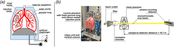
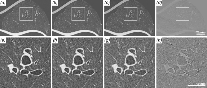
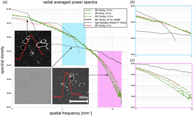
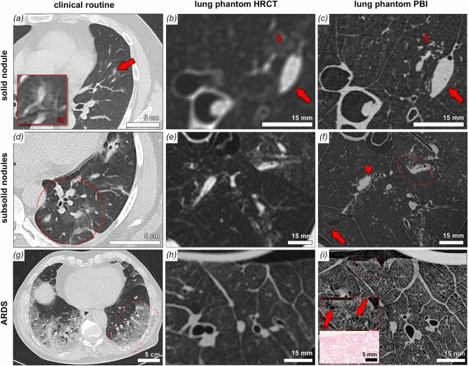
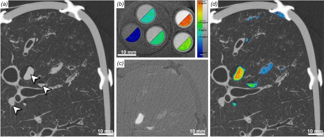
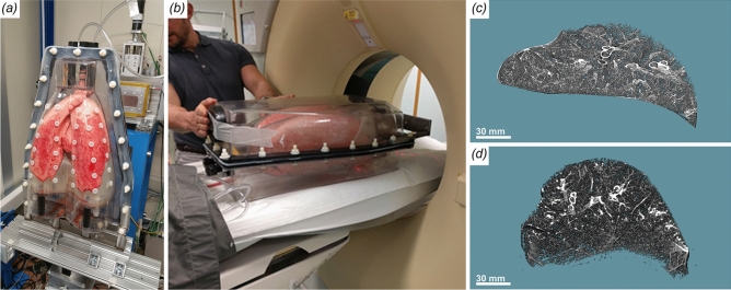
Absorption-based clinical computed tomography (CT) is the current imaging method of choice in the diagnosis of lung diseases. Many pulmonary diseases are affecting microscopic structures of the lung, such as terminal bronchi, alveolar spaces, sublobular blood vessels or the pulmonary interstitial tissue. As spatial resolution in CT is limited by the clinically acceptable applied X-ray dose, a comprehensive diagnosis of conditions such as interstitial lung disease, idiopathic pulmonary fibrosis or the characterization of small pulmonary nodules is limited and may require additional validation by invasive lung biopsies. Propagation-based imaging (PBI) is a phase sensitive X-ray imaging technique capable of reaching high spatial resolutions at relatively low applied radiation dose levels. In this publication, we present technical refinements of PBI for the characterization of different artificial lung pathologies, mimicking clinically relevant patterns in ventilated fresh porcine lungs in a human-scale chest phantom. The combination of a very large propagation distance of 10.7 m and a photon counting detector with [Formula: see text] pixel size enabled high resolution PBI CT with significantly improved dose efficiency, measured by thermoluminescence detectors. Image quality was directly compared with state-of-the-art clinical CT. PBI with increased propagation distance was found to provide improved image quality at the same or even lower X-ray dose levels than clinical CT. By combining PBI with iodine k-edge subtraction imaging we further demonstrate that, the high quality of the calculated iodine concentration maps might be a potential tool for the analysis of lung perfusion in great detail. Our results indicate PBI to be of great value for accurate diagnosis of lung disease in patients as it allows to depict pathological lesions non-invasively at high resolution in 3D. This will especially benefit patients at high risk of complications from invasive lung biopsies such as in the setting of suspected idiopathic pulmonary fibrosis (IPF).


Conflict of interest statement

The authors declare no competing interests.

Figures

Figure 1
Figure 1
Experimental setup. (a) Schematic setup of the human chest phantom. The fresh pig lung is placed in the inner cavity above the artificial diaphragm and attached with the trachea to a tube for external access. One pump (P1) is used to maintain a negative pressure between inner cavity and lung resulting in the inflation of the lung. A second pump (P2) can be used to move the diaphragm, for which different breathing pattern can be programmed. Different outer covers exist that for instance allow to fill the outer cavity with water to mimic the x-ray absorption of the chest. Here we used a single cover with sealed injection ports that allowed injection of artificial tumor nodules in the lung. (b) Shows the phantom mounted up-right on top of the rotary unit in front of the beam outlet and the ionization chamber. The photon counting detector was mounted 10.7 m upstream to provide sufficient sample-to-detector distance to successfully exploit PBI.
Figure 2
Figure 2
Comparison of the image quality of a healthy pig lung at different dose levels. To simulate lower entrance doses reconstructions with less angular projections are shown. (a) Full 8100 projections, entrance dose 10 mGy. (b) Half 4050 projections, entrance dose 5  mGy. (c) Quarter 2025 projections, entrance dose 2.5 mGy. (d) Full 8100 projections, entrance dose 10 mGy, no phase retrieval applied. (e), (f), (g), (h) show zoomed in parts of the images above. No visible difference are found between full and half dose while for a quarter of the dose the image quality diminishes a bit. Without phase retrieval the lung structure can barely be identified.
Figure 3
Figure 3
Comparison of the different imaging setups based on scans of three different healthy pig lungs. (a) High resolution clinical CT, effective pixel size 455μm, entrance dose 13 mGy. (b) SyRMeP synchrotron setup, SDD 2.7 m, effective pixel size 89μm, entrance dose 10 mGy. (c) SyRMeP synchrotron setup, SDD 10.7 m, effective pixel size 67μm, entrance dose 10 mGy. (d), (e), (f) show zoomed in parts of the images above. Clearly, the Synchrotron data sets surpass the image quality of the clinical HRCT scan. Moreover, the data acquired at lower dose at 10.7 m are of better image quality than the one scanned at shorter sample-to-detector distance.
Figure 4
Figure 4
Power spectra. (a) Shows radial averaged power spectra of lung regions of interest from the different measurements (clinical HRCT = black dashed, SR with an SDD of 2.7 m = red, SR with an SDD of 10.7 m at 10, 5 and 2.5 mGy green solid, dashed and dot-dashed subsequently the same data at 10 mGy but without phase retrieval is shown in solid black). Clearly, the phase retrieved synchrotron data demonstrates higher powers at medium spatial frequencies and lower powers at high frequencies (noise region) and has therefore a high image quality, which can also be seen in the inserted images. The red curves in the inserts depicts edge profiles at the positions indicated in yellow over a length of 2.5 mm. Despite the high contrast in the clinical data the limited in plane resolution of 450 µm clearly results in edges with a lower sharpness compared to the synchrotron data set. The highlighted region of the medium spatial frequencies (b) demonstrates that the images taken at 10.7 m surpass the data taken at 2.7 m at even higher dose of 13 mGy. Moreover, it can be seen that the data at 5 mGy does not deviate much from the results at 10 mGy and that only the data at 2.5 mGy shows a visible drop in power. In the enlarged high frequency domain (c) the known effect that phase retrieval acts as a low pass filter and therefore reduces the noise can be seen together with the fact that the noise level depends on the applied dose.
Figure 5
Figure 5
Correlative cHRCT and PBI SRCT with comparative clinical cases. a, d and g show comparative clinical examples of pulmonary damage patterns from cases of viral pneumonia. Patterns of solid nodules (a), subsolid nodules (b) and acute interstitial pneumonitis/acute respiratory distress syndrome (g) are shown. Artificial lesions mimicking those patterns were imaged by clinical HRCT (b, e and h) and the same lung regions were imaged by PBI-based local tomography (c, f and i). For solid nodules PBI-SRCT proofs superior characterization of the lesions borders and substructures (arrow), note the clear depiction of tiny gas enclosures and the orientation of the nodule alongside an interlobular septum. Also a faint ground glass area (§ in b) is unveiled as micro tree-in-bud (§ in c) by PBI-based SRCT. Grouped subsolid nodules can be depicted in detailed context to the surrounding lung parenchyma. In the circle in (f), an extension of the solid component into the interlobular septum can be noted and tiny substructures of the solid component can be depicted (air enclosures indicated by arrowhead in f). Perifocal micro tree-in-bud patterns can be attributed to the ground glass components and a parenchymal laceration can be noted (arrow in f). In cases of acute alveolar damage (gi) substructures of the secondary pulmonary lobule become accessible by PBI-based SRCT, note the heterogeneous distension of individual alveolar air spaces (box in h, arrows) with correlative H&E stained histology of the same specimen.
Figure 6
Figure 6
Iodine k-edge subtraction imaging (KES). (a) Shows a representative cross-section through a scan (33 keV) of a pig lung injected with different concentrations of iodine/agarose gel mixes. The white arrows point to vessels filled with the contrast agent. Differences in iodine concentration can not be observed. (b) CT scan of a phantom with known iodine concentrations (overlaid in pseudo-color), from which an iodine concentration calibration function was retrieved (color bar right). (c) The Subtraction image of two scans at 33.5 and 33 keV demonstrates a high specificity for iodine, which in turn allows to quantify difference in the iodine concentration. (d) Quantification of the iodine concentrations (color coded) using the calibration function (b).
Figure 7
Figure 7
Comparison of synchrotron CT and clinical CT scanning of the human chest phantom. (a) Phantom mounted in up-right position at the synchrotron setup. (b) Phantom placed in horizontal orientation in the clinical CT scanner. (c) Sample lung region extracted from the synchrotron data. (d) The same lung region (from the same lung) extracted from the clinical CT. Clearly, the shape of that region differs dramatically between the two setups.

References

    1. Guo HH, et al. A calibration ct mini-lung-phantom created by 3-d printing and subtractive manufacturing. J. Appl. Clin. Med. Phys. 2021;22:183–190. doi: 10.1002/acm2.13263. - DOI - PMC - PubMed
    1. Kalender, W. A. Computed tomography: fundamentals, system technology, image quality, applications (John Wiley & Sons, 2011).
    1. Lanni TB, et al. Early results from the implementation of a lung cancer screening program: The Beaumont health system experience. Am. J. Clin. Oncol. 2018;41:218–222. doi: 10.1097/COC.0000000000000254. - DOI - PubMed
    1. Mazzone P, Jain P, Arroliga AC, Matthay RA. Bronchoscopy and needle biopsy techniques for diagnosis and staging of lung cancer. Clin. Chest Med. 2002;23:137–158. doi: 10.1016/S0272-5231(03)00065-0. - DOI - PubMed
    1. Park J, et al. Mortality and risk factors for surgical lung biopsy in patients with idiopathic interstitial pneumonia. Eur. J. Cardio-Thoracic Surg. 2007;31:1115–1119. doi: 10.1016/j.ejcts.2007.02.035. - DOI - PubMed