. 2023 Mar 23;13(1):4746.
doi: 10.1038/s41598-023-31780-9.

Differentiation of patient-specific void urine-derived human induced pluripotent stem cells to fibroblasts and skeletal muscle myocytes

Affiliations

Differentiation of patient-specific void urine-derived human induced pluripotent stem cells to fibroblasts and skeletal muscle myocytes

M Kibschull et al. Sci Rep. .

Abstract

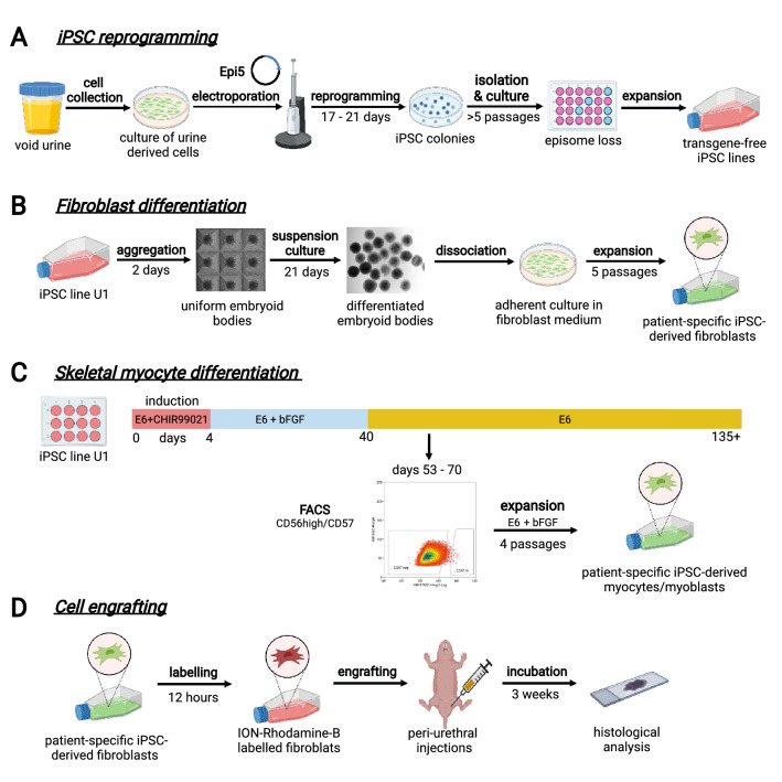
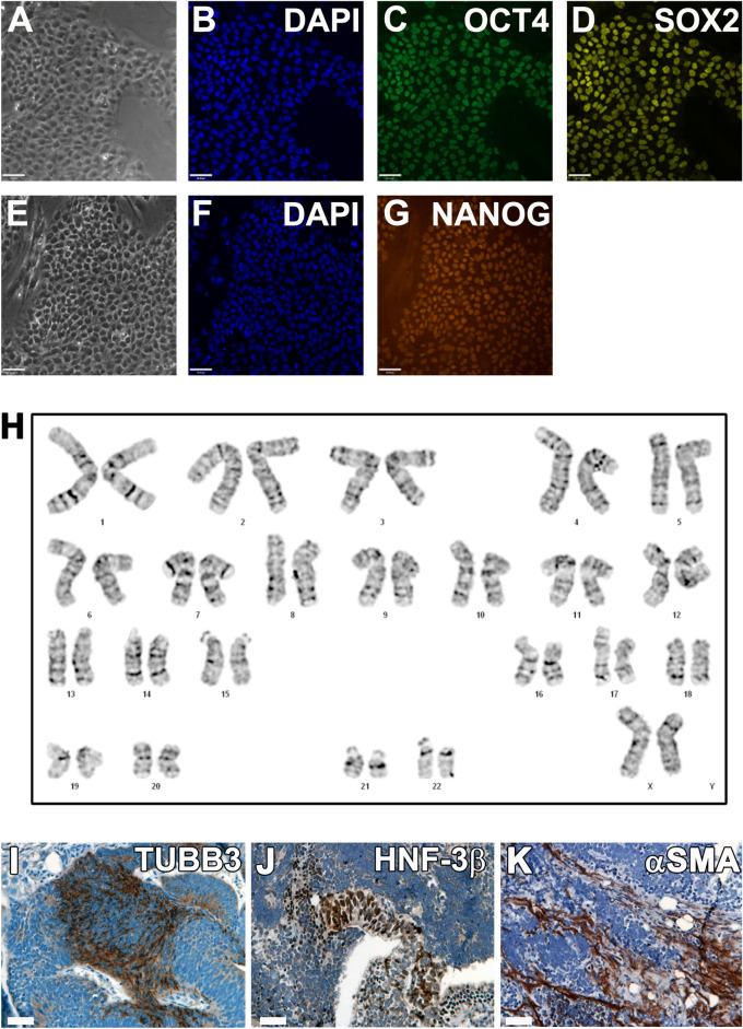
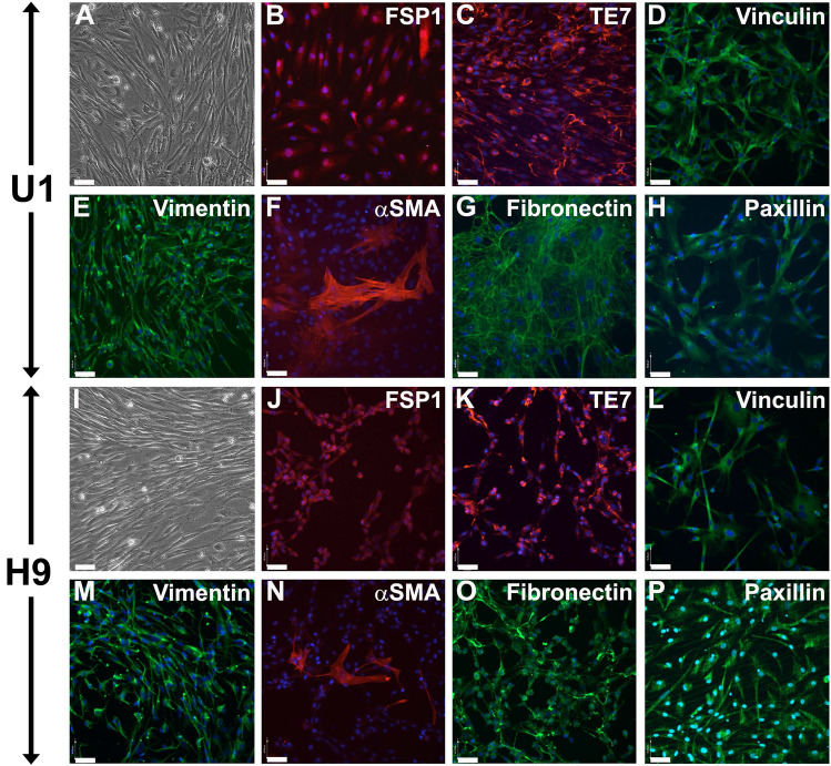
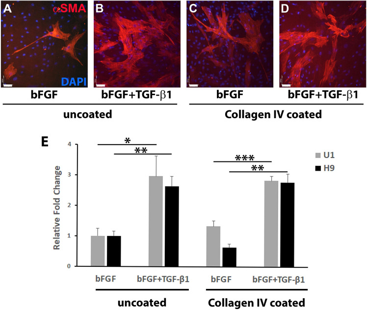
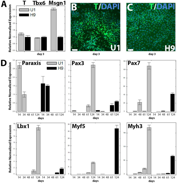
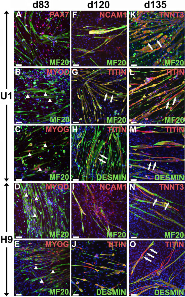
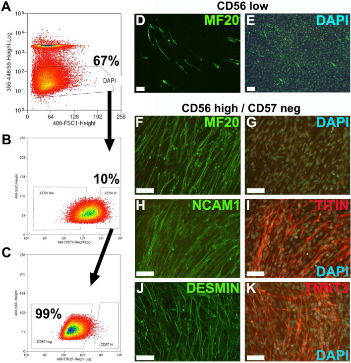
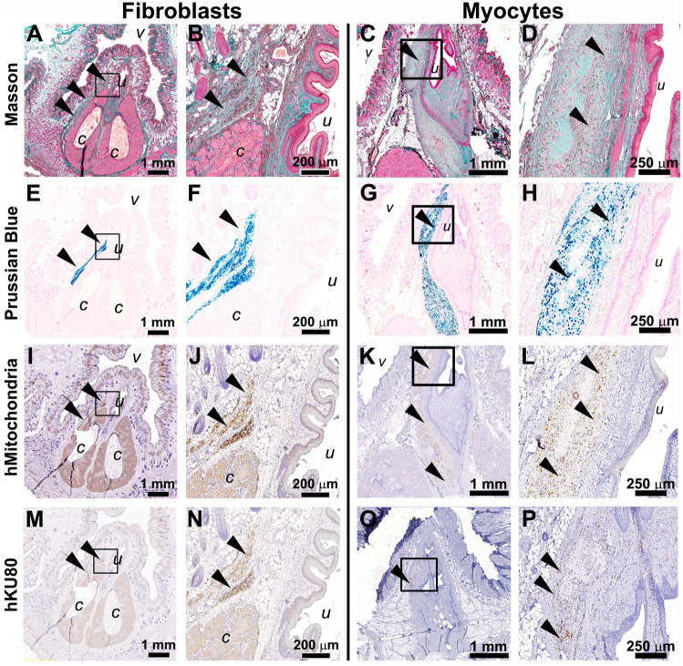
Cell-based therapy is a major focus for treatment of stress urinary incontinence (SUI). However, derivation of primary cells requires tissue biopsies, which often have adverse effects on patients. A recent study used human induced pluripotent stem cells (iPSC)-derived smooth muscle myocytes for urethral sphincter regeneration in rats. Here, we establish a workflow using iPSC-derived fibroblasts and skeletal myocytes for urethral tissue regeneration: (1) Cells from voided urine of women were reprogrammed into iPSC. (2) The iPSC line U1 and hESC line H9 (control) were differentiated into fibroblasts expressing FSP1, TE7, vinculin, vimentin, αSMA, fibronectin and paxillin. (3) Myogenic differentiation of U1 and H9 was induced by small molecule CHIR99021 and confirmed by protein expression of myogenic factors PAX7, MYOD, MYOG, and MF20. Striated muscle cells enriched by FACS expressed NCAM1, TITIN, DESMIN, TNNT3. (4) Human iPSC-derived fibroblasts and myocytes were engrafted into the periurethral region of RNU rats. Injected cells were labelled with ferric nanoparticles and traced by Prussian Blue stain, human-specific nuclear protein KU80, and human anti-mitochondria antibody. This workflow allows the scalable derivation, culture, and in vivo tracing of patient-specific fibroblasts and myocytes, which can be assessed in rat SUI models to regenerate urethral damages and restore continence.


Conflict of interest statement

The authors declare no competing interests.

Figures

Figure 1
Figure 1
Workflow charts of experimental design. (A) iPSCs reprogramming from urine samples. Cells collected from void urine samples of SUI patients were cultured and then electroporated with the Epi5™ episomal reprogramming system. After 17–21 days of culture, emerging iPSC colonies were isolated and individually cultured. During > 5 passages of iPSC sub-culture, episomal reprogramming factors were lost and growth of patient-specific, transgene-free iPSC lines is maintained by endogenous pluripotency markers. (B) Fibroblast differentiation. A single-cell suspension of iPSC was spun into AggreWell multi-well plates to form embryoid bodies (EBs). After 2 days, EBs were transferred to suspension culture dishes for 21 days to induce spontaneous differentiation. EBs were then dissociated, and cells transferred to adherent culture dishes in fibroblast growth medium. After 5 passages, cultures were tested for fibroblast marker expression and purity, expanded, and cryo-preserved. (C) Skeletal myocyte differentiation. iPSCs were seeded in 12-well plates. Mesodermal differentiation was induced by culturing iPSC in E6 medium supplemented with small molecule CHIR99021 for 4 days. Subsequent culture in E6 medium and bFGF led to myogenic cell differentiation and expansion. From day 40 onwards, cells were maintained in E6 medium alone, leading to myocyte differentiation and myotube formation. Between days 53–70, culture samples were dissociated and CD56high/CD57neg myocytes isolated by FACS. Enriched myocyte cultures were expanded for four passages, and cryopreserved. (D) Cell engrafting in rat tissues and histological assessment. Cultures of patient-specific iPSC-derived fibroblasts were labelled with Molday ION Rhodamine B nanoparticles for in vivo cell tracing. Suspensions of labelled fibroblasts were injected into the peri-urethral region of immune-compromised female nude RNU rats. After 3 weeks, urogenital tissues were dissected, fixed, processed, and histologically analyzed.
Figure 2
Figure 2
Characterization of urine cell-derived iPSC line U1. Urine cells were reprogrammed to iPSCs using the Epi5 reprogramming system. (AG) Immunocytochemistry of episome-free U1 line confirms the expression of pluripotency markers in the newly derived iPSC line U1: (A) phase contrast image of a U1 iPSC colony grown on Geltrex in mTeSR, and corresponding co-immunolabeling for (B) DAPI, (C) OCT4, and (D) SOX2. (E) Phase contrast, and co-immunolabeling for (F) DAPI, and (G) NANOG. Bar: 100 µm. (H) G-banded analysis of U1 metaphase chromosomes reveals a normal female karyotype. (I, J, K) Teratoma formation assay of undifferentiated U1 injected into kidney capsule of immune deficient athymic nude mice. Immunohistochemistry of dissected tumors shows tri-lineage differentiation into TUBB3 positive neuroectoderm (I, brown staining), hepatocyte nuclear factor HNF-3β expressing endoderm (J), and alpha smooth muscle actin (αSMA)-positive mesodermal cells (K). Bar: 200 µm.
Figure 3
Figure 3
Characterization of U1 and H9-derived fibroblasts. After 5 passages in monolayer culture, de novo differentiated human fibroblasts from iPSC line U1 (AH) and control hESC line H9 (IP) were characterized by phase contrast (A, I) and immunofluorescence for anti-human fibroblast-specific antibody markers: fibroblast specific protein-1, FSP-1 (B, J); human thymic fibroblast marker, TE7 (C, K); vinculin (D, L); vimentin (E, M); alpha smooth muscle actin, αSMA (F, N); fibronectin (G, O); paxillin (H, P). Nuclear counterstain DAPI, bar: 40 µm.
Figure 4
Figure 4
TGF-β1-induced myofibroblast differentiation of U1 and H9 fibroblasts. (AD) Co-immunolabeling for αSMA and DAPI of fibroblasts cultured on: (A, B) uncoated, or (C, D) recombinant human collagen IV-coated plates; in (A, C) E6 culture medium with 10 ng/mL bFGF only; or (B, D) medium with 10 ng/mL bFGF + 2 ng/mL TGF-β1. Bar: 40 µm. (E) For each culture condition above (AD), total numbers of DAPI-positive cells and αSMA-positive myofibroblasts, were counted in four visual fields. TGF-β1 treatment led to a significant increase of αSMA-positive myofibroblast differentiation as compared to untreated control cultures. In both, the U1 and H9 line, Collagen IV-coating did not have a significant impact on myofibroblast differentiation, independent of TGF-β1 supplementation. *p < 0.05, **p < 0.01, ***p < 0.001.
Figure 5
Figure 5
Mesodermal induction and myogenic differentiation of U1 and H9. (A) At day 3 of induction with CHIR99021, the early mesodermal transcription factors T (T-box T), Tbx6 (T-box 6), and Msgn1 (mesogenin 1) are detected by RT-qPCR. Immunolabeling for T antigen (nuclear counterstain with DAPI) reveals homogenous expression in (B) U1 and (C) H9 cultures at day 3. Bar: 80 µm. (D) RT-qPCR analysis of markers Paraxis, Pax3, Pax7, Lbx1, Myf5 and Myh3 reveals successful differentiation of U1 and H9 along the myogenic lineage into skeletal muscle progenitors and myocytes.
Figure 6
Figure 6
Characterization of U1 and H9 derived myocytes. Co-immunolabelling for myogenic lineage markers at days: d83 (AE), d120 (FJ), and d135 (KO) in differentiating cultures of U1 (AC, FH, KM) and H9 (D, E, I, J, N, O). (A) At d83, U1 cultures show presence of cells expressing the myogenic progenitor marker PAX7 and cells expressing myocyte marker MF20. MF20 positive myocytes co-express MYOD (B, D, arrowheads) and MYOG (C, E, arrowheads). At d120, myocytes co-express MF20, NCAM1, TITIN or DESMIN (FJ). At d135, myocytes co-express MF20, TNNT3, TITIN or DESMIN (KO). Multinucleated cells reveal myotube formation (G, H, KO). Nuclear counterstain with DAPI, bar: 50 µm.
Figure 7
Figure 7
Enrichment of skeletal myocytes by fluorescence-activated cell sorting (FACS) using. Myogenic differentiation cultures of U1 (day 63) were dissociated and labelled with conjugated NCAM/CD56 and HNK-1/CD57 antibodies, and counterstained with DAPI. Gating Strategy: (A) DAPI-negative, intact cells were gated for CD56high myocytes (B) and absence of CD57 expression (C). Sorted cells were cultured for 3 passages and labelled for markers. (D, E) Cultures of CD56low cells show low numbers of myocytes, whereas cultures of CD56high/CD57neg show myocyte enrichment as shown by expression of markers MF20 (F), NCAM1 (H), TITIN (I), DESMIN (J) and TNNT3 (K). DAPI (EG, I, K) was used as counterstain. Bar: 100 µm.
Figure 8
Figure 8
Histologic tracing of U1 fibroblasts and myocytes in the periurethral area of RNU rats. Histological images of the periurethral region of immune-compromised female nude rats injected with U1-derived fibroblasts (A, B, E, F, I, J, M, N) or U1-derived myocytes (C, D, G, H, K, L, O, P) at low and high magnification. (AD) Masson’s trichrome histologic staining of the injection site (periurethral tissue area). (EH) Prussian Blue staining (arrowheads) allows highly sensitive and specific detection of U1-fibroblasts (E, F) or myocytes (G, H) labelled with Molday ION Rhodamine B nanoparticles. (IL) Immunohistochemistry staining with anti-human mitochondrial antigen antibodies, and (MP) anti-human nuclear antigen KU80 antibodies confirm the presence of human fibroblasts or myocyte, respectively, within rat tissues (arrowheads indicate positive brown staining). v, vagina; u, urethra; c, clitoral gland.

References

    1. Maral I, et al. Prevalence of stress urinary incontinence in both sexes at or after age 15 years: A cross-sectional study. J. Urol. 2001;165(2):408–412. doi: 10.1097/00005392-200102000-00013. - DOI - PubMed
    1. Wu JM, et al. Lifetime risk of stress urinary incontinence or pelvic organ prolapse surgery. Obstet. Gynecol. 2014;123(6):1201–1206. doi: 10.1097/AOG.0000000000000286. - DOI - PMC - PubMed
    1. Petros PE, Woodman PJ. The integral theory of continence. Int. Urogynecol. J. Pelvic Floor Dysfunct. 2008;19(1):35–40. doi: 10.1007/s00192-007-0475-9. - DOI - PubMed
    1. Chang J, Lee D. Midurethral slings in the mesh litigation era. Transl. Androl. Urol. 2017;6(Suppl 2):S68–S75. doi: 10.21037/tau.2017.04.06. - DOI - PMC - PubMed
    1. DeLancey JO. Structural support of the urethra as it relates to stress urinary incontinence: The hammock hypothesis. Am. J. Obstet. Gynecol. 1994;170(6):1713–1720. doi: 10.1016/S0002-9378(94)70346-9. - DOI - PubMed

Publication types