Review
. 2023 May;19(5):281-299.
doi: 10.1038/s41581-023-00694-0. Epub 2023 Mar 23.

Regulated cell death pathways in kidney disease

Affiliations
Review

Regulated cell death pathways in kidney disease

Ana B Sanz et al. Nat Rev Nephrol. 2023 May.

Abstract

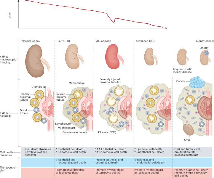
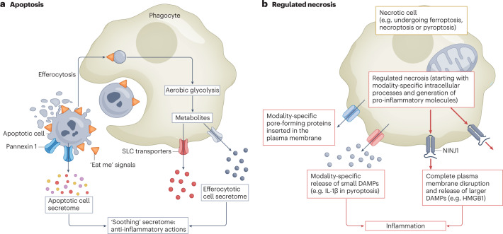
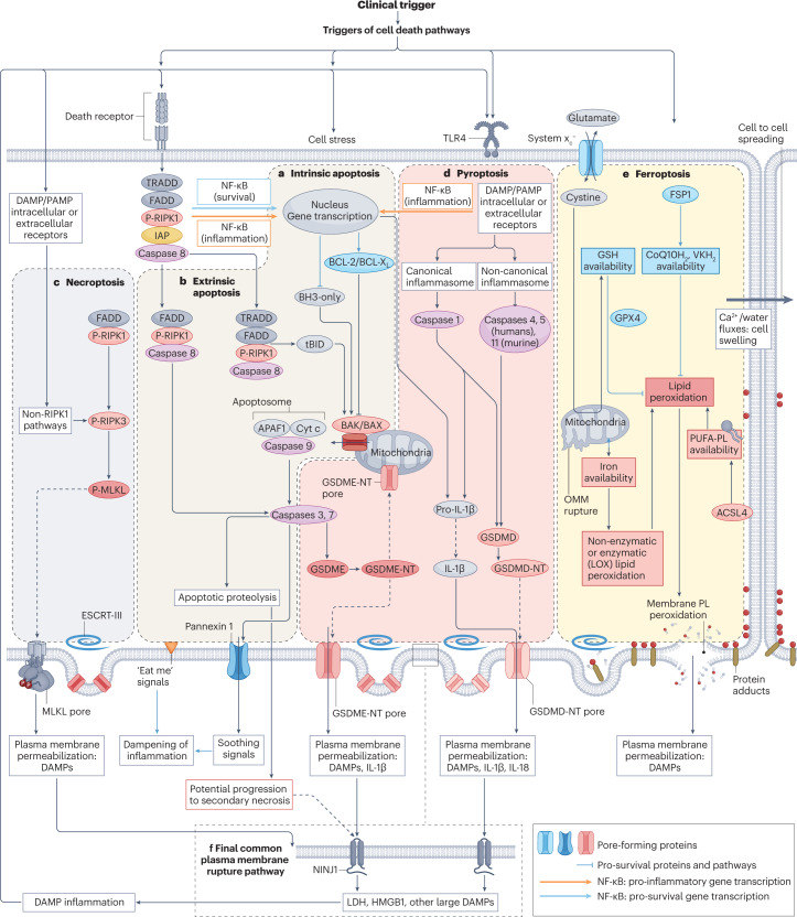
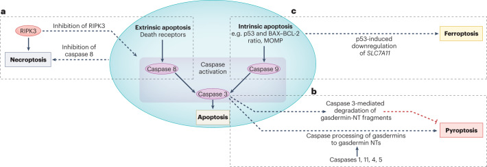
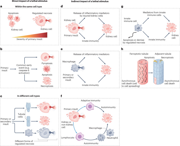
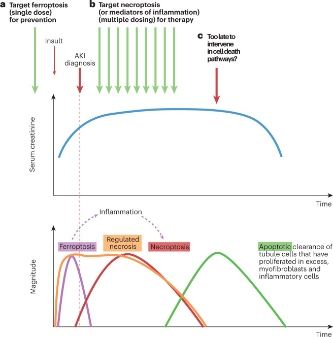
Disorders of cell number that result from an imbalance between the death of parenchymal cells and the proliferation or recruitment of maladaptive cells contributes to the pathogenesis of kidney disease. Acute kidney injury can result from an acute loss of kidney epithelial cells. In chronic kidney disease, loss of kidney epithelial cells leads to glomerulosclerosis and tubular atrophy, whereas interstitial inflammation and fibrosis result from an excess of leukocytes and myofibroblasts. Other conditions, such as acquired cystic disease and kidney cancer, are characterized by excess numbers of cyst wall and malignant cells, respectively. Cell death modalities act to clear unwanted cells, but disproportionate responses can contribute to the detrimental loss of kidney cells. Indeed, pathways of regulated cell death - including apoptosis and necrosis - have emerged as central events in the pathogenesis of various kidney diseases that may be amenable to therapeutic intervention. Modes of regulated necrosis, such as ferroptosis, necroptosis and pyroptosis may cause kidney injury directly or through the recruitment of immune cells and stimulation of inflammatory responses. Importantly, multiple layers of interconnections exist between different modalities of regulated cell death, including shared triggers, molecular components and protective mechanisms.


Conflict of interest statement

A.O. has received grants from Sanofi and consultancy, speaker fees or travel support from Advicciene, Astellas, AstraZeneca, Amicus, Amgen, Fresenius Medical Care, GSK, Bayer, Sanofi-Genzyme, Menarini, Kyowa Kirin, Alexion, Idorsia, Chiesi, Otsuka, Novo-Nordisk and Vifor Fresenius Medical Care Renal Pharma, and is the Director of the Catedra Mundipharma-UAM for diabetic kidney disease and the Catedra Astrazeneca-UAM for chronic kidney disease and electrolytes. A.O. also has stock in Telara Farma. The other authors declare no competing interests.

Figures

Fig. 1
Fig. 1. The natural history of CKD and relationship to cell number and cell death.
The relationship between chronic kidney disease (CKD), acute kidney injury (AKI), acquired kidney cystic disease and kidney cancer can be conceptualized as interrelated processes, characterized by changes in cell number that are driven by the proliferation or death of key cell types. Differences in the dynamics of cell death and turnover represent potential therapeutic targets. AKI is more common in patients with prior CKD and may cause irreversible loss of kidney function or accelerate CKD progression. However, not all episodes of AKI are associated with prior CKD. Of note, AKI is not necessarily characterized by a loss of kidney epithelial cells, but cell death is common in severe episodes of AKI. Acquired kidney cystic disease is usually not clinically relevant although bleeding may occur. However, acquired kidney cystic disease is a risk factor for renal cell carcinoma,. Both acquired kidney cystic disease and kidney cancer are associated with an increase in cell number. In kidney cysts, an excess number of epithelial cells allows cyst growth. Kidney cancer is characterized by an increased number of malignant epithelial cells; the goal of therapy in this context is to kill or extirpate these cells. Renal cell carcinoma may occur in individuals with or without prior CKD. However, CKD is a risk factor for renal cell carcinoma. Of note, acquired cystic kidney disease renal cell carcinoma is considered a distinct renal neoplasm according to the International Society of Urological Pathology–WHO, and is associated with specific somatic mutations. ECM, extracellular matrix; GFR, glomerular filtration rate.
Fig. 2
Fig. 2. Interaction between apoptosis, regulated necrosis and inflammation.
a, The process of apoptosis clears unwanted or damaged cells during development and under homeostatic conditions. Multiple steps within the process of apoptosis convey signals to dampen inflammation. Cells that are undergoing apoptosis display cell surface molecules (‘eat me’ signals) that promote their rapid engulfment (efferocytosis) and clearance by adjacent cells before the plasma membrane ruptures and the pro-inflammatory contents are released. Additionally, caspase-mediated opening of pannexin 1 channels at the plasma membrane of the apoptotic cell facilitates the release of an apoptotic metabolite secretome that comprises AMP, GMP, creatine, spermidine, glycerol-3-phosphate, ATP, fumarate, succinate and other compounds that suppress inflammation in adjacent cells. Furthermore, efferocytosis itself is associated with changes in phagocyte metabolism, including a switch to aerobic glycolysis and the release of glycolytic by-products such as lactate through solute carrier family (SLC) transporters to promote anti-inflammatory responses in adjacent cells. Thus, overall the processes of apoptosis and efferocytosis are associated with a complex programme that promotes the ‘soothing’ of adjacent cells. b, By contrast, regulated necrosis can engage innate and adaptive immune responses at multiple steps through, for example, the lack of early engulfment, the generation of pro-inflammatory molecules (for example, IL-1β in pyroptosis) and the release of small damage-associated molecular patterns (DAMPs) through pore-forming proteins that are specific for each form of regulated necrosis. The final common pathway of these actions is disruption of the plasma membranes through the involvement of NINJ1, which allows the release of larger DAMPs.
Fig. 3
Fig. 3. Key molecular processes and interactions between cell death pathways.
Two main pathways for apoptosis are well characterized. a, In the intrinsic pathway, stressors or the lack of survival signals decrease the balance between pro-survival BCL-2 family proteins and pro-apoptotic BH3-only proteins, favouring the BAK/BAX-dependent mitochondrial outer membrane permeabilization, which results in the release of pro-apoptotic mitochondrial proteins, including cytochrome c, and leads to the formation of a multiprotein structure called the apoptosome that incorporates APAF1 and procaspase 9. This series of events results in activation of caspase 9 — an initiator caspase that subsequently activates executioner caspases such as caspase 3 and caspase 7 that contribute decisively to the dismantling of cell structures. b, In the extrinsic apoptosis pathway, activation of death receptors of the tumour necrosis factor receptor (TNFR) superfamily leads to activation of the initiator caspase 8, which in turn activates executioner caspases and may also process the BH3-only protein BID to tBID, which activates the mitochondrial apoptotic pathway. However, there are alternative outcomes for TNFR activation by TNF, depending on the recruitment of specific proteins to multiprotein complexes. The presence of inhibitor of apoptosis (IAP) proteins activates NF-κB to increase the transcription of anti-apoptotic proteins, although NF-κB can also induce a pro-inflammatory response. In absence of IAPs, caspase 8 is activated. Inhibition of caspase 8 may trigger necroptosis (see below). Apoptotic cells express “eat-me” signals on their surface, which promotes their rapid engulfment (efferocytosis) by adjacent cells. In addition, they secrete anti-inflammatory molecules (soothing signals). However, extensive apoptosis or decreased clearance of apoptotic cells may lead to secondary necrosis, in which the cell membrane is permeabilized through engagement of NINJ1 and damage-associated molecular patterns (DAMPs) released. c, Necroptosis. The core features of necroptosis are receptor-interacting serine/threonine-protein kinase 3 (RIPK3) activation by phosphorylation and the subsequent phosphorylation of mixed lineage kinase domain-like protein (MLKL). Phosphorylated MLKL oligomerizes to form pores that disrupt the plasma membrane, allowing the release of cell contents. Canonical RIPK3 activation is mediated by its interaction with the RIP homotypic interaction motif-containing proteins RIPK1, TIR domain-containing adapter molecule 1 (TICAM1) or Z-DNA-binding protein 1 (ZBP1). Binding of RIPK3 to RIPK1 occurs in response to activation of TNFRs, whereas binding of RIPK3 to TICAM1 is induced by activation of Toll-like receptors (TLRs), and binding to ZBP1 occurs in response to the presence of Z-RNA as a result of viral infection. Non-canonical RIPK3 activation occurs in response to activation of the cell membrane Na+/H+ exchanger 1 and increased intracellular pH (not shown). d, Pyroptosis. The key feature of pyroptosis is the enzymatic processing of gasdermins to amino-terminal (NT) fragments that oligomerize to assemble a plasma membrane pore, allowing the release of intracellular contents and DAMPs. The best-characterized model involves the NLRP3 (canonical) inflammasome-mediated activation of caspase 1 in macrophages, which processes pro-IL-1β to IL-1β, pro-IL-18 to IL-18 and gasdermin D (GSDMD) to GSDMD-NT. However, other inflammasomes and enzymes may process gasdermins in other cell systems and IL-1β may not be a major component of pyroptosis in cell types with low IL-1β gene expression. Additionally, gasdermins may permeabilize mitochondria, which, together with the potential for caspase 3 to cleave gasdermin E (GSDME) and caspase 8 to cleave GSDMD, provides links to apoptosis. e, Ferroptosis. The central event in ferroptosis is the peroxidation of plasma membrane phospholipids (PL) in an iron-dependent manner. The sensitivity of cells to ferroptosis will depend on iron availability (noting that mitochondria may serve to store iron), and on the capacity for enzymatic (for example, mediated by lipoxygenases (LOX)) or non-enzymatic lipid peroxidation as well as on the presence of cell defences against lipid peroxidation, including adequate glutathione (GSH) stores which are maintained through the entry of cystine through the system xc cystine/glutamate antiporter. The enzyme glutathione peroxidase 4 (GPX4) requires GSH for its antioxidant function. Additional antioxidant systems include ferroptosis suppressor protein 1 (FSP1), which maintains vitamin K (VK) and coenzyme Q10 (CoQ10; also known as ubiquinone) in a reduced state (VKH2 and CoQ10H2, respectively). The main consequence of ferroptosis is peroxidation of membrane PL, shown as red dots in the figure, that results in membrane protein adducts and membrane rupture. Following some triggers, ferroptosis may spread from cell to cell in a wave-like pattern via a process that involves volume shifts and calcium fluxes, as has been described in kidney tubules. f, Shared features between the different cell death modalities include the ability of the repair machinery, endosomal sorting complexes required for transport III (ESCRT-III), to repair membranes and the need for NINJ1 in the final step of plasma membrane fragmentation, which leads to the release of the larger DAMPs and proteins. However, NINJ1 is not required for plasma membrane rupture during ferroptosis. Of note, although all molecular pathways in the figure are represented in the same cell to emphasize the interconnections between pathways, not all cell types have the intracellular machinery or microenvironment required for all forms of regulated cell death to proceed. For pyroptosis, the increased transcription, processing and release of IL-1β have been mainly characterized in macrophages, whereas epithelial cells have a more limited capacity to release this interleukin. ACSL, long-chain fatty acid–CoA ligase 4; OMM, outer mitochondrial membrane; PAMP, pathogen-associated molecular pattern; PUFA, polyunsaturated fatty acid; PUFA-PL, PUFA-containing phospholipids.
Fig. 4
Fig. 4. Examples of interactions between apoptosis and regulated necrosis.
The molecular pathways that lead to activation of extrinsic or intrinsic apoptosis may also modulate or trigger different forms of regulated necrosis. Thus, interventions aimed at inhibiting apoptosis may trigger or modulate modalities of regulated necrosis, potentially affecting their safety and efficacy. a, Following the activation of death receptors, such as TNF receptor, inhibition of caspase 8 promotes necroptosis over apoptosis. Conversely, inhibitors of receptor-interacting serine/threonine-protein kinase 3 (RIPK)3 or certain kinase-dead RIPK3 mutants will prevent necroptosis but result in the assembly of multimeric protein complexes that activate caspase 8 and induce apoptotic cell death. b, Caspases are among the enzymes than can cleave gasdermins to generate amino-terminal (NT) fragments that oligomerize at the plasma membrane to form protein pores and trigger pyroptosis. In pyroptosis, canonical activation of the NLRP3 inflammasome results in activation of caspase 1, whereas non-canonical inflammasomes will activate murine caspase 11 (caspase 4 and caspase 5 in humans). In apoptosis, the executioner caspase 3 is activated by either caspase 8 (extrinsic apoptosis pathway) or caspase 9 (intrinsic apoptosis pathway). Caspases that are activated during apoptosis, such as caspase 8 and caspase 3 may also cleave gasdermins to gasdermin-NT fragments, as exemplified here for caspase 3. Additionally, caspase 3 may degrade NT fragments from gasdermins B and D, potentially protecting from pyroptosis. A more detailed representation of the interaction of caspases with gasdermins and gasdermin NTs is shown in Supplementary Fig. 4b. c, Upstream regulators of apoptosis may also modulate ferroptosis. As an example, the tumour-suppressive transcription factor p53 promotes apoptosis by upregulating mediators of apoptosis, such as BAX, and downregulating anti-apoptotic molecules such as BCL-2 (refs. ,), but also sensitizes cells to ferroptosis by downregulating SLC7A11, which encodes a subunit of the system xc cystine–glutamate antiporter, and consequently decreasing cystine uptake. MOMP, mitochondrial outer membrane permeabilization.
Fig. 5
Fig. 5. Interactions between different types of regulated necrosis: conceptual models that may explain the involvement of multiple forms of cell death in the same kidney disease.
The interconnectivity of different modes of cell death suggests that multiple modes of cell death probably concurrently or sequentially coexist within the diseased kidney. The mechanisms underlying the initiation of different cell death pathways are complex and may involve various processes. Primary insults are triggers of injury (for example, cisplatin or ischaemia–reperfusion) and secondary insults are the mediators released in response to cell stress or cell death, such as TNF superfamily cytokines. a, The severity of insult may affect the activation of different cell death pathways. Of note, necrosis is not necessarily associated with a greater severity of insult. c, A primary insult might activate several cell death programmes in the same cell that are mutually exclusive so that inhibition of one will result in death from the other. PANoptosis triggered by caspase 6 activation is one example of such an event, and the interaction between extrinsic apoptosis and necroptosis illustrated in Fig. 4a is another example. c, Primary or secondary insults may differentially impact the mode of cell death in different cell types. d, A primary insult (the cause of the kidney disease) may result in the release of inflammatory mediators by injured kidney cells (secondary insults) and the release of danger-associated molecular signals that trigger an innate immune response that, in turn, amplifies cell death. e, Regulated necrosis of innate immune cells might result in the release of inflammatory mediators and the activation of an innate immune response that, in turn, causes a different form of cell death in kidney epithelial cells. f, A primary insult might result in immunogenic cell death, and the resultant adaptive immune response might further cause cell death and tissue injury via antibodies or cytotoxic T cells. g, Cell death might clear innate immune cells and limit the inflammatory response. In this scenario, inhibition of cell death may result in persistent or amplified inflammation that triggers the death of kidney epithelial cells. h, Ferroptosis might induce synchronized renal tubule cell death following the intercellular propagation of cell death in a wave-like pattern, potentially affecting whole nephrons. By contrast, apoptosis and necroptosis occur in isolated cells. Inflammatory cytokines released by cells recruited in response to ferroptosis may, in turn, induce apoptosis and/or necroptosis of individual tubule cells located in nearby tubules.
Fig. 6
Fig. 6. Interactions between different forms of cell death and their impact on the design of therapeutic approaches for different contexts of use in AKI.
a, In a hypothetical scenario in which an insult causes an initial wave of cell death (for example, an early wave of ferroptosis-induced tubule cell death), the specific form of cell death may be targeted using a preventive approach; for example, with prophylactic administration of an inhibitor. This preventive intervention may avoid the initial wave of cell death and all subsequent consequences, including the development of acute kidney injury (AKI). b, If unchecked, the initial wave of regulated necrosis might result in the release of damage-associated molecular patterns (DAMPs) that triggers inflammation and a second wave of inflammatory cytokine-mediated regulated necrosis, such as necroptosis. In this scenario, a single preventive administration of a cytokine or necroptosis inhibitor will not prevent AKI, as it will not prevent the original wave of cell death caused by ferroptosis. However, necroptosis or other forms of inflammation-related cell death may be amenable to treatment, for example, through initiation of therapy after injury has occurred. This scenario represents the usual clinical situation, in which interventions are prescribed after diagnosis of AKI. Therapy would be expected to be maintained over several days, since DAMPs will be continuously released by ongoing cell death. Successful treatment will not prevent AKI but will accelerate recovery by interfering with the amplification loop of inflammation-related cell death. Biomarkers of regulated necrosis will be needed to assess whether cell death is still ongoing and if so, what is the predominant active mode of kidney cell death. c, In advanced stages of AKI, apoptotic cell death acts to clear excess myofibroblasts and leukocytes as well as excess tubule cells; at this stage, interference with apoptosis might be deleterious while targeting other forms of cell death might be futile. This conceptual representation is based on observations in a mouse model of AKI induced by a folic acid overdose,, and on descriptions of morphological apoptosis during the recovery phase of human AKI. The role of ferroptosis in the early stages of this preclinical model and of the contribution of necroptosis to longer term outcomes has been validated in several studies,. However, validation of this conceptual representation for forms of human AKI requires further studies and clinical trials.

References

    1. Foreman KJ, et al. Forecasting life expectancy, years of life lost, and all-cause and cause-specific mortality for 250 causes of death: reference and alternative scenarios for 2016-40 for 195 countries and territories. Lancet. 2018;392:2052–2090. doi: 10.1016/S0140-6736(18)31694-5. - DOI - PMC - PubMed
    1. Ortiz A. RICORS2040: the need for collaborative research in chronic kidney disease. Clin. Kidney J. 2021;15:372–387. doi: 10.1093/ckj/sfab170. - DOI - PMC - PubMed
    1. Chapman, A. B., Rahbari-Oskoui, F. F & Bennett, W. M. Acquired cystic disease of the kidney in adults. UpToDatehttps://www.uptodate.com/contents/acquired-cystic-disease-of-the-kidney-... (2023).
    1. Denton MD, et al. Prevalence of renal cell carcinoma in patients with ESRD pre-transplantation: a pathologic analysis. Kidney Int. 2002;61:2201–2209. doi: 10.1046/j.1523-1755.2002.00374.x. - DOI - PubMed
    1. Shah A, et al. Acquired cystic kidney disease-associated renal cell carcinoma (ACKD-RCC) harbor recurrent mutations in KMT2C and TSC2 genes. Am. J. Surg. Pathol. 2020;44:1479–1486. doi: 10.1097/PAS.0000000000001530. - DOI - PubMed

Publication types

LinkOut - more resources