. 2023 Jan 31;29(1):9-17.
doi: 10.5152/dir.2022.21640. Epub 2022 Dec 21.

Urethrocystography: a guide for urological surgery?

Affiliations

Urethrocystography: a guide for urological surgery?

Patrícia S Freitas et al. Diagn Interv Radiol. .

Abstract

Urethrocystography remains the gold-standard technique for urethral pathology diagnosis. Nowadays, of the various indications for performing urethrocystography, the most common is due to a clinical suspicion of urethral stricture. Due to the high prevalence of strictures and their substantial impact on a patient's quality of life, the examination must allow the location, exclusion of multifocality, and assessment of the extent of the stricture to influence surgical planning. This article intends to demonstrate that the radiologist's role, by performing and interpreting the modality of urethrocystography, influences and is crucial for the urologic therapeutic decision and that the patients who were submitted to reconstruction by urethroplasty had a better success rate. The authors aim to review the radiological anatomy of the male urethra, discuss the modalities of choice for imaging the urethra (retrograde urethrography and voiding cystourethrography), provide an overview of the different indications for performing the study, examine the different etiologies for urethral strictures, understand the relevance of the different appearances of urethral pathology, and identify the surgical options, especially in the treatment of urethral strictures. Simultaneously, the study exposes cases of urethral trauma, fistulas, diverticulum, and congenital abnormalities.

Keywords: Male urethra; radiology,; surgery; urethral stenosis; urethrocystography; urology.


Conflict of interest statement

Conflict of interest disclosure

The authors declared no conflicts of interest.

Figures

Figure 1
Figure 1
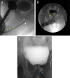
RUG (a) and VCUG (b) images show penile urethra (orange bracket, a); penoscrotal junction (white arrow, a); bulbar urethra (yellow bracket, a); bulbomembranous junction assuming conical shape (yellow arrow, a); membranous urethra (purple line, a); prostatic urethra (blue bracket, a) and bladder neck opening (blue arrow, b). RUG, retrograde urethrocystography; VCUG, voiding cystourethrography.
Figure 2
Figure 2
RUG demonstrating incomplete traction of the penis gland leading to a kinking artifact. RUG, retrograde urethrocystography.
Figure 3
Figure 3
RUG (a) and inverted RUG (b) showing transitory filling defects caused by air bubbles artifacts due to an incorrectly purged Foley catheter, which may mimic a urethral stone (yellow arrow, a). RUG, retrograde urethrocystography.
Figure 4
Figure 4
(a, b) RUG showing non-pathological filling defects in the prostatic urethra caused by a normal anatomical structure. Verumontanum as a filling defect (blue arrow, a) and normal tapering of the transition of the bulbar urethra to the posterior urethra due to the contraction of the constrictor nudae muscle (purple arrows, b). This must not be reported as a focal stricture since the posterior urethra only distends properly in the VCUG. VCUG, voiding cystourethrography.
Figure 5
Figure 5
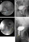
(a–c) RUG (a, b) and VCUG (c) show opacified Littré glands (green arrows, a) and opacified Cowper gland (white arrow a), contrast reflux to seminal vesicles (yellow arrows, b) and opacified left prostatic duct (blue arrow, c). RUG, retrograde urethrocystography; VCUG, voiding cystourethrography.
Figure 6
Figure 6
RUG shows opacification of the venous system due to high resistance during the urethrogram study (white arrows). RUG, retrograde urethrocystography.
Figure 7
Figure 7
(a, b) Two different patients with balanitis xerotica obliterans. The urethrogram shows a filiform anterior urethra due to multiple strictures (a) and a long-segment penile urethra stricture (orange bracket, b). Both patients were proposed for BMG urethroplasty. BMG, buccal mucosal graft.
Figure 8
Figure 8
Bulbar urethral strictures demonstrated in RUG (a, c) and VCUG (b, d). RUG shows proximal bulbar stricture resulting from a straddle injury in a 45-year-old patient with a post-pelvic trauma status (blue arrow, a). VCUG reveals ring stricture of the proximal bulbar urethra (red arrow, b), causing dilatation of the upstream segments in a patient with gonococcal urethritis. Bladder diverticula (orange arrow, b). Long-segment and irregular bulbar stricture in a 72-year-old patient with a clinical history of previous urethritis (yellow bracket, c). The enlarged caliber of the prostatic urethra (yellow arrow, d) due to the high pressure of the long-segment bulbar stricture. RUG, retrograde urethrocystography; VCUG, voiding cystourethrography.
Figure 9
Figure 9
Anterior urethral strictures before (a) and after (b) treatment. The 72-year-old patient shows in the RUG multiple ring strictures of the anterior urethra (orange arrow, a) with a Cowper gland opacification due to high pressure (white arrow, a). Three years later, the patient underwent IU, and the RUG showed a favorable evolution of the previous strictures (b). IU, internal urethrotomy; RUG, retrograde urethrocystography.
Figure 10
Figure 10
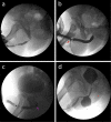
RUG showing post-operative complications in four different patients. Recurrent penile urethra stricture in a patient who has already undergone five IUs (a). Penile urethral trauma already submitted to urethroplasty; as a post-surgery complication, a urethral fistula developed at the healing wound (red arrow, b). Post-urethroplasty diverticulum (purple arrow, c); air bubble artifact (*, c). Status post-artificial urinary sphincter, showing a diverticular image upstream of the stenotic segment (29 × 30 mm), which communicates with the penile urethra, suggestive of a pre-stenotic diverticulum (d). RUG, retrograde urethrocystography; IU, internal urethrotomy.
Figure 11
Figure 11
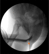
A 40-year-old man with a bulbar urethral fracture after trauma (orange arrow).
Figure 12
Figure 12
(a, b) Status post-hypospadias surgery. Dilatation of terminal urethra (blue arrow, a and b) and ventral penile fistula (yellow arrow, a).

References

    1. Patel DN, Fok CS, Webster GD, Anger JT. Female urethral injuries associated with pelvic fracture: a systematic review of the literature. BJU Int. 2017;120(6):766–773. - PubMed
    1. Waterloos M, Verla W. Female urethroplasty: a practical guide emphasizing diagnosis and surgical treatment of female urethral stricture disease. Biomed Res Int. 2019;2019:6715257. - PMC - PubMed
    1. Kawashima A, Sandler CM, Wasserman NF, Le- Roy AJ, King BF Jr, Goldman SM. Imaging of urethral disease: a pictorial review. Radiographics. 2004;24 Suppl 1:S195–S216. - PubMed
    1. Eaton J, Richenberg J. Imaging of the urethra: Current status. Imaging. 2005;17(2):139–149.
    1. Sandler CM, Corriere JN Jr. Urethrography in the diagnosis of acute urethral injuries. Urol Clin North Am. 1989;16(2):283–289. - PubMed