Review
. 2023 Mar 7:17:1141913.
doi: 10.3389/fnins.2023.1141913. eCollection 2023.

A novel feature of the ancient organ: A possible involvement of the subcommissural organ in neurogenic/gliogenic potential in the adult brain

Affiliations
Review

A novel feature of the ancient organ: A possible involvement of the subcommissural organ in neurogenic/gliogenic potential in the adult brain

Hitoshi Inada et al. Front Neurosci. .

Abstract

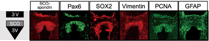
The subcommissural organ (SCO) is a circumventricular organ highly conserved in vertebrates from Cyclostomata such as lamprey to mammals including human. The SCO locates in the boundary between the third ventricle and the entrance of the aqueduct of Sylvius. The SCO functions as a secretory organ producing a variety of proteins such as SCO-spondin, transthyretin, and basic fibroblast growth factor (FGF) into the cerebrospinal fluid (CSF). A significant contribution of the SCO has been thought to maintain the homeostasis of CSF dynamics. However, evidence has shown a possible role of SCO on neurogenesis in the adult brain. This review highlights specific features of the SCO related to adult neurogenesis, suggested by the progress of understanding SCO functions. We begin with a brief history of the SCO discovery and continue to structural features, gene expression, and a possible role in adult neurogenesis suggested by the SCO transplant experiment.

Keywords: Pax6; Sox2; neural stem progenitor cell; neurogenesis; subcommissural organ.


Conflict of interest statement

The authors declare that the research was conducted in the absence of any commercial or financial relationships that could be construed as a potential conflict of interest.

Figures

FIGURE 1
FIGURE 1
Location of the SCO in the adult human and rodent brains. (A) Sagittal section of human brain. (B) Sagittal and coronal sections of rodent brains. The SCO locates at the boundary between the third ventricle (3V) and the entrance of the aqueduct of Sylvius (Aq). The SCO is composed of an ependymal cell layer(s) lining the third ventricular side of the posterior commissure. In the coronal section, the SCO appears as an inverted U-shape underneath the posterior commissure (PC). 3V, third ventricle; Aq, aqueduct of Sylvius; PC, posterior commissure; SCO, subcommissural organ (Corales et al., 2022). (C) Illustration of “a pair of ciliated grooves”. Ar.T., arachnoidal tissue; C.G., ciliated groove; Ch.PL, choroid plexus; Com.P., posterior commissure; M.C., connective tissue brain case. Used with permission of The Royal Society (U.K.), from Dendy (1902); permission conveyed through Copyright Clearance Center, Inc. (D) The secretory feature of the SCO ependymal cell. Scheme of an SCO secretory ependymal cell. The secretory proteins such as SCO-spondin and transthyretin are stored in the endoplasmic reticulum (ER), modified in the Golgi apparatus (G), and released both apically into the CSF and basally into the matrix of the posterior commissure. The SCO-spondin released into the CSF forms the RF. BP, basal process; Ci, cilia; ER, endoplasmic reticulum; G, Golgi apparatus; Mt, mitochondria; N, nucleus; PC, posterior commissure; RF, Reisner’s fiber; ZA, zonula adherens.
FIGURE 2
FIGURE 2
Histological structure of the SCO. (A) The SCO region of mouse brain. The SCO is composed of an ependymal cell layer(s) with an inverted U-shape lining the third ventricular side of the posterior commissure (Allen Brain Atlas: Mouse Brain, https://atlas.brain-map.org/). (B) The SCO region of human brain (BrainSpan Atlas of the Developing Human Brain, https://atlas.brain-map.org/). The SCO regions are surrounded by red dotted lines. (C) Immunostaining of SCO-spondin in the SCO region of the adult mouse and rat brain. The SCO-spondin staining pattern in the adult rat SCO shows a weak staining pattern in the nuclear region compared to the adult mouse SCO allowing better visualization of hypendymal cells between the ependymal layer and the posterior commissure. Arrows indicate hypendymal cells. Scale bars: 50 μm (Corales et al., 2022). 3V, third ventricle; Ep, ependymal cells; NR, nuclear region; PC, posterior commissure.
FIGURE 3
FIGURE 3
Structure of SCO-spondin and its downstream signaling pathways. (A) A cartoon of SCO-spondin structure. EMI, elastin microfibril interface; vWF-D, von Willebrand factor type-D; FA5/8C, Factor V/Factor VIII type C; LDLrA, low-density lipoprotein receptor class A; TIL, trypsin inhibitor-like; TSR thrombospondin type I repeat; vWF-C, von Willebrand factor type-C; EGF, epidermal growth factor; CTCK, C-terminal cystine knot-like. (B) Possible interactions of domains contained in SCO-spondin with soluble factors. FGF2, fibroblast growth factor 2; TGF-β, transforming growth factor-β; VEGF, vascular endothelial growth factor; HSPG, heparan sulfate proteoglycan; LPR, LDLr-related protein.
FIGURE 4
FIGURE 4
Expression of NSPC markers in the SCO. Immunostaining of NSPC markers in the mouse SCO (Corales et al., 2022).

References

    1. Aboitiz F., Montiel J. F. (2021). The enigmatic Reissner’s fiber and the origin of chordates. Front. Neuroanat. 15:703835. 10.3389/fnana.2021.703835 - DOI - PMC - PubMed
    1. Alshehri B., D’Souza D. G., Lee J. Y., Petratos S., Richardson S. J. (2015). The diversity of mechanisms influenced by transthyretin in neurobiology: development, disease and endocrine disruption. J. Neuroendocrinol. 27 303–323. 10.1111/jne.12271 - DOI - PubMed
    1. Baas D., Meiniel A., Benadiba C., Bonnafe E., Meiniel O., Reith W., et al. (2006). A deficiency in RFX3 causes hydrocephalus associated with abnormal differentiation of ependymal cells. Eur. J. Neurosci. 24 1020–1030. 10.1111/j.1460-9568.2006.05002.x - DOI - PubMed
    1. Bach A., Lallemand Y., Nicola M. A., Ramos C., Mathis L., Maufras M., et al. (2003). Msx1 is required for dorsal diencephalon patterning. Development 130 4025–4036. 10.1242/dev.00609 - DOI - PubMed
    1. Blackshear P. J., Graves J. P., Stumpo D. J., Cobos I., Rubenstein J. L., Zeldin D. C. (2003). Graded phenotypic response to partial and complete deficiency of a brain-specific transcript variant of the winged helix transcription factor RFX4. Development 130 4539–4552. 10.1242/dev.00661 - DOI - PubMed