Review
. 2023 Mar 8;25(4):163.
doi: 10.3892/ol.2023.13749. eCollection 2023 Apr.

Prostate cancer bone metastases biology and clinical management (Review)

Affiliations
Review

Prostate cancer bone metastases biology and clinical management (Review)

Emily Archer Goode et al. Oncol Lett. .

Abstract

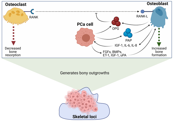
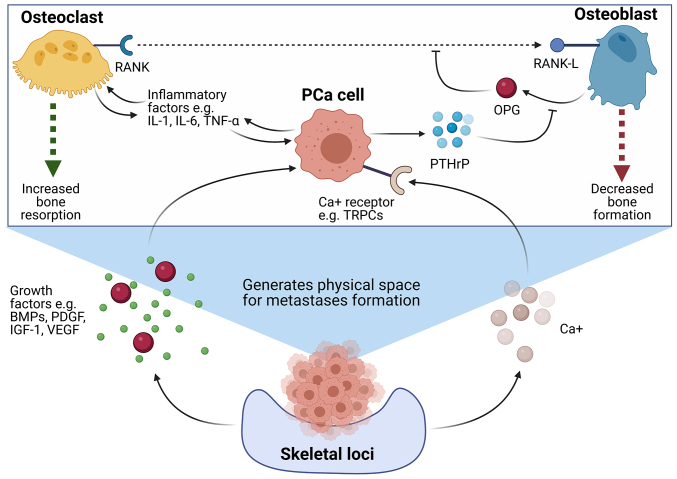
Prostate cancer (PCa) is one of the most prominent causes of cancer-related mortality in the male population. A highly impactful prognostic factor for patients diagnosed with PCa is the presence or absence of bone metastases. The formation of secondary tumours at the bone is the most commonly observed site for the establishment of PCa metastases and is associated with reduced survival of patients in addition to a cohort of life-debilitating symptoms, including mobility issues and chronic pain. Despite the prevalence of this disease presentation and the high medical relevance of bone metastases, the mechanisms underlying the formation of metastases to the bone and the understanding of what drives the osteotropism exhibited by prostate tumours remain to be fully elucidated. This lack of in-depth understanding manifests in limited effective treatment options for patients with advanced metastatic PCa and culminates in the low rate of survival observed for this sub-set of patients. The present review aims to summarise the most recent promising advances in the understanding of how and why prostate tumours metastasise to the bone, with the ultimate aim of highlighting novel treatment and prognostic targets, which may provide the opportunity to improve the diagnosis and treatment of patients with PCa with bone metastases.

Keywords: biomarker; bone metastases; osteoblasts; osteoclasts; prostate cancer; treatment.


Conflict of interest statement

The authors declare they have no competing interests.

Figures

Figure 1.
Figure 1.
A summary of the key molecular interactions between metastatic PCa cells and osteoblasts in the formation of osteoblastic bone metastases. PCa cells and osteoblasts secrete OPG, which acts as a soluble decoy receptor to inhibit the interaction between RANK and its ligand, RANK-L. The inhibition of this interaction prevents the maturation of osteoclasts, and promotes the activation of osteoblasts, thereby facilitating bone reformation. Furthermore, PCa cells have also been shown to secrete PAP. PAP has been demonstrated to stimulate osteoblast maturation through modulating the RANK/RANK-L/OPG axis, and promotes collagen synthesis and alkaline phosphatase production to facilitate further bone formation. In addition to this, PCa cells secrete a number of additional factors which stimulate the maturation and activity of osteoblasts, such as FGFs, BMPs, ET-1, IGF-1 and uPA. Mature osteoblasts have similarly been shown to secrete a number of factors which further support the survival and growth of localised PCa cells, such as IGF-1, IL-6 and IL-8. The net increase in osteoblast activity results in the formation of bony outgrowths which are colonised by sclerotic bone metastatic PCa cells (–43,132). Created with BioRender.com. BMPs, bone morphogenic proteins; ET-1, endothelin-1; FGFs, fibroblast growth factors; IGF-1, insulin-like growth factor 1; IL, interleukin; OPG, osteoprotegerin; PAP, prostatic acid phosphatase; PCa, prostate cancer; RANK, receptor activator of nuclear factor κB; RANK-L, RANK-ligand; uPA, urokinase-type plasminogen activator.
Figure 2.
Figure 2.
Molecular interactions between osteoclasts and PCa cells leading to the formation of lytic bone metastases. PCa cells with the ability to give rise to lytic bone metastases have been shown to secrete a number of factors which promote osteoclast maturation and activity, and concurrently, downregulate osteoblast maturation and activity, in order to generate a supportive niche for the growth of a bone lytic tumour. PCa cells secrete PTHrP, which inhibits the release of osteoblast-derived OPG, an inhibitor of the interaction between RANK and its ligand, RANK-L. This facilitates the binding of RANK-L to RANK on the surface of pre-osteoclasts, which initiates an intracellular signalling cascade resulting in osteoclast maturation. In addition to this, it has been demonstrated that PCa cells secrete a number of inflammatory factors, including IL-1, IL-6 and TNFα, which act to further stimulate osteoclast maturation and activity. Mature osteoclasts have been demonstrated to further promote the growth of PCa cells through the secretion of a similar subset of pro-inflammatory factors. The heightened levels of bone resorption results in a net loss in bone tissue, and generates a ‘vicious cycle’ in which the degradation of bone leads to the release of a number of growth factors, including BMPs, PDGFs, IGF-1 and VEGF, which act to further promote the survival and development of localised PCa cells. Furthermore, the excessive breakdown of bone leads to elevated levels of calcium in the tumour microenvironment, which has been demonstrated to interact with calcium ion receptors expressed on the surface of PCa cells, in order to further facilitate cancer cell survival and growth (,–63,180). Created with BioRender.com. BMPs, bone morphogenic proteins; IGF-1, insulin-like growth factor 1; IL, interleukin; OPG, osteoprotegerin; PCa, prostate cancer; PDGFs, platelet-derived growth factors; PTHrP, parathyroid hormone-related peptide; RANK, receptor activator of nuclear factor κB; RANK-L, RANK-ligand; TRPC, transient receptor potential canonical; VEGF, vascular endothelial growth factor.
Figure 3.
Figure 3.
Clinical management options for patients diagnosed with metastatic prostate cancer, depending on the clinical presentation of disease being hormone-naïve or castration-resistant (first-line), or as a second line (/post-docetaxel treatment) therapeutic intervention according to European Society for Medical Oncology clinical practice guidelines (104). Created with BioRender.com. ADT, androgen deprivation therapy; RT, radiotherapy.

References

    1. Rawla P. Epidemiology of prostate cancer. World J Oncol. 2019;10:63–89. doi: 10.14740/wjon1191. - DOI - PMC - PubMed
    1. Pernar CH, Ebot EM, Wilson KM, Mucci LA. The epidemiology of prostate cancer. Cold Spring Harb Perspect Med. 2018;8:a030361. doi: 10.1101/cshperspect.a030361. - DOI - PMC - PubMed
    1. Vietri MT, D'Elia G, Caliendo G, Resse M, Casamassimi A, Passariello L, Albanese L, Cioffi M, Molinari AM. Hereditary prostate cancer: Genes related, target therapy and prevention. Int J Mol Sci. 2021;22:3753. doi: 10.3390/ijms22073753. - DOI - PMC - PubMed
    1. Ahmed HU, El-Shater Bosaily A, Brown LC, Gabe R, Kaplan R, Parmar MK, Collaco-Moraes Y, Ward K, Hindley RG, Freeman A, et al. Diagnostic accuracy of multi-parametric MRI and TRUS biopsy in prostate cancer (PROMIS): A paired validating confirmatory study. Lancet. 2017;389:815–822. doi: 10.1016/S0140-6736(16)32401-1. - DOI - PubMed
    1. Descotes JL. Diagnosis of prostate cancer. Asian J Urol. 2019;6:129–136. doi: 10.1016/j.ajur.2018.11.007. - DOI - PMC - PubMed