Review
. 2023 Jan 31;29(1):146-154.
doi: 10.5152/dir.2022.21193. Epub 2023 Jan 11.

A comprehensive review of transvenous obliteration techniques in the management of gastric varices

Affiliations
Review

A comprehensive review of transvenous obliteration techniques in the management of gastric varices

Irfan Masood et al. Diagn Interv Radiol. .

Abstract

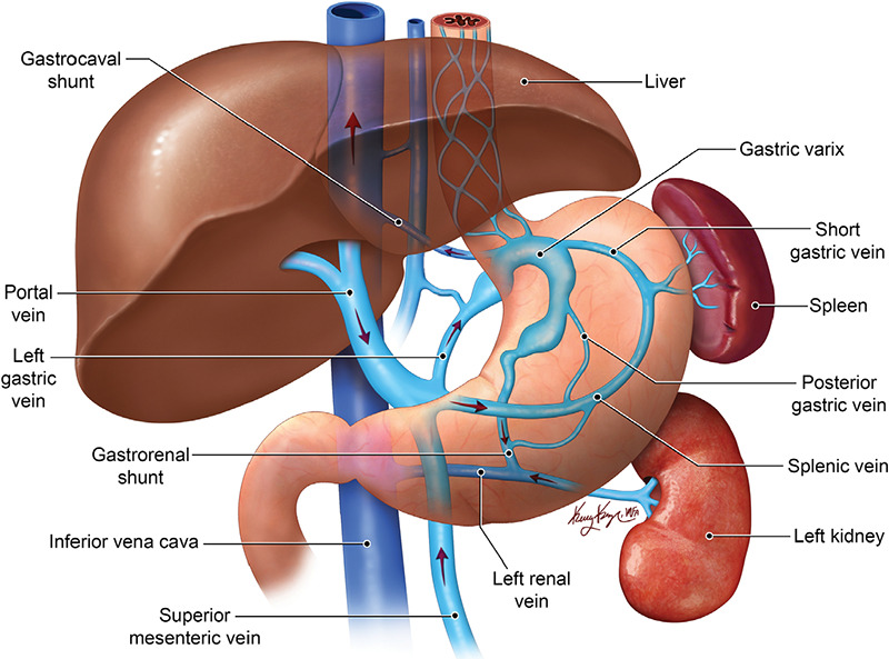
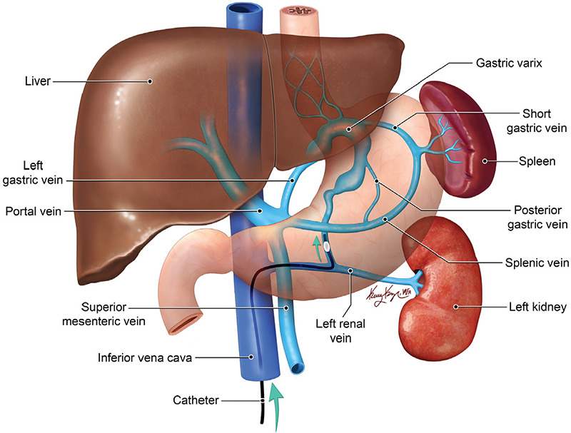
Bleeding gastric varices (GVs) is a life-threatening complication of portal hypertension, with higher morbidity and mortality rates compared with bleeding esophageal varices (EVs). The endovascular techniques for the management of GVs are mainly transjugular intrahepatic portosystemic shunt (TIPS) and transvenous obliteration of the GVs. Transvenous obliteration techniques can be an alternative or an adjunct to TIPS for treatment of GVs, depending on the clinical scenario, and are less invasive than TIPS. However, these procedures are associated with increased portal pressure and related complications, mainly worsening of the EVs. In this article, the different techniques of transvenous obliteration of GVs, their indications, contraindications, and outcomes are discussed.

Keywords: Cirrhosis; TIPS; gastric varices; transvenous obliteration; upper GI bleeding.


Conflict of interest statement

Conflict of interest disclosure

The authors declared no conflicts of interest.

Figures

Figure 1
Figure 1
Anatomy of gastric variceal complex. Graphic showing that the afferent veins of GVal complex are mainly formed by the left gastric vein, posterior gastric vein, and short gastric vein. The efferent system is formed by the gastrorenal shunt into the left renal vein and less commonly via gastrocaval shunt.
Figure 2
Figure 2
Classification of gastric varices on the basis of afferent veins. Type I has a single afferent vein, the left gastric vein in the graphic. Type II is supplied by two afferent veins: the left gastric vein and posterior gastric vein. Type III has a separate vein draining into the gastrorenal shunt without communication with the remaining afferents.
Figure 3
Figure 3
Classification of gastric varices on the basis of efferent veins. Type A has a single draining shunt. Type B has a single draining shunt with single or multiple collaterals. Type C has both gastrorenal shunt and gastrocaval shunt. Type D does not have a shunt and drains via small collaterals.
Figure 4
Figure 4
Balloon-occluded retrograde transvenous obliteration (BRTO). Graphic shows a BRTO procedure for gastric varices that drain via the gastrorenal shunt. A balloon catheter is inserted into the outlet of the gastrorenal shunt (in this drawing) or gastrocaval shunt via femoral venous access. Following balloonoccluded venography, sclerosant is then infused through the balloon catheter to fill the entire variceal complex.
Figure 5
Figure 5
Vascular plug-assisted retrograde transvenous obliteration. A 68-year-old female with cirrhosis presented with upper gastrointestinal bleed and type 1 isolated gastric varices on endoscopy. A contrastenhanced axial CT image (a) prior to the procedure showing fundal gastric varices (black*). Fluoroscopic images (b, c) showing vascular plug placement in the left adrenal vein (white arrow) via the right femoral vein access site with embolization of the gastrorenal shunt and fundal varices (white*) using a thick slurry of Gelfoam mixed with saline and contrast. Contrast-enhanced axial CT image (d) after the procedure showing complete obliteration of the fundal varices. CT, computed tomography.
Figure 6
Figure 6
Balloon-occluded antegrade transvenous obliteration (BATO). Subtype: trans-transjugular intrahepatic portosystemic shunt (TIPS) BATO: BATO involves embolization of the varices from the portal venous side. The graphic shows a subclassification of the BATO procedure known as trans-TIPS obliteration, in which the portal vein, and subsequently the portal venous side of varices, is accessed via indwelling TIPS.
Figure 7
Figure 7
Balloon-occluded antegrade transvenous obliteration (BATO). Subtype: percutaneous transhepatic obliteration. BATO involves embolization of the varices from the portal venous side. The graphic shows a subclassification of the BATO procedure known as percutaneous transhepatic obliteration, in which the portal vein, and subsequently the portal venous side of varices, is accessed percutaneously.

Similar articles

Cited by

References

    1. de Franchis R, Primignani M. Natural history of portal hypertension in patients with cirrhosis. Clin Liver Dis. 2001;5(3):645–663. - PubMed
    1. Sarin SK, Lahoti D, Saxena SP, Murthy NS, Makwana UK. Prevalence, classification and natural history of gastric varices: a long-term follow-up study in 568 portal hypertension patients. Hepatology. 1992;16(6):1343–1349. - PubMed
    1. Garcia-Tsao G, Abraldes JG, Berzigotti A, Bosch J. Portal hypertensive bleeding in cirrhosis: risk stratification, diagnosis, and management: 2016 practice guidance by the American Association for the study of liver diseases. Hepatology. 2017;65(1):310–335. - PubMed
    1. Ferral H, Gamboa P, Postoak DW, et al. Survival after elective transjugular intrahepatic portosystemic shunt creation: prediction with model for end-stage liver disease score. Radiology. 2004;231(1):231–236. - PubMed
    1. Montgomery A, Ferral H, Vasan R, Postoak DW. MELD score as a predictor of early death in patients undergoing elective transjugular intrahepatic portosystemic shunt (TIPS) procedures. Cardiovasc Intervent Radiol. 2005;28(3):307–312. - PubMed

MeSH terms

LinkOut - more resources