. 2023 Sep 15;44(5):779-818.
doi: 10.1210/endrev/bnad009.

Molecular and Clinical Spectrum of Primary Hyperparathyroidism

Affiliations

Molecular and Clinical Spectrum of Primary Hyperparathyroidism

Smita Jha et al. Endocr Rev. .

Abstract

Recent data suggest an increase in the overall incidence of parathyroid disorders, with primary hyperparathyroidism (PHPT) being the most prevalent parathyroid disorder. PHPT is associated with morbidities (fractures, kidney stones, chronic kidney disease) and increased risk of death. The symptoms of PHPT can be nonspecific, potentially delaying the diagnosis. Approximately 15% of patients with PHPT have an underlying heritable form of PHPT that may be associated with extraparathyroidal manifestations, requiring active surveillance for these manifestations as seen in multiple endocrine neoplasia type 1 and 2A. Genetic testing for heritable forms should be offered to patients with multiglandular disease, recurrent PHPT, young onset PHPT (age ≤40 years), and those with a family history of parathyroid tumors. However, the underlying genetic cause for the majority of patients with heritable forms of PHPT remains unknown. Distinction between sporadic and heritable forms of PHPT is useful in surgical planning for parathyroidectomy and has implications for the family. The genes currently known to be associated with heritable forms of PHPT account for approximately half of sporadic parathyroid tumors. But the genetic cause in approximately half of the sporadic parathyroid tumors remains unknown. Furthermore, there is no systemic therapy for parathyroid carcinoma, a rare but potentially fatal cause of PHPT. Improved understanding of the molecular characteristics of parathyroid tumors will allow us to identify biomarkers for diagnosis and novel targets for therapy.

Keywords: PTH; calcium; genetics of hyperparathyroidism; parathyroid adenomas; parathyroid cancer; parathyroid tumors.


Figures

Graphical Abstract
Graphical Abstract
Figure 1.
Figure 1.
(A) Parathyroid glands are derived from the endoderm of the third and fourth pharyngeal pouches. The third pharyngeal pouch gives rise to the inferior parathyroid glands and thymus while the fourth pharyngeal pouch gives rise to the superior parathyroid gland. Note the longer path of descent of inferior parathyroid glands and increased likelihood of its ectopic location. The inferior parathyroid glands may descend lower into the thymus or chest or leave residual tissue along the path of descent. (B) Microscopically, the parathyroid gland is enveloped by a thin fibrous capsule that extends into the parenchyma dividing the gland into multiple lobules. The gland consists of the following parenchymal cells: chief, oxyphil, clear, and transitional. Both chief and oxyphil cells are eosinophilic, express calcium-sensing receptor, and secrete parathyroid hormone. Clear cells decrease with age while oxyphil and transitional oxyphil cells increase with age. The stromal fat content of the gland (∼50%) varies with nutritional status and weight of the patient.
Figure 2.
Figure 2.
(A) PTH is an 84 amino acid peptide with its biologic activity attributed to the first 7 amino acids in its amino terminal. The first 34 amino acids of the peptide are as potent as the intact native hormone, with signaling and receptor binding domains residing within N-terminal and C-terminal portions of PTH(1-34), respectively. PTH(7-34) peptide analogs function as potent antagonists as these fragments are devoid of signaling activity but can bind to the PTH receptor. The PTH(15-34) fragment is the shortest length peptide of native sequence that exhibits detectable binding to the PTH receptor while PTH(1-14) is the shortest length N-terminal peptide that exhibits cyclic adenosine monophosphate signaling activity (although its potency is 5 times weaker, likely from the absence of receptor binding domain). PTH-related peptide is a 141 amino acid peptide with variants that are 139 or 173 amino acids long. Structurally, the region of homology with PTH runs around the N-terminal sequence with the first 13 residues being identical (36). (B) The evolution of PTH assays can be divided into 3 stages: competitive immunoassays (first generation), “sandwich” immunometric assays (second and third generation). In the first-generation assays, a single polyclonal antibody competed for labeled PTH and the serum forms. In comparison, second-generation assays are based on recognition of 2 distinct antibodies (typically monoclonal): 1 carboxyl terminal and the other amino terminal specific (targeting antigenic site located around amino acids 20-25). However, all these assays also measure forms other than intact PTH(1-84); for instance, PTH(7-84), predominantly an issue in patients with chronic kidney disease.
Figure 3.
Figure 3.
Upon ligand (PTH and PTH-related peptide) activation, the PTH receptor undergoes a series of conformational changes resulting in coupling to the stimulatory G-protein located on the inner, cytoplasmic surface of the cell membrane. Gαs activates adenylate cyclase, which results in synthesis of cyclic adenosine monophosphate (cAMP). cAMP binds to the regulatory 1A subunits (R) of protein kinase A (PKA), the primary effector of cAMP. On activation, the catalytic subunits (C) dissociate from the R subunits and phosphorylate several transcription factors, including cAMP-responsive element-binding (CREB) protein which interacts with cAMP-responsive element (CRE) in the nucleus and regulates transcription of target genes. Created with Biorender.com.
Figure 4.
Figure 4.
CASR binds to many physiological ligands including cations such as magnesium, l-amino acids, polyamines, and γ-glutamyl peptides like glutathione. CASR has a large extracellular domain that consists of a bilobed Venus flytrap module and a cysteine-rich domain. The Venus flytrap has 3 distinct extracellular calcium (Ca2+e) binding sites. CASR exists as a dimer through interactions at the amino terminal of the Venus flytrap lobe. The transmembrane domain of the protein is typical of other GPCRs and consists of 7 hydrophobic helical domains connected by 3 extracellular and intracellular loops each. Signal transduction on activation of CASR propagates through Gq/11 family proteins that activate phospholipase C-beta, resulting in the release of 2 second messengers, diacylglycerol (DAG) and inositol 14,5-triphosphate (IP3). DAG activates protein kinase C, which activates extracellular signal–regulated kinases 1 and 2 (mitogen activated protein kinases [MAPKs] signaling). IP3 binds to its receptors on the endoplasmic reticulum and raises intracellular calcium by releasing calcium from the reticulum. CASR activates the Gi/o proteins, which results in suppression of adenylate cyclase–mediated cAMP production eventually resulting in decreased PTH secretion and increased urinary calcium excretion. CASR can also activate MAPK signaling via a G-protein–independent mechanism involving β-arrestins. CASR demonstrates biased signaling in showing preferential activation of distinct intracellular signaling responses in different tissues. The mechanism for this functional selectivity is unclear. Ligand-induced insertional signaling drives cell surface expression of CASR by anterograde trafficking. The σ subunit of adaptor protein 2 (AP2σ) mediates the endocytosis and trafficking of CASR in combination with clathrin and β-arrestin. Mutations in CASR, Gq/11 family (GNA11) and AP2σ (AP2S1) can cause familial hypercalcemic hypocalciuria and autosomal dominant hypocalcemia. Created with Biorender.com.
Figure 5.
Figure 5.
Upon binding to 1,25(OH)2 vitamin D, VDR heterodimerizes with other nuclear hormone receptors, particularly the family of retinoid x receptors (RXRs), and this complex then binds to vitamin D response elements (VDREs) within the promoters of a large number of genes it regulates, modulating their transcription and subsequent effects in a ligand-dependent manner. Coactivators and corepressors are additional proteins that complex with VDR to regulate transcription. Created with Biorender.com.
Figure 6
Figure 6
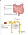
(A) Course of calcium absorption and contribution of active and passive calcium absorption over the course of jejunum, ileum, and colon. Most of the total calcium absorption (65%) takes place in ileum because transit time through the ileum is almost ten times longer than through duodenum. Created with Biorender.com. (B) The adult kidney has a glomerular filtration rate (GFR) of ∼100 mL/min and produces >8000 mg of calcium in the GFR/24 hours; ∼98% of the filtered calcium is reabsorbed, with only about 200 mg/24 hours of calcium appearing in the urine. Sixty to 70% of the filtered calcium is reabsorbed at the proximal convoluted tubule mainly by passive diffusion. However, 10% to 15% of total proximal tubule calcium reabsorption occurs via active transport and is mainly regulated by parathyroid hormone and calcitonin. No calcium reabsorption occurs within the thin segment of the loop of Henle. Twenty percent of the filtered calcium is reabsorbed in the cortical thick ascending limb, 10% in the distal convoluted tubule, and another 3% to 10% in the connecting tubule. PHP, pseudohypoparathyroidism. Created with Biorender.com.
Figure 7.
Figure 7.
Vitamin D3 is transported via vitamin D binding protein (DBP) to the liver for synthesis of 25-hydroxyvitamin D3 [25(OH)D3], the major circulating form of vitamin D. Subsequently, 25(OH)D3 is then transported via DBP to the kidney where it is taken up by the tubular epithelial cells. Low calcium sensed through the calcium sensing receptor (CASR) in parathyroid cells stimulates release of parathyroid hormone (PTH). PTH hormone acts via PTHR1 expressed predominantly in osteoblasts and osteocytes (induces release of calcium from bone) and renal proximal (decreases rate of phosphorus reabsorption, stimulates the rate of transcription of CYP27B1, encoding 25(OH)D 1-α-hydroxylase) and distal (increases rate of calcium reabsorption) tubule cells. 25(OH)D 1-α-hydroxylase converts 25(OH)D3 to 1,25(OH)2D3, the functionally active form of vitamin D. The primary function of 1,25(OH)2D3 and VDR is intestinal calcium absorption, which although most rapid in the duodenum occurs primarily in the distal segments of the intestine (only ∼10% in duodenum). 1,25(OH)2D3 in turn suppresses PTH synthesis directly at the level of transcription of PTH gene (through vitamin D response element (VDRE) within PTH) and indirectly by increasing serum calcium and upregulating the expression and transcription of CASR (by binding to VDRE within the CASR promoter). 1,25(OH)2D3 regulates its own synthesis by inhibiting CYP27B1. 25(OH)D3 (and 1,25(OH)2D3 acting as the preferred substrate) can also be converted to 24,25(OH)2D3, products targeted for excretion by the enzyme 24,25-α-hydroxylase (encoded by CYP24A1, present in all cells containing VDR). This process helps in regulation of levels of circulating and, possibly, intracellular 1,25(OH)2D3. The regulation of CYP24A1 is opposite to CYP27B1—stimulated by 1,25(OH)2D3 and inhibited by low calcium and PTH. 1,25(OH)2D3 and elevations in serum phosphate stimulate production of fibroblast growth factor-23 (FGF23), a “phosphate-wasting” glycoprotein produced by osteoblasts and osteocytes. FGF23 and its coreceptor, α-klotho, suppress 1-α-hydroxylase and induce 24,25-α-hydroxylase. In addition to the parathyroid gland, CASR is expressed at a low level in the proximal convoluted tubule (regulates expression of 25(OH)D 1-α-hydroxylase and inhibits PTH-mediated phosphate excretion); distal convoluted tubule (increases calcium reabsorption via transient receptor potential cation channel subfamily V member 5 (TRPV5) channel when tubular fluid calcium concentration is high); and is highly expressed in the thick ascending limb, where it has a PTH-independent key role in maintaining calcium homeostasis (senses increases in calcium and promotes calcium excretion via Claudin 14 tight junction protein), and in renal collecting ducts (prevents development of hypercalciuria-mediated nephrocalcinosis by increasing urinary acidification and water excretion).
Figure 8.
Figure 8.
Spectrum of disorders leading to primary hyperparathyroidism (PHPT). The heritable forms of the disease are followed by the gene causing the disease enclosed in brackets. Similar to sporadic PHPT, heritable forms of PHPT can also be classified based on histological findings as parathyroid adenoma, carcinoma, or parathyromatosis.
Figure 9.
Figure 9.
Clinical manifestations in syndromic forms of primary hyperparathyroidism with its reported frequency. (A) Multiple endocrine neoplasia type 1 (MEN1). (B) Multiple endocrine neoplasia type 2 (MEN2). (C) Multiple endocrine neoplasia type 4 (MEN4). (D) Multiple endocrine neoplasia type 5 (MEN5); *comprehensive phenotype evolving. (E) Hyperparathyroidism-jaw tumor syndrome (HPT-JT). Frequency reported in parenthesis.
Figure 10.
Figure 10.
Genes implicated in parathyroid tumorigenesis. The thick outlined boxes represent the genes known to cause parathyroid tumors when mutated in the germline and somatic. PA, parathyroid adenoma; PC, parathyroid carcinoma. Created with Biorender.com.

References

    1. Collip J, Clark E. Further studies on the physiological action of a parathyroid hormone. J Biol Chem. 1925;64(2):485‐507.
    1. Brewer HB J, Ronan R. Bovine parathyroid hormone: amino acid sequence. Proc Natl Acad Sci U S A. 1970;67(4):1862‐1869. - PMC - PubMed
    1. Niall HD, Keutmann H, Sauer R, et al. . The amino acid sequence of bovine parathyroid hormone I. Hoppe Seylers Z Physiol Chem. 1970;351(12):1586‐1588. - PubMed
    1. Summers GW. Parathyroid update: a review of 220 cases. Ear Nose Throat J. 1996;75(7):434‐439. - PubMed
    1. Thompson NW, Eckhauser FE, Harness JK. The anatomy of primary hyperparathyroidism. Surgery. 1982;92(5):814‐821. - PubMed

Publication types

MeSH terms