. 2023 Apr 5;145(13):7361-7375.
doi: 10.1021/jacs.2c13732. Epub 2023 Mar 24.

Enzyme-Responsive Double-Locked Photodynamic Molecular Beacon for Targeted Photodynamic Anticancer Therapy

Affiliations

Enzyme-Responsive Double-Locked Photodynamic Molecular Beacon for Targeted Photodynamic Anticancer Therapy

Leo K B Tam et al. J Am Chem Soc. .

Abstract

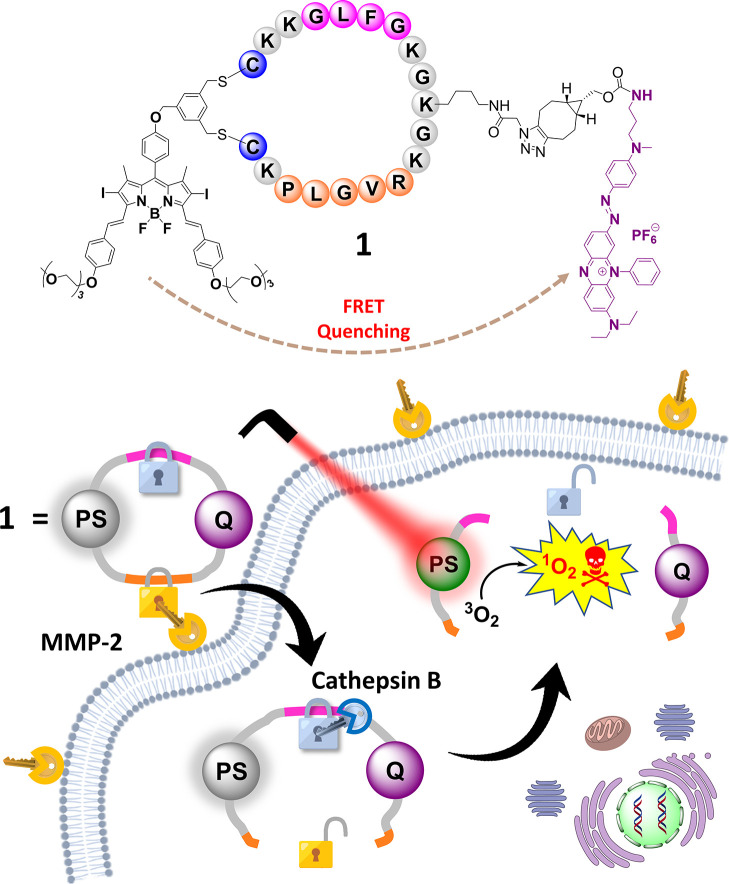
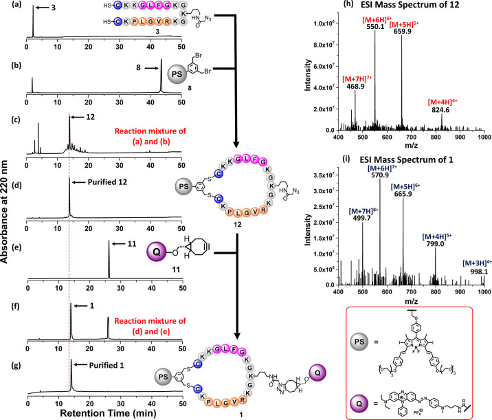
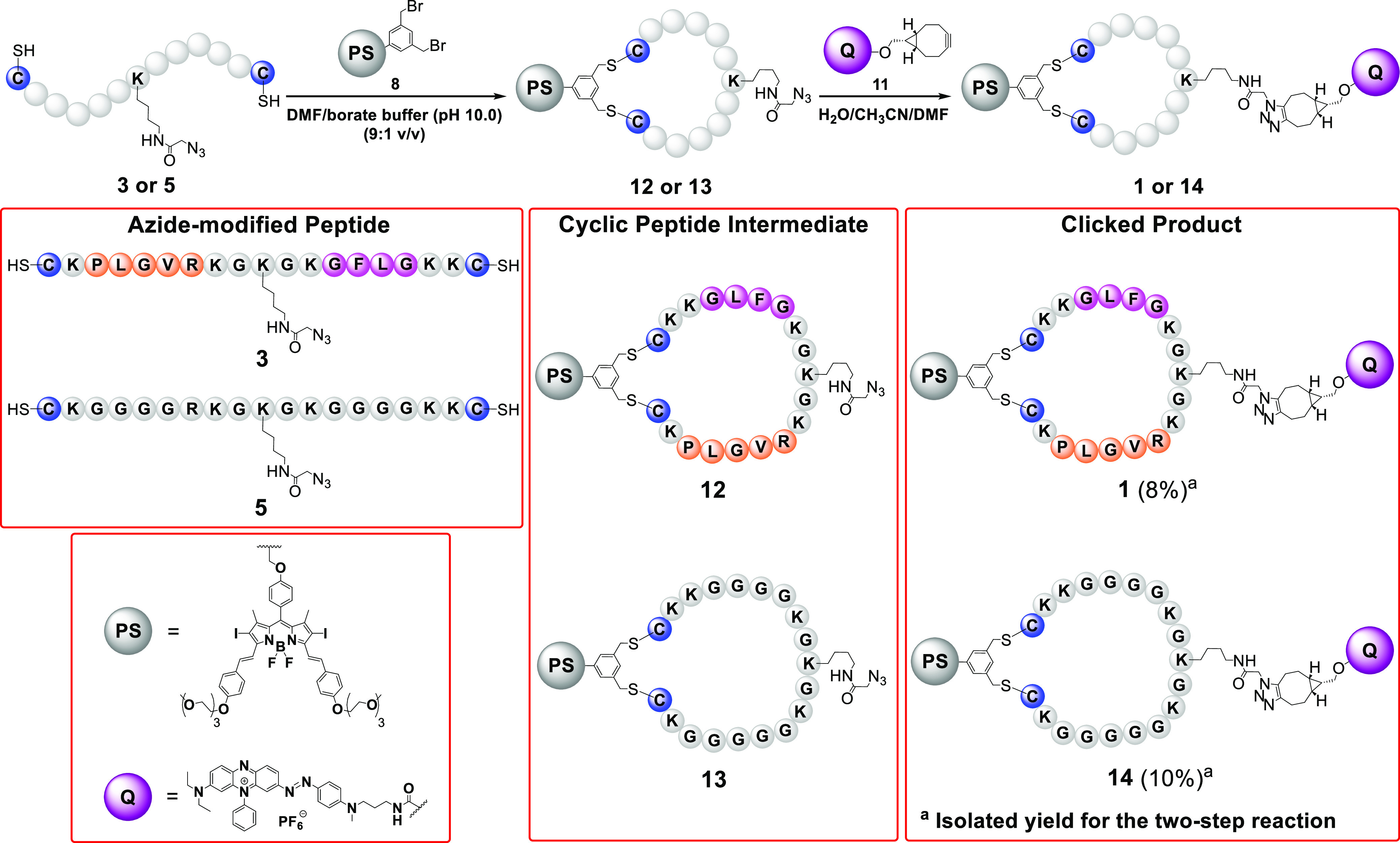
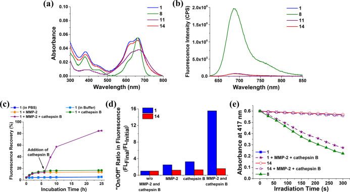
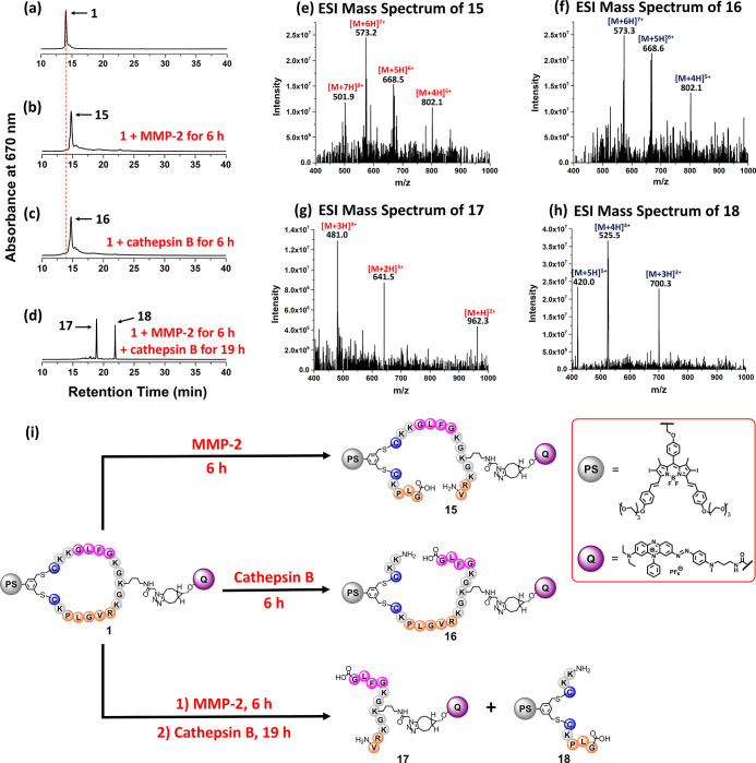
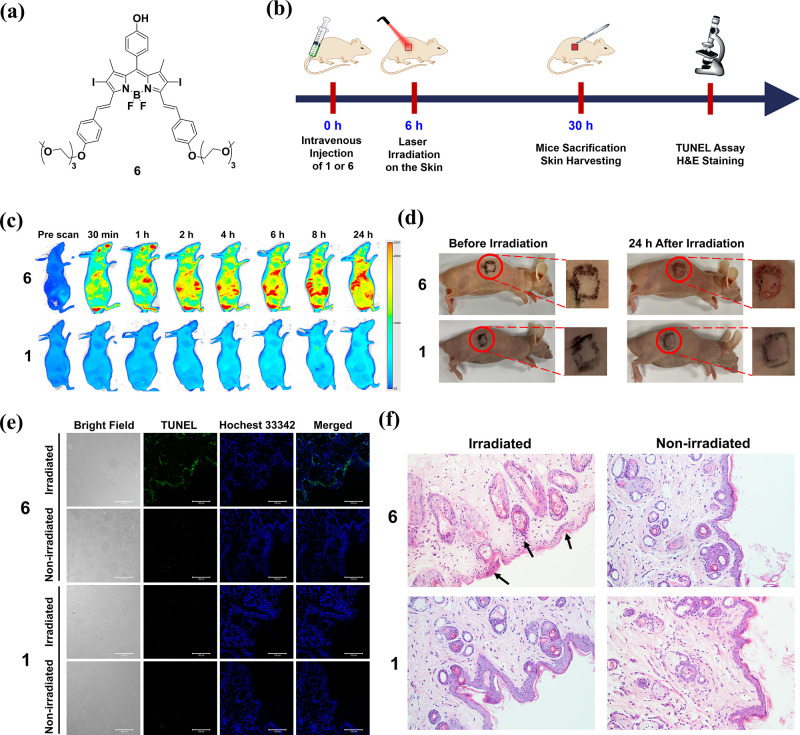
An advanced photodynamic molecular beacon (PMB) was designed and synthesized, in which a distyryl boron dipyrromethene (DSBDP)-based photosensitizer and a Black Hole Quencher 3 moiety were connected via two peptide segments containing the sequences PLGVR and GFLG, respectively, of a cyclic peptide. These two short peptide sequences are well-known substrates of matrix metalloproteinase-2 (MMP-2) and cathepsin B, respectively, both of which are overexpressed in a wide range of cancer cells either extracellularly (for MMP-2) or intracellularly (for cathepsin B). Owing to the efficient Förster resonance energy transfer between the two components, this PMB was fully quenched in the native form. Only upon interaction with both MMP-2 and cathepsin B, either in a buffer solution or in cancer cells, both of the segments were cleaved specifically, and the two components could be completely separated, thereby restoring the photodynamic activities of the DSBDP moiety. This PMB could also be activated in tumors, and it effectively suppressed the tumor growth in A549 tumor-bearing nude mice upon laser irradiation without causing notable side effects. In particular, it did not cause skin photosensitivity, which is a very common side effect of photodynamic therapy (PDT) using conventional "always-on" photosensitizers. The overall results showed that this "double-locked" PMB functioned as a biological AND logic gate that could only be unlocked by the coexistence of two tumor-associated enzymes, which could greatly enhance the tumor specificity in PDT.


Conflict of interest statement

The authors declare no competing financial interest.

Figures

Figure 1
Figure 1
Molecular structure of the double-locked PMB 1 and its dual-enzyme-unlocked mechanism.
Figure 2
Figure 2
Monitoring the course of the formation of PMB 1 using LC–MS.
Scheme 1
Scheme 1. Synthetic Route of PMB 1 and Its Non-cleavable Analogue 14
Figure 3
Figure 3
(a) Electronic absorption and (b) fluorescence (λex = 610 nm) spectra of 1, 8, 11, and 14 (all at 1 μM) in PBS at pH 7.4 with 0.1% Tween 80 (v/v). (c) Fluorescence recovery for 1 (1 μM) in the absence of MMP-2 and cathepsin B, upon treatment with either MMP-2 (2 μg mL–1) or cathepsin B (1 unit mL–1) at 37 °C over a period of 25 h or upon treatment with MMP-2 (2 μg mL–1) for 6 h and then with cathepsin B (1 unit mL–1) for a further 19 h at 37 °C. The solvent was either PBS at pH 7.4 (for the study involving MMP-2) or buffer solution (pH 5.0, 25 mM NaOAc, 1 mM EDTA, 500 μM GSH) (for the study involving cathepsin B), both in the presence of 0.1% Tween 80 (v/v). (d) On/off ratios for 1 and 14 under different conditions as determined by dividing the corresponding fluorescence intensity at 25 h by the initial fluorescence intensity. (e) Comparison of the rates of decay of DPBF (initial concentration = 30 μM) sensitized by 1 and 14 (both at 1 μM), both with and without the sequential treatment with MMP-2 and cathepsin B as described above, followed by light irradiation (λ > 610 nm) for 300 s. The results for 8 (1 μM) in the buffer are also included for comparison.
Figure 4
Figure 4
HPLC chromatograms of (a) 1 and the reaction mixtures of (b) 1 after the treatment with MMP-2 for 6 h, (c) 1 after the treatment with cathepsin B for 6 h, and (d) 1 after the treatment with MMP-2 for 6 h and then with cathepsin B for 19 h. ESI mass spectra of (e) 15, (f) 16, (g) 17, and (h) 18. (i) Enzymatic reactions of 1 with MMP-2 and/or cathepsin B.
Figure 5
Figure 5
(a) Bright field, fluorescence, and the merged images of A549, U-87 MG, HeLa, and HEK-293 cells after incubation with 1 (2 μM) in a serum-free medium for 1 h, followed by incubation in the neat medium for a further 6 h. (b) Mean intracellular fluorescence intensities of A549, U-87 MG, HeLa, and HEK-293 cells under these conditions as determined by flow cytometry. Data are expressed as the mean ± standard error of the mean (SEM) of three independent experiments. (c) Bright field, fluorescence, and the merged images of A549 and U-87 MG cells after incubation in a serum-free medium in the absence or presence of SB-3CT (10 μM) and/or CA-074 Me (25 μM) for 2 h, and then with 1 (2 μM) for 1 h, followed by post-incubation in the medium for a further 6 h or incubation with 14 (2 μM) for 1 h, followed by post-incubation in the medium for a further 6 h. (d) Mean intracellular fluorescence intensities of A549 and U-87 MG cells under these conditions as determined by flow cytometry. Data are expressed as the mean ± SEM of three independent experiments. ***p < 0.001 as calculated by the Student’s t-test.
Figure 6
Figure 6
(a) Intracellular ROS generation as reflected by the intracellular fluorescence intensity of DCF. A549 and HeLa cells were incubated in a serum-free medium in the absence or presence of SB-3CT (10 μM) and/or CA-074 Me (25 μM) for 2 h, and then with 1 (2 μM) for 1 h, or simply with 14 (2 μM) for 1 h. After post-incubation in the medium for 6 h, the cells were incubated with H2DCFDA (10 μM) for 30 min, followed by dark or light (λ > 610 nm, 23 mW cm–2, 14 J cm–2) treatment. (b) Dark and photo (λ > 610 nm, 23 mW cm–2, 28 J cm–2) cytotoxicity of 1 and 14 against A549, U-87 MG, HeLa, and HEK-293 cells under the different conditions as described. Data are expressed as the mean ± SEM of three independent experiments, each performed in quadruplicate. **p < 0.01 and ***p < 0.001 as calculated by the Student’s t-test.
Figure 7
Figure 7
(a) Fluorescence images of A549 tumor-bearing nude mice before and after intratumoral injection of 1 or 14 over a period of 24 h (excitation wavelength = 680 nm, emission wavelength ≥700 nm). (b) Change in fluorescence intensity per unit area of the tumor for the mice being treated with 1 or 14 over 24 h. (c) Photographs of the A549 tumor-bearing nude mice before and after intratumoral injection with 1 or 14 followed by laser irradiation (680 nm, 0.3 W cm–2, 180 J cm–2) or with PBS without laser irradiation over 14 days. (d) Tumor-growth curves for A549 tumor-bearing nude mice after the aforementioned treatments. (e) Change in body weight of the mice being treated as above over 14 days. (f) H&E-stained slices of the tumor and major organs of the mice on Day 14 after the above treatments. The drug dose was 20 nmol in 20 μL of distilled water containing 7.5% DMSO and 0.5% Tween 80 (v/v) for all cases. For (b,d,e), data are expressed as the mean ± standard deviation for n = 4. ***p < 0.001 as calculated by the Student’s t-test.
Figure 8
Figure 8
(a) Molecular structure of 6. (b) Timeline for the investigation of the in vivo photodynamic effect of 1 and 6 on the skin of the mice. (c) Fluorescence images of the nude mice before and after intravenous injection of 1 or 6 [20 nmol in 200 μL of distilled water containing 3% DMSO (v/v) and 0.5% Tween 80 (v/v)] over a period of 24 h (excitation wavelength = 680 nm, emission wavelength ≥700 nm). (d) Photographs of the nude mice before and after laser irradiation (680 nm, 0.3 W cm–2) for 10 min at 6 h post-injection taken after 24 h. (e) TUNEL-stained slices of the skins of the mice at 24 h after the above treatments. The cell nuclei were stained with Hoechst 33342. (f) H&E-stained slices of the skins of the mice at 24 h after the above treatments (original magnification 200×).

References

    1. Brown S. B.; Brown E. A.; Walker I. The present and future role of photodynamic therapy in cancer treatment. Lancet Oncol. 2004, 5, 497–508. 10.1016/s1470-2045(04)01529-3. - DOI - PubMed
    1. Correia J. H.; Rodrigues J. A.; Pimenta S.; Dong T.; Yang Z. Photodynamic therapy review: principles, photosensitizers, applications, and future directions. Pharmaceutics 2021, 13, 1332.10.3390/pharmaceutics13091332. - DOI - PMC - PubMed
    1. Algorri J. F.; Ochoa M.; Roldán-Varona P.; Rodríguez-Cobo L.; López-Higuera J. M. Photodynamic therapy: a compendium of latest reviews. Cancers 2021, 13, 4447.10.3390/cancers13174447. - DOI - PMC - PubMed
    1. Mishchenko T.; Balalaeva I.; Gorokhova A.; Vedunova M.; Krysko D. V. Which cell death modality wins the contest for photodynamic therapy of cancer?. Cell Death Dis. 2022, 13, 455.10.1038/s41419-022-04851-4. - DOI - PMC - PubMed
    1. Castano A. P.; Mroz P.; Hamblin M. R. Photodynamic therapy and anti-tumour immunity. Nat. Rev. Cancer 2006, 6, 535–545. 10.1038/nrc1894. - DOI - PMC - PubMed

Publication types