. 2023 Apr 5;145(13):7123-7135.
doi: 10.1021/jacs.2c09387. Epub 2023 Mar 24.

High Accuracy Prediction of PROTAC Complex Structures

Affiliations

High Accuracy Prediction of PROTAC Complex Structures

Mikhail Ignatov et al. J Am Chem Soc. .

Abstract

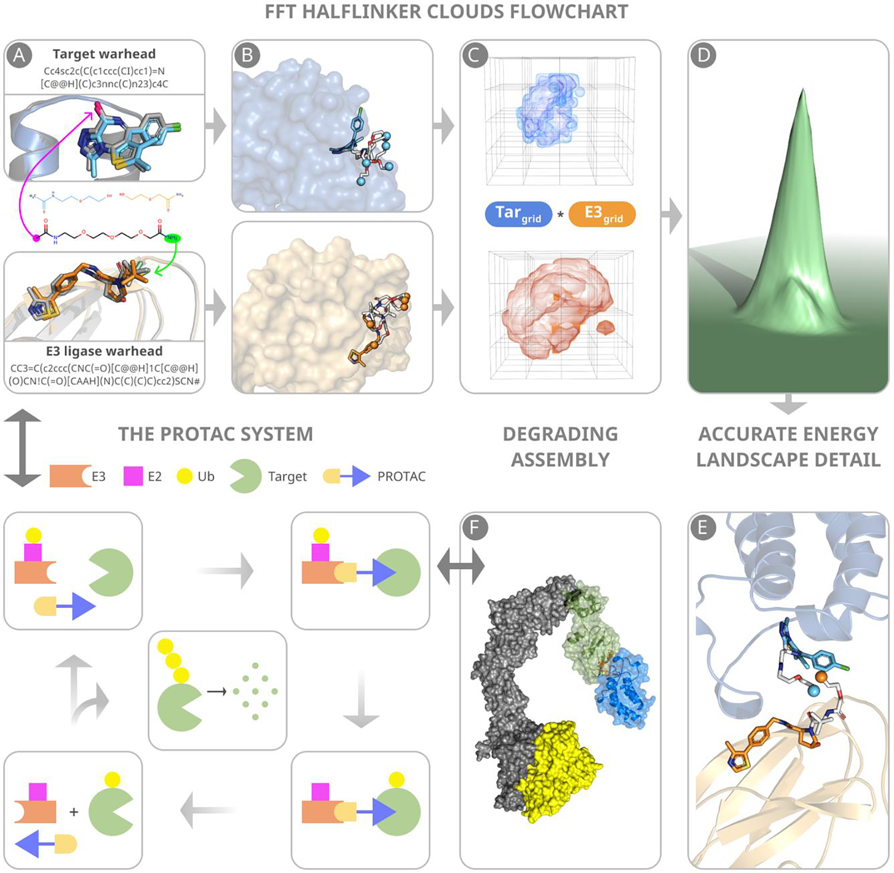
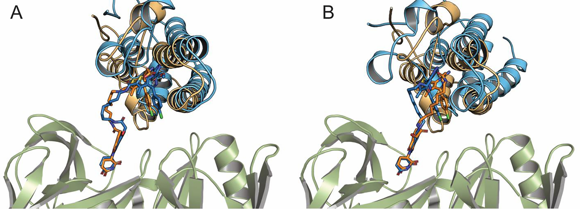
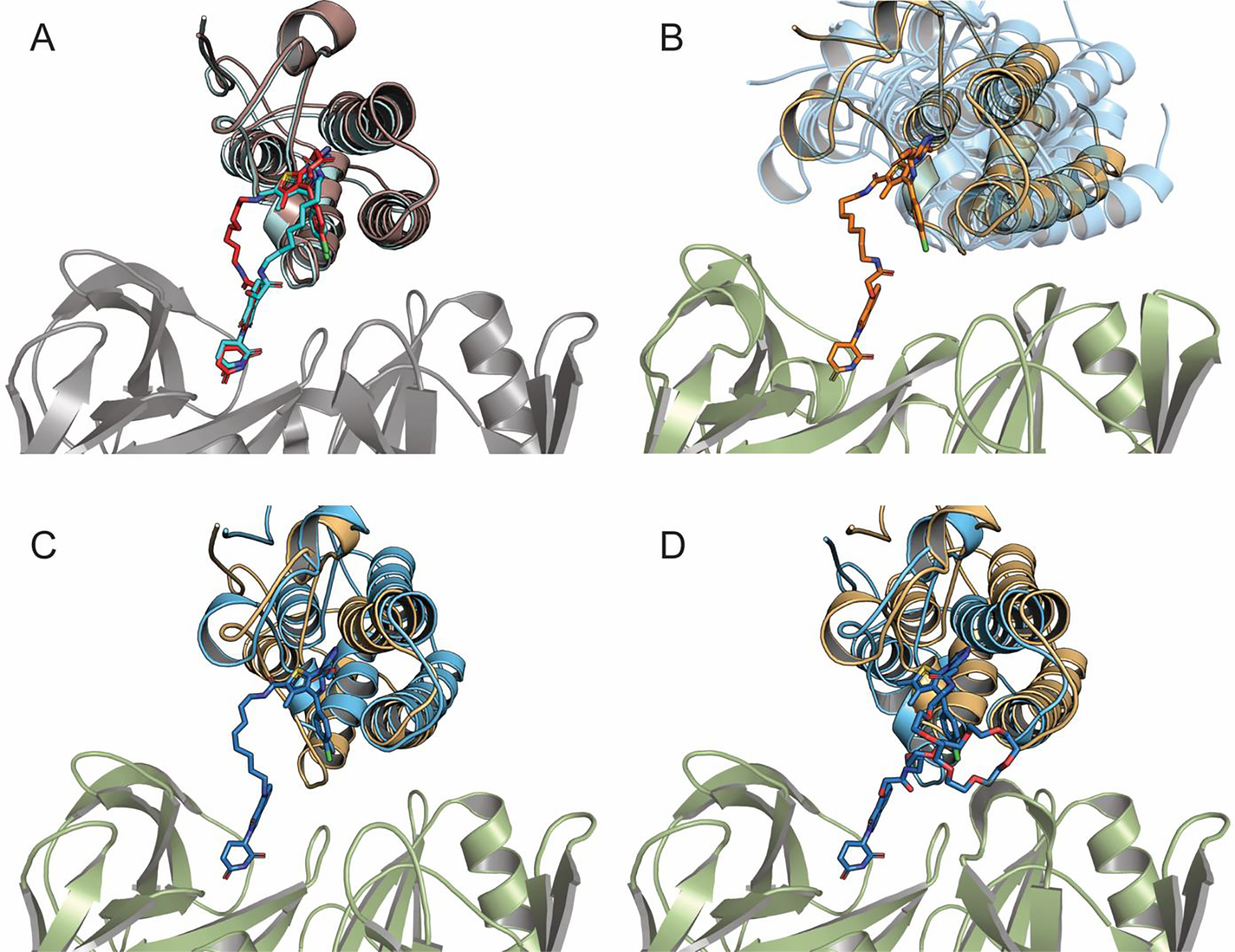
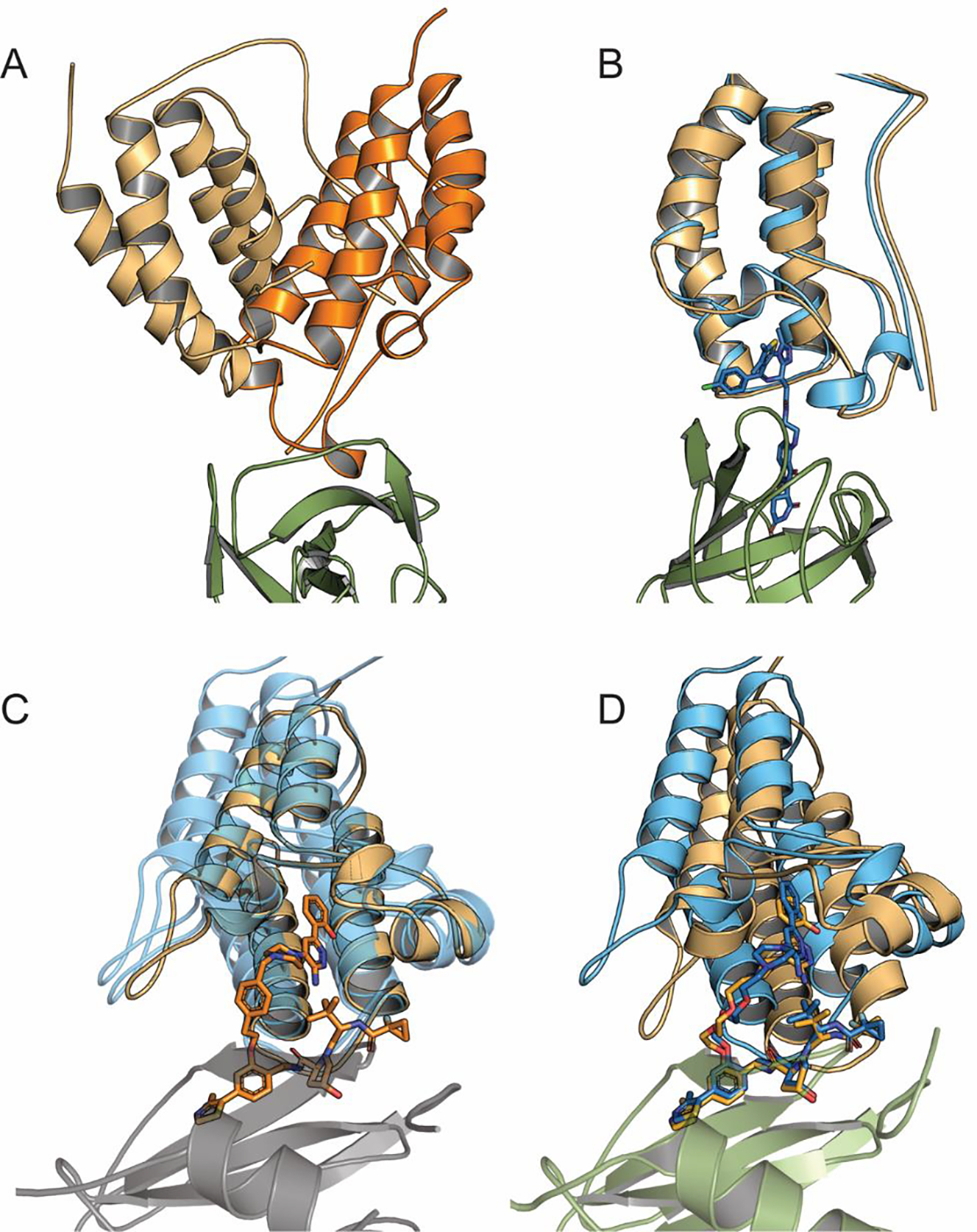
The design of PROteolysis-TArgeting Chimeras (PROTACs) requires bringing an E3 ligase into proximity with a target protein to modulate the concentration of the latter through its ubiquitination and degradation. Here, we present a method for generating high-accuracy structural models of E3 ligase-PROTAC-target protein ternary complexes. The method is dependent on two computational innovations: adding a "silent" convolution term to an efficient protein-protein docking program to eliminate protein poses that do not have acceptable linker conformations and clustering models of multiple PROTACs that use the same E3 ligase and target the same protein. Results show that the largest consensus clusters always have high predictive accuracy and that the ensemble of models can be used to predict the dissociation rate and cooperativity of the ternary complex that relate to the degrading activity of the PROTAC. The method is demonstrated by applications to known PROTAC structures and a blind test involving PROTACs against BRAF mutant V600E. The results confirm that PROTACs function by stabilizing a favorable interaction between the E3 ligase and the target protein but do not necessarily exploit the most energetically favorable geometry for interaction between the proteins.


Conflict of interest statement

The authors declare no competing financial interest.

Figures

Figure 1.
Figure 1.
Main steps of predicting PROTAC structures. (A) Warheads are docked to component proteins. Green and magenta arrows indicate attachment points to the E3 ligase and target warheads, respectively. (B) Half-linker conformations are generated and attached to each protein-bound warhead. The small colored spheres represent the half-linker end points. In the VHL:MZ1:BRD4BD2 (PDB ID: 5T35) complex shown, the BRD4 warhead and the end points of the attached half-linkers are depicted in cyan, and the VHL warhead and the end points of the attached half-linkers are in orange. (C) Generating favorable protein-protein poses that have half-linker ends placed sufficiently close to each other. (D) Selection of low energy poses. (E) A resulting pose with half-linker end points in close proximity before connecting the half-linkers. (F) The VHL–ElonginC–ElonginB–Cul2–Rbx1 structural assembly with the CRL2VHL complex shown in blue. The assembly includes a ubiquitin-like protein (ULP), shown in yellow, separated by a favorable distance to facilitate transfer of ubiquitin to the target. The figure also include a schematic outline of the PROTAC system.
Figure 2.
Figure 2.
Accuracy of some predicted structures. (A) Model 3 of the CRBN-dBET23-BRD4 BD1 complex (blue), superimposed on its X-ray structure (orange, PDB ID: 6BN7). (B) Model 5 of the CRBN-dBET6-BRD4 BD1 complex (blue), superimposed on its X-ray structure (orange, PDB ID: 6BOY)
Figure 3.
Figure 3.
Structures and models of CRBN-PROTAC-BRD4 BD1 ternary complexes. (A) Superimposing the X-ray structures of CRBN-dBET6-BRD4 BD1 (light-blue/cyan, PDB ID: 6BOY) and of CRBN-dBET23-BRD4 BD1 (red/orange, PDB ID: 6BN7). (B) Superimposing the best model CRBN-dBET6-BRD4BD1 at the center of the consensus cluster (orange) and the ternary complexes with dBET23, dBET55, and dBET70 (all shown as transparent blue). (C) Superimposing the consensus model (model 1) of CRBN-dBET70-BRD4 BD1 (blue) and its X-ray structure (light orange, PDB ID: 6BN9). (D) Superimposing the consensus model (model 1) of CRBN-dBET55-BRD4 BD1 (blue) and its X-ray structure (light orange, PDB ID: 6BN8).
Figure 4.
Figure 4.
Models of CRBN–dBET57– BRD4BD1 and VHL- PROTAC1/2-SMARCA2/4 complexes. (A) Superposing the best model (model 5) of CRBN–dBET57– BRD4 BD1 (light orange) and the consensus prediction of CRBN–dBET23– BRD4 BD1 (bright orange) to show that they substantially differ. (B) Model 5 of CRBN–dBET57– BRD4 BD1 (blue), superimposed on its X-ray structure (orange, PDB ID: 6BNB). (C) Superimposing the best model (model 2) of VHL- PROTAC2-SMARCA2 (orange) at the center of the consensus cluster and the consensus models of VHL- PROTAC1-SMARCA2 and VHL- PROTAC2-SMARCA4, both shown in transparent blue color. (D) Superimposing the consensus prediction (model 5) of VHL- PROTAC1-SMARCA2 (blue) and its X-ray structure (orange, PDB ID: 5NVX)
Figure 5.
Figure 5.
Using the weighted sum of acceptable models for predicting degradation activity and selectivity. (A) Weighted sum values for the series VHL- PROTAC1-BTK (denoted as P1) through VHL- PROTAC10-BTK (denoted as P10), considered here as predictions of degrading activity. (B) Weighted sum of predicted structures that satisfy the restraints posed by the linker for CRBN-MT-802-BTK (denoted as 802) and CRBN-MT-794-BTK (denoted as 794) ternary complexes. (C) Chemical structures of PROTACS MT-802 and MT-794. (D) PROTAC9 (yellow stick model) has limited interactions with CRBN (shown as green cartoon) apart from the ligand binding site, but interacts extensively with the BTK, shown as surface model on the right. (E) Weighted sums of acceptable models for the CRBN-ZXH-3–26-BDR4 BD1, BRD2 BD1, BRD3 BD1, BRD2 BD2, and BRD3 BD2 complexes, demonstrating some level of selective degradation of BRD4 BD1.
Figure 6.
Figure 6.
In cell BRAF degradation analysis. (A) Chemical structures of P4B and compounds 28–30. (B) Relative degrading activity of PROTACs 28, 29 and 30 and P4B as predicted using our computational method. Value is the weighted number of predicted structures that satisfy the restraints posed by the linker, considered as the prediction of degrading activity.

References

    1. Neklesa TK, Winkler JD & Crews CM Targeted protein degradation by PROTACs. Pharmacol. Ther. 174, 138–144 (2017). - PubMed
    1. Pettersson M & Crews CM PROteolysis TArgeting Chimeras (PROTACs) - Past, present and future. Drug Discov. Today Technol 31, 15–27 (2019). - PMC - PubMed
    1. Bondeson DP et al. Lessons in PROTAC Design from Selective Degradation with a Promiscuous Warhead. Cell Chem. Biol. 25, 78–87 e75 (2018). - PMC - PubMed
    1. Fisher SL & Phillips AJ Targeted protein degradation and the enzymology of degraders. Curr. Opin. Chem. Biol. 44, 47–55 (2018). - PubMed
    1. Gadd MS et al. Structural basis of PROTAC cooperative recognition for selective protein degradation. Nat. Chem. Biol. 13, 514–521 (2017). - PMC - PubMed

Publication types