. 2023 Apr 11;18(4):829-840.
doi: 10.1016/j.stemcr.2023.02.013. Epub 2023 Mar 23.

Generation of functional thymic organoids from human pluripotent stem cells

Affiliations

Generation of functional thymic organoids from human pluripotent stem cells

Stephan A Ramos et al. Stem Cell Reports. .

Abstract

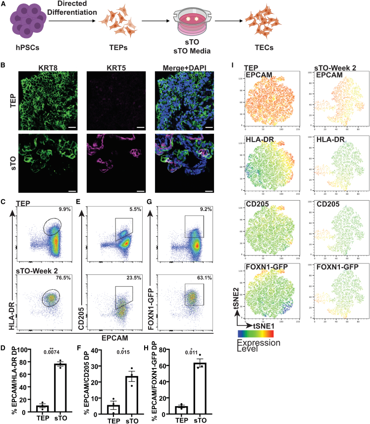
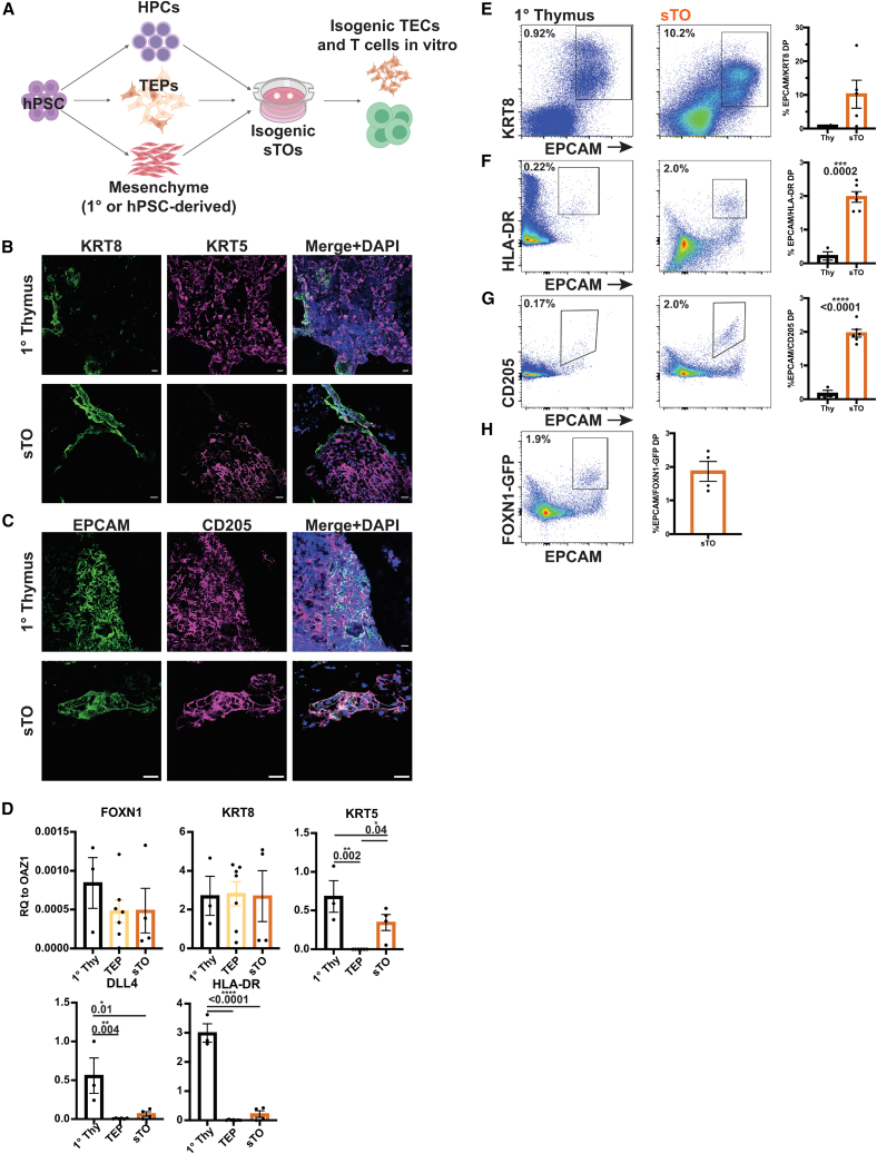
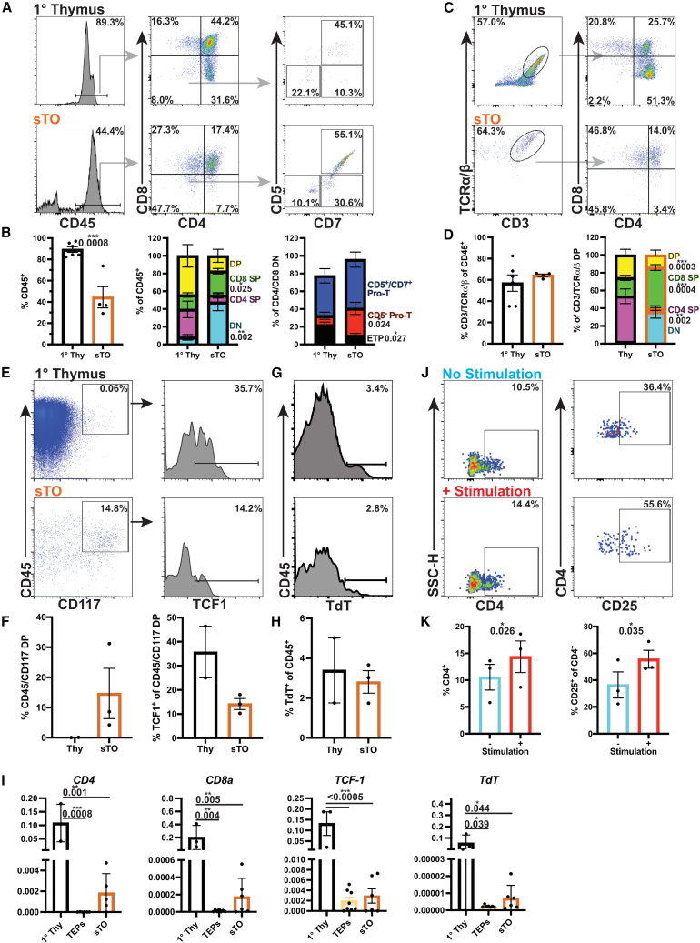
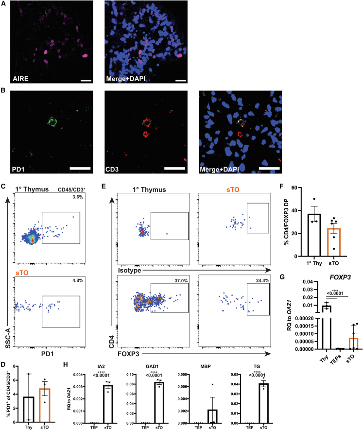
The thymus is critical for the establishment of a functional and self-tolerant adaptive immune system but involutes with age, resulting in reduced naive T cell output. Generation of a functional human thymus from human pluripotent stem cells (hPSCs) is an attractive regenerative strategy. Direct differentiation of thymic epithelial progenitors (TEPs) from hPSCs has been demonstrated in vitro, but functional thymic epithelial cells (TECs) only form months after transplantation of TEPs in vivo. We show the generation of TECs in vitro in isogenic stem cell-derived thymic organoids (sTOs) consisting of TEPs, hematopoietic progenitor cells, and mesenchymal cells, differentiated from the same hPSC line. sTOs support T cell development, express key markers of negative selection, including the autoimmune regulator (AIRE) protein, and facilitate regulatory T cell development. sTOs provide the basis for functional patient-specific thymic organoid models, allowing for the study of human thymus function, T cell development, and transplant immunity.

Keywords: cell therapy; human T cell development; human pluripotent stem cells; positive and negative selection; stem cell derived thymic organoids; thymic epithelial cells.


Conflict of interest statement

Conflict of interests A.J. consults for SuviCa and owns stock options in SuviCa and Champions Oncology. H.A.R. was a SAB member at Sigilon Therapeutics and Prellis Biologics and consulted for Eli Lilly and Minutia. S.A.R., L.H.A., J.J.M., A.J., and H.A.R. filed patent applications based on this work.

Figures

Figure 1
Figure 1
Generation of hPSC-derived TECs in vitro (A) Schematic of TEP-only sTOs. (B) Representative IF images of KRT8 (green) and KRT5 (magenta) in day 16–22 TEPs and week 2 TEP-only sTOs (TEPs, n = 2 indepdendent experiment (ind. exp.), 2 hPSC lines; sTOs, n = 3 ind. exp., 2 hPSC lines). (C–H) FC plots and quantification of EPCAM/HLA-DR (C and D), EPCAM/CD205 (E and F), and EPCAM/GFP (G and H) expression in day 16–22 TEPs and week 2 TEP-only sTOs. (I) tSNE plots of EPCAM, HLA-DR, CD205, and GFP expression in 16–22 TEPs and week 2 TEP-only sTOs, n = 3 ind. exp., 1 hPSC line. Plots show percentage of the mean; data are shown as mean ± SEM.
Figure 2
Figure 2
sTOs support TEC development (A) Schematic of multicellular, isogenic sTOs. (B and C) Representative IF images of KRT8 and KRT5 (B) or EPCAM and CD205 (C) in 1° thymus and sTOs (thymi, n = 3; sTOs, n = 9 ind. exp., 2 hPSC lines). (D) qPCR analysis of TEC markers in day 17–30 TEPs and week 4 CD45 sTO cells (thymi, n = 3; TEPs, n = 7 ind. exp., 2 hPSC lines; sTOs, n = 4 ind. exp., 2 hPSC lines). p values were determined by t test. (E–H) CyTOF (E) and FC (FH) plots and quantification of TEC markers in 1° thymus and sTOs (CyTOF: thymi, n = 2; sTOs, n = 3–6 ind. exp., 3 hPSC lines; FC: thymi, n = 4, sTOs, n = 4–6 ind. exp., 2 hPSC lines). Plots show percentage of the mean; data are shown as mean ± SEM.
Figure 3
Figure 3
sTOs support isogenic T cell development (A–D) FC plots (A and C) and quantification (B and D) of thymocytes in 1° thymus and week 4 sTOs. (E–H) CyTOF plots (E and G) and quantification (F and H) of thymocyte markers in 1° thymus and week 4 sTOs (thymi, n = 6; sTOs, n = 4, 2 hPSC lines). (I) qPCR analysis of CD4, CD8, and TdT in 1° thymus, TEPs, and sTOs (thymi, n = 3; TEPs, n = 6–7, 4 hPSC lines; sTOs, n = 5–6, 4 hPSC lines). (J and K) FC plots (J) and quantification (K) of CD4 and CD25 expression on stimulated T cells from week 4 sTOs (n = 3 ind. exp., 1 hPSC line). Plots show percentage of the mean; data are shown as mean ± SEM; p values were determined by t test.
Figure 4
Figure 4
Potential negative selection and Treg development in sTOs (A) Representative IF image of AIRE in week 4 sTOs (n = 2). (B) Representative IF image of PD-1 and CD3 in week 4 sTOs (n = 2). (C and D) FC plots (C) and quantification (D) of PD-1 expression in CD45+/CD3+ cells in 1° thymi and week 4 sTOs (thymi, n = 2; sTOs, n = 3 ind. exp., 1 hPSC line). (E and F) FC plots (E) and quantification (F) of FOXP3 expression in CD25+/CD4+ cells in 1° thymi and sTOs (thymi, n = 3; sTOs, n = 6 ind. exp., 2 hPSC lines). (G and H) qPCR analysis of FOXP3 (G) and TRAs (H) in 1° thymus, TEPs, and week 4 CD45 sTO cells (G: thymi, n = 3; TEPs, n = 6–7 ind. exp., 4 hPSC lines; sTOs, n = 5–6 ind. exp., 4 hPSC lines; H: TEPs, n = 5 ind. exp., 1 hPSC line; sTOs, n = 3 ind. exp., 1 hPSC line). Plots show percentage of the mean; data are shown as mean ± SEM; p values were determined by t test.

References

    1. Anderson G., Jenkinson E.J., Moore N.C., Owen J.J.T. MHC class II-positive epithelium and mesenchyme cells are both required for T-cell development in the thymus. Nature. 1993;362:70–73. doi: 10.1038/362070a0. - DOI - PubMed
    1. Anderson M.S., Venanzi E.S., Klein L., Chen Z., Berzins S.P., Turley S.J., von Boehmer H., Bronson R., Dierich A., Benoist C., Mathis D. Projection of an immunological self shadow within the thymus by the Aire protein. Science. 2002;298:1395–1401. doi: 10.1126/science.1077464. - DOI - PubMed
    1. Auerbach R. Morphogenetic interactions in the development of the mouse thymus gland. Dev. Biol. 1960;2:271–284. - PubMed
    1. Awong G., Singh J., Mohtashami M., Malm M., La Motte-Mohs R.N., Benveniste P.M., Serra P., Herer E., Van Den Brink M.R., Zúñiga-Pflücker J.C. Human proT-cells generated in vitro facilitate hematopoietic stem cell-derived T-lymphopoiesis in vivo and restore thymic architecture. Blood. 2013;122:4210–4219. doi: 10.1182/blood. - DOI - PMC - PubMed
    1. Baldwin T.A., Hogquist K.A. Transcriptional analysis of clonal deletion in vivo. J. Immunol. 2007;179:837–844. doi: 10.4049/jimmunol.179.2.837. - DOI - PubMed

Publication types

LinkOut - more resources