

Structure and RAF family kinase isoform selectivity of type II RAF inhibitors tovorafenib and naporafenib
- PMID: 36963492
- PMCID: PMC10149214
- DOI: 10.1016/j.jbc.2023.104634
Structure and RAF family kinase isoform selectivity of type II RAF inhibitors tovorafenib and naporafenib
Abstract
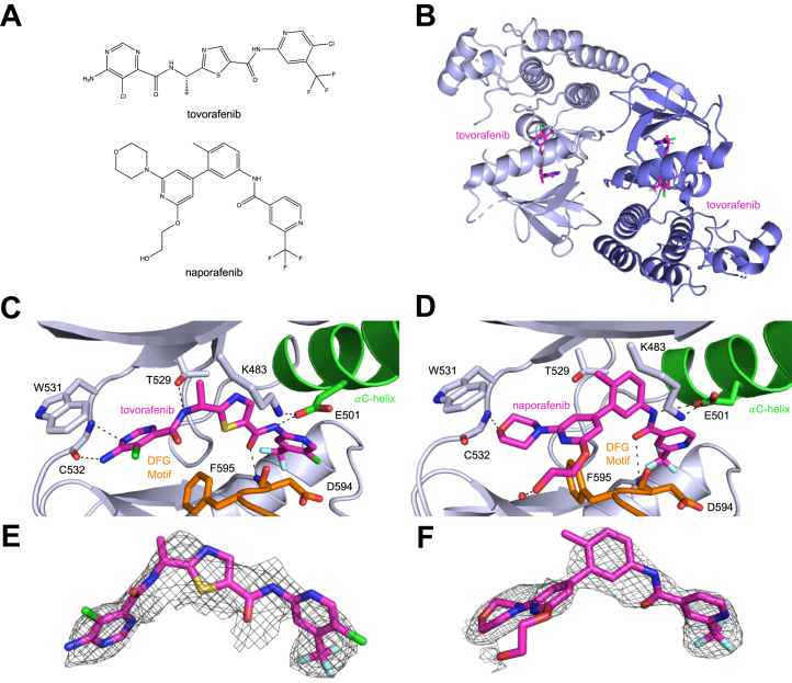
Upon activation by RAS, RAF family kinases initiate signaling through the MAP kinase cascade to control cell growth, proliferation, and differentiation. Among RAF isoforms (ARAF, BRAF, and CRAF), oncogenic mutations are by far most frequent in BRAF. The BRAFV600E mutation drives more than half of all malignant melanoma and is also found in many other cancers. Selective inhibitors of BRAFV600E (vemurafenib, dabrafenib, encorafenib) are used clinically for these indications, but they are not effective inhibitors in the context of oncogenic RAS, which drives dimerization and activation of RAF, nor for malignancies driven by aberrantly dimerized truncation/fusion variants of BRAF. By contrast, a number of "type II" RAF inhibitors have been developed as potent inhibitors of RAF dimers. Here, we compare potency of type II inhibitors tovorafenib (TAK-580) and naporafenib (LHX254) in biochemical assays against the three RAF isoforms and describe crystal structures of both compounds in complex with BRAF. We find that tovorafenib and naporafenib are most potent against CRAF but markedly less potent against ARAF. Crystal structures of both compounds with BRAFV600E or WT BRAF reveal the details of their molecular interactions, including the expected type II-binding mode, with full occupancy of both subunits of the BRAF dimer. Our findings have important clinical ramifications. Type II RAF inhibitors are generally regarded as pan-RAF inhibitors, but our studies of these two agents, together with recent work with type II inhibitors belvarafenib and naporafenib, indicate that relative sparing of ARAF may be a property of multiple drugs of this class.
Keywords: RAF kinase; cooperativity; drug action; fluorescence resonance energy transfer (FRET); inhibitor mechanism; x-ray crystallography.
Copyright © 2023 The Authors. Published by Elsevier Inc. All rights reserved.
Conflict of interest statement
Conflict of interest M. J. E. is a consultant for Novartis and receives sponsored research support from Novartis Institutes for Biomedical Research and Springworks Therapeutics. All other authors declare that they have no conflicts of interest with the contents of this article.
Figures
References
-
- Raman M., Chen W., Cobb M.H. Differential regulation and properties of MAPKs. Oncogene. 2007;26:3100–3112. - PubMed
Publication types
MeSH terms
Substances
Grants and funding
LinkOut - more resources
Full Text Sources
Research Materials
Miscellaneous
