. 2023 Nov;193(11):1809-1816.
doi: 10.1016/j.ajpath.2023.02.016. Epub 2023 Mar 22.

Histopathology and SARS-CoV-2 Cellular Localization in Eye Tissues of COVID-19 Autopsies

Collaborators, Affiliations

Histopathology and SARS-CoV-2 Cellular Localization in Eye Tissues of COVID-19 Autopsies

H Nida Sen et al. Am J Pathol. 2023 Nov.

Abstract

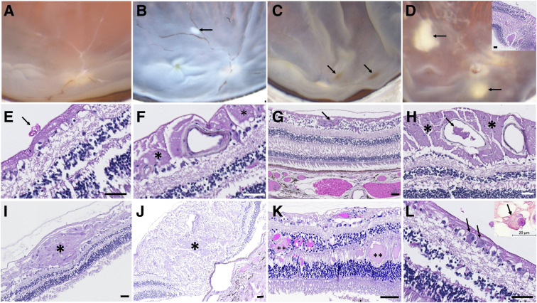
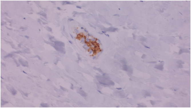
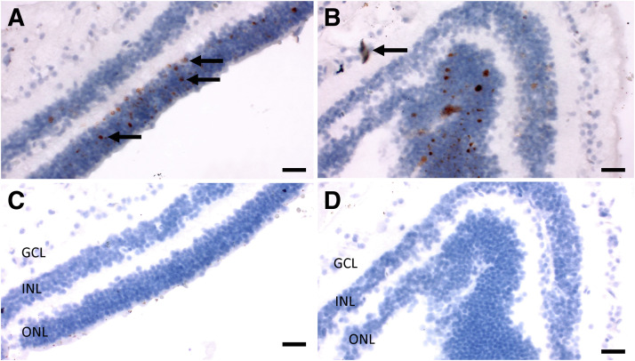
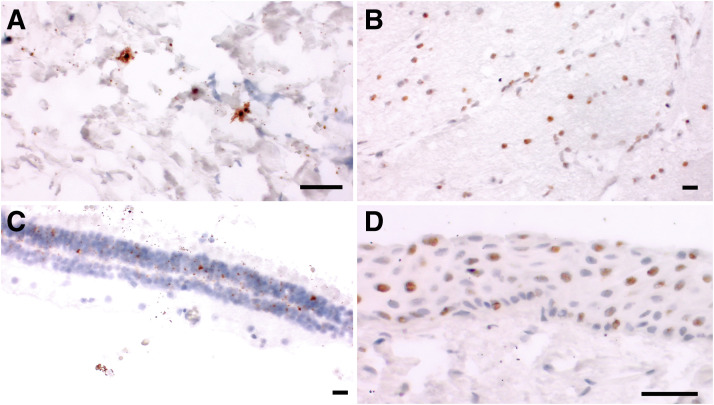
Ophthalmic manifestations and tissue tropism of severe acute respiratory syndrome coronavirus 2 (SARS-CoV-2) have been reported in association with coronavirus disease 2019 (COVID-19), but the pathology and cellular localization of SARS-CoV-2 are not well characterized. The objective of this study was to evaluate macroscopic and microscopic changes and investigate cellular localization of SARS-CoV-2 across ocular tissues at autopsy. Ocular tissues were obtained from 25 patients with COVID-19 at autopsy. SARS-CoV-2 nucleocapsid gene RNA was previously quantified by droplet digital PCR from one eye. Herein, contralateral eyes from 21 patients were fixed in formalin and subject to histopathologic examination. Sections of the droplet digital PCR-positive eyes from four other patients were evaluated by in situ hybridization to determine the cellular localization of SARS-CoV-2 spike gene RNA. Histopathologic abnormalities, including cytoid bodies, vascular changes, and retinal edema, with minimal or no inflammation in ocular tissues were observed in all 21 cases evaluated. In situ hybridization localized SARS-CoV-2 RNA to neuronal cells of the retinal inner and outer layers, ganglion cells, corneal epithelia, scleral fibroblasts, and oligodendrocytes of the optic nerve. In conclusion, a range of common histopathologic alterations were identified within ocular tissue, and SARS-CoV-2 RNA was localized to multiple cell types. Further studies will be required to determine whether the alterations observed were caused by SARS-CoV-2 infection, the host immune response, and/or preexisting comorbidities.


Figures

Figure 1
Figure 1
Histopathologic findings in postmortem eyes of patients with coronavirus disease 2019. AD: Macroscopic views of the opened autopsy eyes. EL: Hematoxylin and eosin–stained tissue sections. A: Vitreous haze and retinal vascular sheathing with focal sites of vascular occlusion in case E18. B: A focal whitish cotton-wool spot (arrow) superior to the macula in case E11. C: Multiple small dots and flame retinal hemorrhages (arrows) in the posterior pole in case E8. D: Vitreous opacity and several large and small patches of whitish subretinal infiltrates (arrows) in case E19. Inset illustrates microscopic view of the infiltrates composed of severe retinal edema and disorganization, serious fluids in the outer plexiform layer, and vitreous strands and debris. E: Small neovascular tuft (arrow) on retinal surface and focal loss of photoreceptor cells in case E18. F: Sclerotic retinal vessel and cytoid bodies (asterisks) due to microinfarction in the nerve fiber layer in case E18. G: Small obstructive retinal vessel (arrow) in case E18. H: Large cytoid bodies (asterisks) in the nerve fiber layer and retinal vascular thrombi (arrow) in case E18. I: Large cytoid body (asterisk) due to microinfarction in the nerve fiber layer and focal thinning of outer and inner nuclear layers in case E7. J: Several loci of cytoid bodies (asterisk) on the optic nerve head, mild optic nerve edema, and focal loss of photoreceptor cells in case E4. K: Serous retinal edema (double asterisks) in retinal outer plexiform and inner nuclear layers and focal loss of photoreceptor cells in case E8. Focal retinal hemorrhage and retinal vascular congestion are also seen. L: Viral inclusions (arrows) in cytoplasm and nucleus in a few retinal ganglion cells in case E18. Inset illustrates a higher magnification of a ganglion cell containing numerous viral inclusions (arrow). Scale bars: 50 nm (D inset and E–L); 20 μm (Linset).
Figure 2
Figure 2
Identification of platelet microthrombi with anti-CD61 immunostain. Platelet microthrombi were identified as clusters of CD61-positive platelets within small vessels. In this vessel, the microthrombus is associated with several mononuclear cells. Original magnification, ×400.
Figure 3
Figure 3
Detection of severe acute respiratory syndrome coronavirus 2 (SARS-CoV-2) spike RNA by in situ hybridization (ISH) in the retina. A–D: ISH for SARS-CoV-2 spike RNA (brown dots) primarily located in the outer nuclear layer of the retina (arrows, A) in case E22. Fewer positive signals are noted in the inner nuclear layer (A and B) and a retinal ganglion cell in case E23 (arrow, B). Panels A and B are with the SARS-CoV-2 spike RNA probe, and panels C and D are negative control probe for DapB. Scale bars = 20 μm. GCL, ganglion cell layer; INL, inner nuclear layer; ONL, outer nuclear layer.
Figure 4
Figure 4
Detection of severe acute respiratory syndrome coronavirus 2 (SARS-CoV-2) spike RNA by in situ hybridization (ISH) in the sclera, optic nerve, retina, and cornea. A–D: ISH for SARS-CoV-2 spike RNA in scleral fibroblasts in case E24 (A), optic nerve oligodendrocytes in case E25 (B), retinal outer and inner nuclear layers in case E25 (C), and corneal epithelium in case E25 (D). Scale bars = 20 μm.

Similar articles

Cited by

References

    1. Stein S.R., Ramelli S.C., Grazioli A., Chung J.-Y., Singh M., Yinda C.K., Winkler C.W., Dickey J., Ylaya K., Ko S.H., Platt A., Burbelo P.D., Quezado M., Pittaluga S., Purcell M., Munster V.J., Belinky F., Ramos-Benitez M.J., Boritz E.A., Herr D.L., Rabin J., Saharia K.K., Madathil R.J., Tabatabai A., Soherwardi S., McCurdy M.T., NIH COVID-19 Autopsy Consortium, Peterson K.E., Cohen J.I., de Wit E., Vannella K.M., Hewitt S.M., Kleiner D.E., Chertow D.S. SARS-CoV-2 infection and persistence throughout the human body and brain. Nature. 2022;612:758–763. - PMC - PubMed
    1. Sawant O.B., Singh S., Wright R.E., 3rd, Jones K.M., Titus M.S., Dennis E., Hicks E., Majmudar P.A., Kumar A., Mian S.I. Prevalence of SARS-CoV-2 in human post-mortem ocular tissues. Ocul Surf. 2021;19:322–329. - PMC - PubMed
    1. Casagrande M., Fitzek A., Püschel K., Aleshcheva G., Schultheiss H.-P., Berneking L., Spitzer M.S., Schultheiss M. Detection of SARS-CoV-2 in human retinal biopsies of deceased COVID-19 patients. Ocul Immunol Inflamm. 2020;28:721–725. - PubMed
    1. Casagrande M., Fitzek A., Spitzer M.S., Püschel K., Glatzel M., Krasemann S., Nörz D., Lütgehetmann M., Pfefferle S., Schultheiss M. Presence of SARS-CoV-2 RNA in the cornea of viremic patients with COVID-19. JAMA Ophthalmol. 2021;139:383–388. - PMC - PubMed
    1. Penkava J., Muenchhoff M., Badell I., Osterman A., Delbridge C., Niederbuchner F., Soliman S., Rudelius M., Graf A., Krebs S., Blum H., Ulbig M., Baumann C., Zapp D., Maier M., Keppler O.T., Lohmann C.P., Ledderose S. Detection of SARS-CoV-2-RNA in post-mortem samples of human eyes. Graefes Arch Clin Exp Ophthalmol. 2022;260:1789–1797. - PMC - PubMed

Publication types