Review
. 2023 Mar;10(1):e002244.
doi: 10.1136/openhrt-2022-002244.

Pathophysiology, emerging techniques for the assessment and novel treatment of aortic stenosis

Affiliations
Review

Pathophysiology, emerging techniques for the assessment and novel treatment of aortic stenosis

Syed Muneeb Shah et al. Open Heart. 2023 Mar.

Abstract

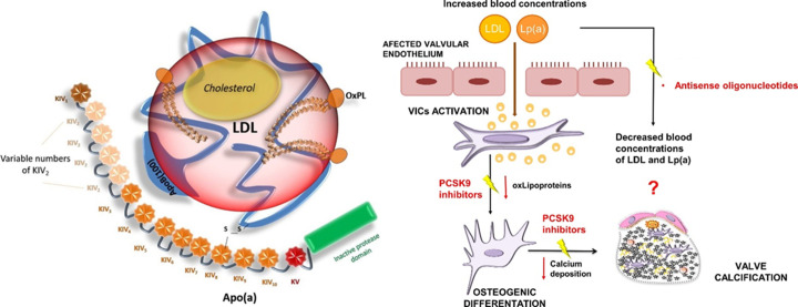
Our perspectives on aortic stenosis (AS) are changing. Evolving from the traditional thought of a passive degenerative disease, developing a greater understanding of the condition's mechanistic underpinning has shifted the paradigm to an active disease process. This advancement from the 'wear and tear' model is a result of the growing economic and health burden of AS, particularly within industrialised countries, prompting further research. The pathophysiology of calcific AS (CAS) is complex, yet can be characterised similarly to that of atherosclerosis. Progressive remodelling involves lipid-protein complexes, with lipoprotein(a) being of particular interest for diagnostics and potential future treatment options.There is an unmet clinical need for asymptomatic patient management; no pharmacotherapies are proven to slow progression and intervention timing varies. Novel approaches are developing to address this through: (1) screening with circulating biomarkers; (2) development of drugs to slow disease progression and (3) early valve intervention guided by medical imaging. Existing biomarkers (troponin and brain natriuretic peptide) are non-specific, but cost-effective predictors of ventricular dysfunction. In addition, their integration with cardiovascular MRI can provide accurate risk stratification, aiding aortic valve replacement decision making. Currently, invasive intervention is the only treatment for AS. In comparison, the development of lipoprotein(a) lowering therapies could provide an alternative; slowing progression of CAS, preventing left ventricular dysfunction and reducing reliance on surgical intervention.The landscape of AS management is rapidly evolving. This review outlines current understanding of the pathophysiology of AS, its management and future perspectives for the condition's assessment and treatment.

Keywords: AORTIC VALVE DISEASE; Aortic Valve Stenosis; Biomarkers; Diagnostic Imaging.


Conflict of interest statement

Competing interests: None declared.

Figures

Figure 1
Figure 1
Diagram showing the anatomy of the heart and stenosis of the aortic valve during the cardiac cycle. During systole, as the valve opens, the flow of blood from the left ventricle to the aorta is reduced due to the progressive narrowing of the aortic valve. Furthermore, thickening of the valve can reduce its mobility to close fully during diastole, leading to mixed aortic valve disease in some patients. Image from Zakkar et a.
Figure 2
Figure 2
Schematic diagram portraying the pathophysiology of CAS. (A) Diagram showing the histological structure of the aortic valve. Initial endothelial damage promotes the uptake of LDLs which in turn activate an inflammatory cascade leading to subsequent calcification. Of note is the abundance of pathological processes occurring within the fibrosa microenvironment The two stages of CAS progression are initiation and propagation. Initiation is associated with inflammation, mediated by immune cells, whereas propagation involves fibrocalcification. (B) On a local, valvular level, morphological change is visualised by calcium deposits narrowing the valve. (C) As a result of valvular narrowing, Doppler velocities increase leading to maladaptive ventricular remodelling. Image from Iung and Vahanian. Ang II, angiotensin II; BMP-2, bone morphogenic protein 2; CAS, calcific aortic stenosis; LDL, low-density lipoprotein; Lrp5: low-density lipoprotein receptor-related protein 5; TGF- β: transforming growth factor beta; TNF- α: tumour necrosis factor alpha.
Figure 3
Figure 3
Diagram illustrating the morphological pathology and use of multiple markers within the pathological timeline of CAS. Morphological change is demonstrated by myocardial remodelling. Hypertrophy is followed by fibrosis which leads to the decompensatory dilation of the heart, visualised through imaging modalities. Specific markers include the CMR techniques of T1 mapping, which can quantitatively analyse diffuse fibrosis and late gadolinium enhancement. The red arrow illustrates an area of irreversible, replacement myocardial fibrosis. Both troponin and BNP are non-specific biomarkers for the assessment of CAS. Troponin detects myocyte injury and fibrosis. Whereas BNP measures the degree of ventricular stretch as a result of the fluid overload exhibited in heart failure. Image adapted from Everett et al. BNP, brain natriuretic peptide; CAS, calcific aortic stenosis; CMR, cardiovascular MR.
Figure 4
Figure 4
(A) Three-chamber cine demonstrating restrictive aortic valve at peak systole with a dephasing artefact at the level of accelerated flow through the restrictive aortic valve. (B, C) T1-mapping and extracellular volume (ECV) mapping demonstrating a rise in ECV with increased afterload associated fibrotic changes. (D) Late gadolinium enhancement imaging shows no evidence of any ischaemic scar. (E, F) Four-dimensional flow mapping demonstrating in 3D the peak velocity (red zones) in 3D (E) and in two orthogonal planes (F). The peak velocity was 4.3 m/s, which is consistent with severe AS. 3D, three dimensions; AS, aortic stenosis.
Figure 5
Figure 5
Diagram illustrating the structure of Lp(a) and drug mechanisms for its treatment. Left panel: The characteristic disulphide bridge between apolipoprotein(b) and apolipoprotein(a) in Lp(a) differentiates it from low-density lipoprotein. Variance within the KIV-2 domain is due to genetics and this affects its molecular weight. Individuals with smaller isoforms are thought to have a greater amount of circulating Lp(a). Right panel: Proposed mechanisms for the action of Lp(a) lowering drugs. PCKS9 reduces both Lp(a) and LDL concentrations (with a greater impact on the latter) whereas antisense oligonucleotides decrease Lp(a) by targeting apolipoprotein(a). Images adapted from Fusco et al and Natorska et al. Apo(a), apolipoprotein(a); ApoB, apolipoprotein(b); KIV, kringle IV domain; KV, kringle V domain; LDL, low-density lipoprotein; oxLipoproteins, oxidised lipoproteins; PCSK9, proprotein convertase subtilisin/kexin type 9; S-S, disulphide bridge.

References

    1. Baumgartner H, Falk V, Bax JJ, et al. . 2017 ESC/EACTS guidelines for the management of valvular heart disease. Eur Heart J 2017;38:2739–91. 10.1093/eurheartj/ehx391 - DOI - PubMed
    1. Stewart RL, Chan KL. Management of asymptomatic severe aortic stenosis. Curr Cardiol Rev 2009;5:29–35. 10.2174/157340309787048103 - DOI - PMC - PubMed
    1. Bohbot Y, Renard C, Manrique A, et al. . Usefulness of cardiac magnetic resonance imaging in aortic stenosis. Circ: Cardiovascular Imaging 2020;13. 10.1161/CIRCIMAGING.119.010356 - DOI - PubMed
    1. Danielsen R, Aspelund T, Harris TB, et al. . The prevalence of aortic stenosis in the elderly in iceland and predictions for the coming decades: the AGES-reykjavík study. Int J Cardiol 2014;176:916–22. 10.1016/j.ijcard.2014.08.053 - DOI - PMC - PubMed
    1. Joseph J, Naqvi SY, Giri J, et al. . Aortic stenosis: pathophysiology, diagnosis, and therapy. Am J Med 2017;130:253–63. 10.1016/j.amjmed.2016.10.005 - DOI - PubMed