Genomic profile of two Brazilian choroid plexus tumors by whole-exome sequencing
- PMID: 36963804
- PMCID: PMC10111795
- DOI: 10.1101/mcs.a006245
Genomic profile of two Brazilian choroid plexus tumors by whole-exome sequencing
Abstract
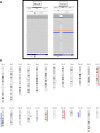
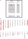
Choroid plexus tumors (CPTs) are rare intracranial neoplasms, representing <1% of all brain tumors, yet they represent 20% of first-year pediatric brain tumors. Although these tumors have been linked to TP53 germline mutations in the context of Li-Fraumeni syndrome, their somatic driver alterations remain poorly understood. In this study, we report two cases of lateral ventricle tumors: 3-yr-old male diagnosed with an atypical choroid plexus papilloma (aCPP), and a 6-mo-old female diagnosed with a choroid plexus carcinoma (CPC). We performed whole-exome sequencing of paired blood and tumor tissue in both patients, categorized somatic variants, and determined copy-number alterations. Our analysis revealed a tier II variant (Association for Molecular Pathology [AMP] criteria) in BRD1, a H3 and TP53 acetylation agent, in the aCPP. In addition, we detected copy-number gains on Chromosomes 12, 18, and 20 and copy-number losses on Chromosomes 13q and 22q (BRD1 locus) in this tumor. The CPC tumor had only a pathogenic germline TP53 variant, based on American College of Medical Genetics (ACMG) criteria, with a clinical and familiar history of Li-Fraumeni syndrome. The CPC patient presented loss of heterozygosity (LoH) of TP53 loci and hyperdiploid genome. Both tumors were microsatellite-stable. This is the first study performing whole-exome sequencing in Brazilian choroid plexus tumors, and in line with the literature, we corroborate the absence of recurrent somatic mutations in these tumors. Further studies with larger sample sizes are necessary to confirm our findings and better understand the underlying biology of these tumors.
Keywords: neoplasm of the nervous system.
© 2023 Garcia et al.; Published by Cold Spring Harbor Laboratory Press.
Figures


Similar articles
-
Li-Fraumeni and Li-Fraumeni-like syndrome among children diagnosed with pediatric cancer in Southern Brazil.Cancer. 2013 Dec 15;119(24):4341-9. doi: 10.1002/cncr.28346. Epub 2013 Oct 7. Cancer. 2013. PMID: 24122735
-
Molecular insights into malignant progression of atypical choroid plexus papilloma.Cold Spring Harb Mol Case Stud. 2021 Feb 19;7(1):a005272. doi: 10.1101/mcs.a005272. Print 2021 Feb. Cold Spring Harb Mol Case Stud. 2021. PMID: 33608379 Free PMC article.
-
TP53 alterations determine clinical subgroups and survival of patients with choroid plexus tumors.J Clin Oncol. 2010 Apr 20;28(12):1995-2001. doi: 10.1200/JCO.2009.26.8169. Epub 2010 Mar 22. J Clin Oncol. 2010. PMID: 20308654
-
Correlation between choroid plexus carcinoma and Li-Fraumeni syndrome: implications of TP53 mutations and management strategies-a case-based narrative review.Childs Nerv Syst. 2024 Jun;40(6):1699-1705. doi: 10.1007/s00381-024-06313-y. Epub 2024 Feb 6. Childs Nerv Syst. 2024. PMID: 38316675 Review.
-
Epigenetics impacts upon prognosis and clinical management of choroid plexus tumors.J Neurooncol. 2020 May;148(1):39-45. doi: 10.1007/s11060-020-03509-5. Epub 2020 Apr 28. J Neurooncol. 2020. PMID: 32342334 Free PMC article. Review.
Cited by
-
Cancer genomics and bioinformatics in Latin American countries: applications, challenges, and perspectives.Front Oncol. 2025 Jul 9;15:1584178. doi: 10.3389/fonc.2025.1584178. eCollection 2025. Front Oncol. 2025. PMID: 40703551 Free PMC article. Review.
References
-
- Alonso M, Hamelin R, Kim M, Porwancher K, Sung T, Parhar P, Miller DC, Newcomb EW. 2001. Microsatellite instability occurs in distinct subtypes of pediatric but not adult central nervous system tumors. Cancer Res 61: 2124–2128. - PubMed
-
- Campanella NC, Berardinelli GN, Scapulatempo-Neto C, Viana D, Palmero EI, Pereira R, Reis RM. 2014. Optimization of a pentaplex panel for MSI analysis without control DNA in a Brazilian population: correlation with ancestry markers. Eur J Hum Genet 22: 875–880. 10.1038/ejhg.2013.256 - DOI - PMC - PubMed
Publication types
MeSH terms
LinkOut - more resources
Full Text Sources
Research Materials
Miscellaneous
