. 2023 Mar 24;8(1):139.
doi: 10.1038/s41392-023-01376-w.

Mechanisms of obesity- and diabetes mellitus-related pancreatic carcinogenesis: a comprehensive and systematic review

Affiliations

Mechanisms of obesity- and diabetes mellitus-related pancreatic carcinogenesis: a comprehensive and systematic review

Rexiati Ruze et al. Signal Transduct Target Ther. .

Abstract

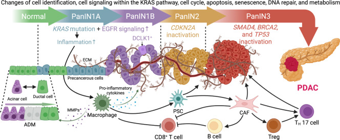
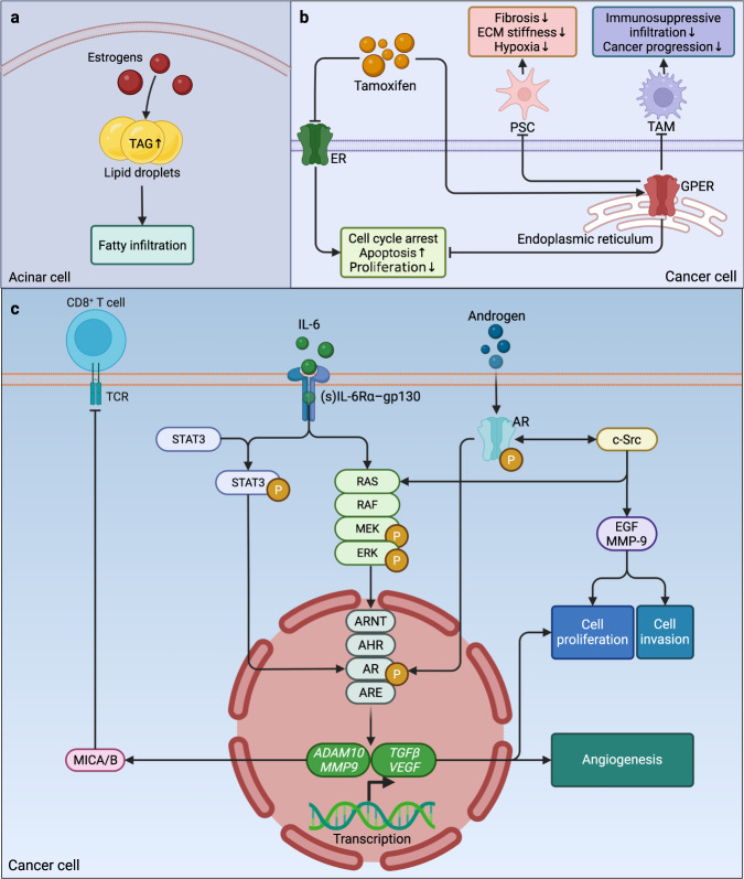
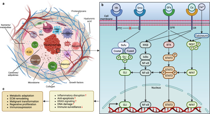
Research on obesity- and diabetes mellitus (DM)-related carcinogenesis has expanded exponentially since these two diseases were recognized as important risk factors for cancers. The growing interest in this area is prominently actuated by the increasing obesity and DM prevalence, which is partially responsible for the slight but constant increase in pancreatic cancer (PC) occurrence. PC is a highly lethal malignancy characterized by its insidious symptoms, delayed diagnosis, and devastating prognosis. The intricate process of obesity and DM promoting pancreatic carcinogenesis involves their local impact on the pancreas and concurrent whole-body systemic changes that are suitable for cancer initiation. The main mechanisms involved in this process include the excessive accumulation of various nutrients and metabolites promoting carcinogenesis directly while also aggravating mutagenic and carcinogenic metabolic disorders by affecting multiple pathways. Detrimental alterations in gastrointestinal and sex hormone levels and microbiome dysfunction further compromise immunometabolic regulation and contribute to the establishment of an immunosuppressive tumor microenvironment (TME) for carcinogenesis, which can be exacerbated by several crucial pathophysiological processes and TME components, such as autophagy, endoplasmic reticulum stress, oxidative stress, epithelial-mesenchymal transition, and exosome secretion. This review provides a comprehensive and critical analysis of the immunometabolic mechanisms of obesity- and DM-related pancreatic carcinogenesis and dissects how metabolic disorders impair anticancer immunity and influence pathophysiological processes to favor cancer initiation.


Conflict of interest statement

The authors declare no competing interests.

Figures

Fig. 1
Fig. 1
Progression and microenvironment of PanINs that favor the formation of PDAC. Ductal cells can transdifferentiate into acinar cells under normal conditions as a compensatory regenerative process to maintain the proper function of the pancreas. Meanwhile, the highly plastic acinar cells can also be turned into ductal cells through the metaplastic process called ADM when stimulated by inflammatory macrophages via the secretion of MMPs, and they maintain their ductal phenotype in the presence of oncogenic KRAS mutation, followed by enhanced EGFR signaling and sequential inactivation of tumor-suppressive genes, such as CDKN2A, TP53, BRCA2, and SMAD4, to form a carcinoma in situ. Initially, oncogenic KRAS magnifies proinflammatory signaling in macrophages and promotes ADM by producing MMPs while secreting inflammatory cytokines into the microenvironment. Some serine/threonine-protein kinase DCLK1+ cells of acinar origin are also formed during low-grade PanIN lesions, such as PanIN1A, PanIN1B, and PanIN2, putatively serving as progenitor cells with cancer stem cell functions. Meanwhile, macrophages activate PSCs and change their morphology into CAFs, which enhance the desmoplastic reaction and ECM production, increasing tissue tension and creating a hypoxic microenvironment within the PanINs that is made up of abundant precancerous metaplastic epithelia and tuft cells. Furthermore, CAFs can activate immunosuppressive B cells, Tregs, and TH17 cells and collaboratively sabotage the anticancer immunity of CD8+ T cells with macrophages. During this process, precancerous cells are transformed by strengthened KRAS signaling. The aberrance of the cell cycle, apoptosis, senescence, DNA repair, and metabolism in this immunosuppressive microenvironment jointly favors the formation of PDAC. ADM acinar-to-ductal metaplasia, CAF cancer-associated fibroblast, CDKN2A cyclin-dependent kinase inhibitor 2A, DCLK1 doublecortin-like kinase 1, ECM extracellular matrix, EGFR epidermal growth factor receptor, KRAS Kirsten rat sarcoma viral oncogene homolog, MMPs matrix metalloproteinases, PanIN pancreatic intraepithelial neoplasia, PDAC pancreatic ductal adenocarcinoma, PSC pancreatic stellate cell, SMAD4 SMAD family member 4, TP53 tumor suppressor p53, Tregs T helper cells, TH helper T. This figure was adapted from a previous publication
Fig. 2
Fig. 2
The roles of AGEs and RAGEs in pancreatic carcinogenesis. The production of AGEs is drastically increased in obesity and DM, and the binding of AGEs to RAGEs activates MAPK and NF-κB signaling and increases the transcription of HIF-1α and NF-κB, which prevents cell death from oxidative stress and creates a hypoxic microenvironment while promoting proinflammatory signaling to exacerbate inflammatory reactions and recruit immunosuppressive MDSCs to diminish anticancer immunity. In addition to the decreased apoptosis due to the decline in the transcription of TP53 following the activation of KRAS signaling, the enhanced PI3K-AKT signaling and the direct activation of mTOR by RAGEs mitigate autophagy to improve the proliferation and survival of cancer cells, thereby promoting pancreatic carcinogenesis. AGEs advanced glycation end products, AKT protein kinase B, DM diabetes mellitus, ERK extracellular signal-regulated kinase, HIF-1α hypoxia-inducible factor 1 subunit α, IKKβ inhibitor of nuclear factor-κB (NF-κB) kinase subunit β, Ikβ inhibitor of NF-κB subunit β, KRAS Kirsten rat sarcoma viral oncogene homolog, MAPK mitogen-activated protein kinase, MDSCs myeloid-derived suppressor cells, MEK mitogen extracellular kinase, mTOR mammalian target of rapamycin, NF-κB nuclear factor-κB, P phosphorylation, PI3K phosphatidylinositol-3-kinase, RAF Raf proto-oncogene, RAGE receptor of AGEs, TP53 tumor suppressor p53
Fig. 3
Fig. 3
Cyclical relationships between obesity and dysregulated sex hormone levels in both sexes. The excessive accumulation and expansion of adipose tissue disrupts the secretion of metabolic and inflammatory adipokines and cytokines, eventually causing systemic inflammation, insulin resistance, and hyperglycemia. a In men, metabolic disorders along with increased aromatase activity and estrogen levels induce an inhibitory effect on the secretion of gonadotropin from the hypothalamus and pituitary gland, suppressing the production of androgen from the testes and resulting in MOSH and the exacerbation of obesity. b In contrast, the inhibitory effect of the hypothalamus and pituitary gland on the ovaries and the influence of systemic inflammation and metabolic disorders on the adrenals and ovaries lead to the elevation of androgen, resulting in PCOS and hyperandrogenism in women. MOSH male obesity-associated secondary hypogonadism, PCOS polycystic ovary syndrome
Fig. 4
Fig. 4
Sex hormones and pancreatic carcinogenesis and cancer progression. a In the cytoplasm of pancreatic acinar cells, the increased estrogen levels lead to the elevation of TAGs and total lipids in the pancreas, contributing to fatty infiltration. b Tamoxifen can play an anticancer role by antagonizing estrogen receptors and agonizing GPER, and the latter can mitigate fibrosis and hypoxia in the TME by targeting PSCs, while it also ameliorates the immunosuppressive infiltration of macrophages and hinders cancer progression. c According to the description of Kanda et al. , the tumorigenic cytokine IL-6 can activate both STAT3 and MAPK signaling in PC cells, while extracellular androgen and oncogenic c-Src can also enhance AR and MAPK signaling and trigger the transactivation of nuclear ARs. Meanwhile, AHR, ARNT, and ARE interact with AR in a testosterone-dependent manner and translocate into the nucleus to increase the transcription of ADAM10, MMP9, TGFβ, and VEGF. ADAM10 and MMP-9 increase the expression of MICA and MICB and hamper the immune response of NK cells and T cells against cancer cells. In combination with the enhanced cell proliferation and invasion favored by the activation of EGF and MMP-9, TGF-β and VEGF also jointly promote angiogenesis and cell proliferation. ADAM10 a disintegrin and metalloprotease 10, AHR aryl hydrocarbon (or dioxin) receptor, AR androgen receptor, ARE androgen-responsive element, ARNT AHR nuclear translocator, ECM extracellular matrix, EGF epidermal growth factor, ERK extracellular signal-regulated kinase, GPER G-protein-coupled estrogen receptor, IL-6 interleukin 6, MAPK mitogen-activated protein kinase, MEK mitogen extracellular kinase, MICA/B major histocompatibility complex class I chain-related gene A/B, MMP-9 matrix metalloprotease 9, NK natural killer, PC pancreatic cancer, PSC pancreatic stellate cell, RAF Raf proto-oncogene, STAT3 signal transducer and activator of transcription 3, TAGs triglycerides, TAM tumor-associated macrophage, TCR T-cell receptor, TGF-β transforming growth factor β, TME tumor microenvironment, VEGF vascular endothelial growth factor. Panel c in this figure was adapted from a previous publication
Fig. 5
Fig. 5
Microbes and pancreatic carcinogenesis. Upper left panel: Beyond their distant impact and transfer of their carcinogenic products, microbes from different regions of the GI tract may migrate to the pancreas via retrograde transfer through the opening of the sphincter of Oddi and contribute to pancreatic carcinogenesis. Marks a, b, and b (corresponding to panels a, b, and c, respectively) indicate the possible distant influence and transfer of oral, gastric, and GM carcinogenic products or their migration to the pancreas. a Distinct effect of different oral microbiome species on the risk of PC. b Two hypothetical theories on pancreatic carcinogenesis related to Hp infection. c Some metabolites of the GM, such as lipopolysaccharide (LPS), can enhance chronic inflammation by activating multiple carcinogenic pathways and increasing the secretion of proinflammatory components. In contrast, altered levels of carcinogenic metabolites (e.g., BAs) can promote pancreatic carcinogenesis by accelerating the senescence-associated secretory phenotype and increasing DNA damage and genomic instability. Some viruses from the GM community (e.g., HBV and HCV) are also suggested to increase inflammation-induced DNA damage and carcinogenesis. Later, microbe-induced inflammation can be carcinogenic and initiate the activation of KRAS, which also exacerbates inflammatory reactions in return. The enhancement of oncogenic signaling subsequently triggers other factors that promote the progression of carcinogenesis, such as oxidative stress, cell cycle disruption, suppressed apoptosis, and the immune response. BAs bile acids, GI gastrointestinal, GM gut microbiome, HB (C) V hepatitis B (C) virus, Hp Helicobacter pylori, KRAS Kirsten rat sarcoma viral oncogene homolog, PC pancreatic cancer
Fig. 6
Fig. 6
Correlations between the ATME and pancreatic carcinogenesis. Beyond an immediate increase in the development of low-grade chronic inflammation via the enhanced secretion of proinflammatory cytokines, the excessively expanded adipose tissue exacerbates metabolic disorders and magnifies the negative impact of adipokines. Meanwhile, this hypertrophic expansion inflicts stress on adipocytes and increases the production and release of substantial proinflammatory cytokines into the ATME, promoting the recruitment and proliferation of inflammatory cells and exacerbating oxidative stress, fibrosis, hypoxia, and lipolysis, which collaboratively have toxic, diabetogenic, and carcinogenic influences on the pancreas. ATMs adipose tissue macrophages, ATME adipose tissue microenvironment, NK natural killer, ROS reactive oxygen species
Fig. 7
Fig. 7
The impacts of the inflammatory microenvironment on pancreatic carcinogenesis. a The desmoplastic and inflammatory pancreatic microenvironment in obesity and DM during carcinogenesis comprises transformed cells, stromal cells (mainly CAFs and PSCs), adipocytes, diverse immune cells, dead cells, microbes, metabolites, ROS, RNS, and substantial proinflammatory cytokines/adipokines and GFs, etc., jointly fueling carcinogenesis via constantly activated inflammatory reactions. b Similar to other cells in this microenvironment, inflammatory stimuli activate various proinflammatory and oncogenic transcription factors in precancerous cells undergoing malignant transformation. The inhibitory effect of the transmembrane protein PTC on Smo is reversed after its binding to Hh, which increases the release of GLI from their inhibitory complex and favors nuclear translocation. Furthermore, the binding of GFs (e.g., EGF, etc.) and cytokines (IL-1, etc.) to calcineurin can enhance the nuclear translocation and transcriptional activity of NF-κB, STAT3, and NFAT. Bidirectional arrows indicate the interactions among transcription factors. c The activation of these transcription factors upregulates the expression of numerous target genes mediating inflammation, cell death, KRAS signaling, DNA damage, and immune surveillance, thereby jointly magnifying the impacts of inflammatory disruption on pancreatic carcinogenesis. BTK Bruton’s tyrosine kinase, Cs cytokines, CR cytokine receptor, DC dendritic cell, ECM extracellular matrix, EGF epidermal growth factor, GFR growth factor receptor, GFs growth factors, GLI glioma-associated oncogene, Hh Hedgehog, IκBα inhibitor of NF-κB subunit α, IKKβ inhibitor of nuclear factor-κB (NF-κB) kinase subunit β, IL-1 interleukin 1, MDSC myeloid-derived suppressor cell, NFAT nuclear factor of activated T cells, NF-κB nuclear factor-κB, P phosphorylation, PSCs pancreatic stellate cells, PTC patched, RNS reactive nitrogen species, ROS reactive oxygen species, Smo Smoothened, STAT3 signal transducer and activator of transcription 3, Sufu suppressor of fused, TH T helper, Treg regulatory T cell. Panel b in this figure was adapted from a previous publication
Fig. 8
Fig. 8
Interactions among cells within the tumor-killing (a) and immunosuppressive (b) TME of PC. Promotive effects are indicated with black arrows, and the suppressive impacts of cells on anticancer immunity are marked with lines in color. Some signaling components are shown near the arrows and lines. CAF cancer-associated fibroblast, CCL C-C motif chemokine ligand, CTLA4 cytotoxic T lymphocyte-associated protein 4, CXCL CXC-chemokine ligand, DC dendritic cell, EVs extracellular vesicles, FGF fibroblast growth factor, GM-CSF granulocyte-macrophage colony-stimulating factor, HGF hepatocyte growth factor, IDO indoleamine 2,3-dioxygenase, IFN interferon, IL interleukin, M1(2) M1(2) macrophage, MCP monocyte chemoattractant protein, MDSC myeloid-derived suppressor cell, MIP-1 macrophage inflammatory protein 1, MMPs matrix metalloproteinases, N2 N2 neutrophil, NE neutrophil elastase, NET neutrophil extracellular trap, NK natural killer, PC pancreatic cancer, PCCs pancreatic cancer cells, PD(-L) programmed death (ligand), PDGF platelet‐derived growth factor, PSC pancreatic stellate cell, RAGE receptor of advanced glycation end products, TGF transforming growth factor, TH T helper (cell), TNF tumor necrosis factor, Treg regulatory T cell, VEGF vascular endothelial growth factor
Fig. 9
Fig. 9
The intricate relationships among obesity, DM, and pancreatic carcinogenesis and the three most important values of relevant research. DM diabetes mellitus

References

    1. World Obesity Federation. World Obesity Atlas 2022 (World Obesity Federation, 2022).
    1. International Diabetes Federation. IDF Diabetes Atlas (International Diabetes Federation, Brussels, Belgium, 2021).
    1. Tsai AG, Bessesen DH. Obesity. Ann. Intern. Med. 2019;170:Itc33–itc48. doi: 10.7326/AITC201903050. - DOI - PubMed
    1. Kumanyika S, Dietz WH. Solving population-wide obesity - progress and future prospects. N. Engl. J. Med. 2020;383:2197–2200. doi: 10.1056/NEJMp2029646. - DOI - PubMed
    1. Blüher M. Obesity: global epidemiology and pathogenesis. Nat. Rev. Endocrinol. 2019;15:288–298. doi: 10.1038/s41574-019-0176-8. - DOI - PubMed

Publication types