. 2023 Mar 24;14(3):209.
doi: 10.1038/s41419-023-05728-w.

Type I interferon signaling in malignant blasts contributes to treatment efficacy in AML patients

Affiliations

Type I interferon signaling in malignant blasts contributes to treatment efficacy in AML patients

Peter Holicek et al. Cell Death Dis. .

Abstract

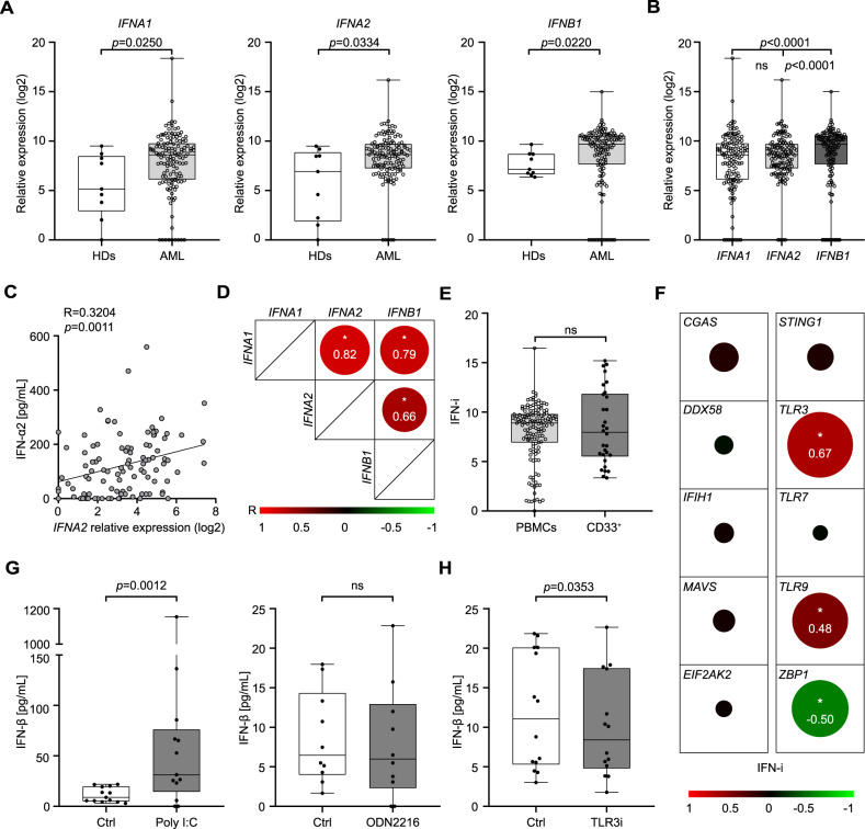
While type I interferon (IFN) is best known for its key role against viral infection, accumulating preclinical and clinical data indicate that robust type I IFN production in the tumor microenvironment promotes cancer immunosurveillance and contributes to the efficacy of various antineoplastic agents, notably immunogenic cell death inducers. Here, we report that malignant blasts from patients with acute myeloid leukemia (AML) release type I IFN via a Toll-like receptor 3 (TLR3)-dependent mechanism that is not driven by treatment. While in these patients the ability of type I IFN to stimulate anticancer immune responses was abolished by immunosuppressive mechanisms elicited by malignant blasts, type I IFN turned out to exert direct cytostatic, cytotoxic and chemosensitizing activity in primary AML blasts, leukemic stem cells from AML patients and AML xenograft models. Finally, a genetic signature of type I IFN signaling was found to have independent prognostic value on relapse-free survival and overall survival in a cohort of 132 AML patients. These findings delineate a clinically relevant, therapeutically actionable and prognostically informative mechanism through which type I IFN mediates beneficial effects in patients with AML.


Conflict of interest statement

LG is/has been holding research contracts with Lytix Biopharma, Promontory and Onxeo, has received consulting/advisory honoraria from Boehringer Ingelheim, AstraZeneca, OmniSEQ, Onxeo, The Longevity Labs, Inzen, Imvax, Sotio, Promontory, Noxopharm, EduCom, and the Luke Heller TECPR2 Foundation, and holds Promontory stock options. All other authors have no conflicts to declare.

Figures

Fig. 1
Fig. 1. TLR3 drives type I IFN secretion from AML blasts.
A, B Relative expression levels of IFNA1, IFNA2, and IFNB1 in peripheral blood mononuclear cells (PBMCs) from 9 healthy donors (HDs) and 132 AML patients (Study Cohort 1) prior to induction chemotherapy, as determined by RT-qPCR. Data are presented as median, quartiles and extremes plus individual data points. p values are reported (Mann-Whitney test). C Correlation between IFNA2 serum levels and IFNA2 expression in 101 AML patients (Study Cohort 1), as determined by Luminex and RT-qPCR, respectively. Spearman correlation coefficient (R) and associated p value are reported. D Correlation matrix for IFNA1, IFNA2 and IFNB1 expression in 132 AML patients from Study Cohort 1. Spearman correlation coefficient (R) is reported; *p < 0.0001. E Relative expression abundance of type I IFN index (IFN-i) in whole PBMCs (n = 132) versus isolated CD33+ malignant blasts (n = 30) from AML patients. Data are reported are presented as median, quartiles and extremes plus individual data points. ns, not significant (Mann-Whitney test). F Correlation matrix between IFN-i and relative expression levels of CGAS, DDX58, IFIH1, MAVS, EIF2AK2, STING1, TLR3, TLR7, TLR9 and ZBP1 in CD33+ leukemic blasts isolated from 30 AML patients (Study Cohort 1). Significant Spearman correlation coefficients (R) are reported; *p < 0.05. G, H IFN-β production by CD33+ blasts 24 h after optional treatment with polyI:C (n = 10), ODN2216 (CpG) (n = 10) G or a TLR3 inhibitor (TLR3i) (n = 10) H, as determined by ELISA. Data are presented as median, quartiles and extremes plus individual data points. Significant p values are reported; ns, not significant (Mann-Whitney test).
Fig. 2
Fig. 2. Type I IFN-driven immunostimulation is suppressed by malignant blasts.
A Relative expression levels of selected genes associated with NK cells, TH1 and TH2 response, T cell activation and cytotoxicity in 12 IFN-iLo and 12 IFN-iHi AML patients from Study Cohort 1 as determined RNAseq (see Supplementary Table 4). B Percentage of circulating CD45+CD3+, CD45+CD3+CD4+, CD45+CD3+CD8+ T cells and CD45+CD3CD56+, CD45+CD3CD56dim and CD45+CD3CD56bright NK cells in 13 IFN-iLo versus 34 IFN-iHi AML patients from Study Cohort 1 prior to induction chemotherapy, as determined by flow cytometry. Data are presented as median, quartiles and extremes plus individual data points. ns, not significant (Mann-Whitney test). C Gating strategy for IFN-γ+ and CD107a+ CD45+CD3+CD8+ T cells and CD45+CD3CD56+ NK cells. The percentage of cells in each gate is reported. D Percentage of IFN-γ+ and CD107a+ CD45+CD3+CD8+ T cells and CD45+CD3CD56+ NK cells upon stimulation with PMA plus ionomycin or K562 cells of peripheral blood mononuclear cells (PBMCs) from 10 IFN-iLo versus 23 IFN-iHi AML patients of Study Cohort 1, as determined by flow cytometry. Data are presented as median, quartiles and extremes plus individual data points. Significant p values are reported; ns, not significant (Mann-Whitney test). E Representative dot plots showing PBMC composition of one AML patient before and after depletion of CD33+ leukemic blasts. F, G Percentage of IFN-γ+ and CD107a+ CD8+ T cells and NK cells upon stimulation with PMA plus ionomycin of PBMCs optionally depleted of CD33+ blasts and optionally exposed to recombinant IFN-α plus IFN-β (rIFNs) from 7 AML patients of Study Cohort 1, as determined by flow cytometry. Data are reported as means ± SEM. *p < 0.05; **p < 0.01; ns, not significant (paired t-test).
Fig. 3
Fig. 3. Recombinant type I IFN mediates direct cytostatic and cytotoxic activity on AML blasts and leukemic stem cells.
A, B Representative dot plots A and percentages B of viable (Annexin V/DAPI) KASUMI-1, MOLM-13 and MV4–11 cells after daunorubicin (DNR) and cytarabine (Ara-C) 24 h treatment with optional recombinant IFN-α plus IFN-β (rIFNs) (500 pg/mL) 3 days pre-incubation, as determined by flow cytometry. Data are reported as means ± SD plus individual data points. *p < 0.05; **p < 0.01, ****p < 0.0001, ns: not significant (paired t-test). C Relative viability of CD33+ leukemic blasts from 10 AML patients (Study Cohort 3) after DNR or Ara-C treatment with optional 3 days pre-incubation with rIFNs (500 pg/mL). Data are reported as means, quartiles and extremes plus individual data points. ***p < 0.001 (paired t-test). D Gating strategy for determination of leukemic stem cells (LSCs) in AML patient PBMCs using flow cytometry. E Relative viability of LSCs isolated from 10 AML patients (Study Cohort 3) after DNR or Ara-C treatment with optional 3 days pre-incubation with rIFNs (500 pg/mL). Data are reported as means, quartiles and extremes plus individual data points. ***p < 0.001 (paired t-test). F Relative viability of CD33+ leukemic blasts from 8 AML patients (Study Cohort 3) 96 h upon exposure to polyI:C in the optional presence of an IFNAR1 blocking antibody, or rIFNs, Data are reported as means, quartiles and extremes plus individual data points. *p < 0.05, **p < 0.01 (paired t-test).
Fig. 4
Fig. 4. Chemosensitizing effects of type I IFN in human AML xenografts.
A Experimental study design of an AML xenograft model using human WT or IFNAR2-/- KASUMI-1 cells in Rag2-/- mice. B Overall survival (OS) of Rag2-/- mice xenografted with WT KASUMI-1 cells and optionally treated with human rIFN-β (IFN), daunorubicin (DNR) or IFN + DNR. C OS of Rag2-/- mice xenografted with IFNAR2-/- KASUMI-1 cells and optionally treated with IFN, DNR or IFN + DNR. Survival curves were estimated by the Kaplan-Meier method, and differences between groups were evaluated using log-rank test. Median OS (days) and p values are reported.
Fig. 5
Fig. 5. Type I IFN levels correlate with improved disease outcome in patients with AML.
AC Relapse-free (RFS) and overall survival (OS) of 132 AML patients from Study Cohort 1 upon median stratification based on IFNA1, IFNA2, IFNB1 expression or type I IFN index (IFN-i). Survival curves were estimated by the Kaplan-Meier method, and differences between groups were evaluated using log-rank test. Number of patients at risk and p values are reported.

References

    1. Kroemer G, Galassi C, Zitvogel L, Galluzzi L. Immunogenic cell stress and death. Nat Immunol. 2022;23:487–500. doi: 10.1038/s41590-022-01132-2. - DOI - PubMed
    1. McNab F, Mayer-Barber K, Sher A, Wack A, O’Garra A. Type I interferons in infectious disease. Nat Rev Immunol. 2015;15:87–103. doi: 10.1038/nri3787. - DOI - PMC - PubMed
    1. Schoggins JW. Interferon-stimulated genes: what do they all do? Annu Rev Virol. 2019;6:567–84. doi: 10.1146/annurev-virology-092818-015756. - DOI - PubMed
    1. Galluzzi L, Humeau J, Buque A, Zitvogel L, Kroemer G. Immunostimulation with chemotherapy in the era of immune checkpoint inhibitors. Nat Rev Clin Oncol. 2020;17:725–41. doi: 10.1038/s41571-020-0413-z. - DOI - PubMed
    1. Petroni G, Buque A, Zitvogel L, Kroemer G, Galluzzi L. Immunomodulation by targeted anticancer agents. Cancer Cell. 2021;39:310–45. doi: 10.1016/j.ccell.2020.11.009. - DOI - PubMed

Publication types