Review
. 2023 Dec;192(6):2881-2885.
doi: 10.1007/s11845-023-03344-3. Epub 2023 Mar 25.

An unusual case of a giant fetal facial tumour and review of the literature

Affiliations
Review

An unusual case of a giant fetal facial tumour and review of the literature

Alex O Start et al. Ir J Med Sci. 2023 Dec.

Abstract

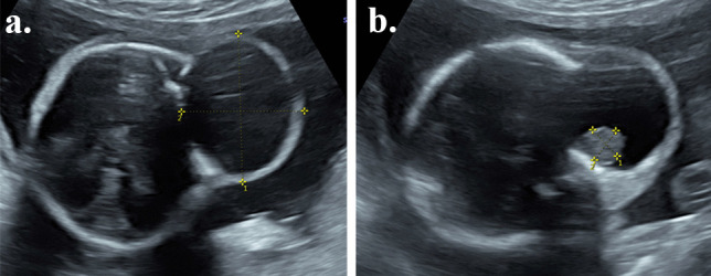
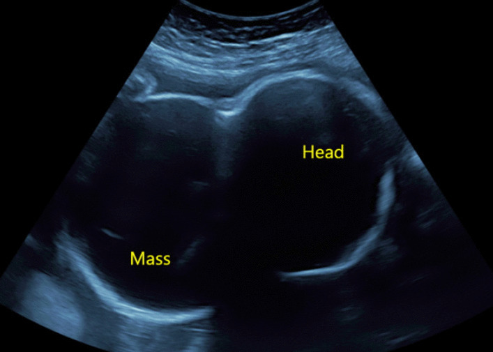
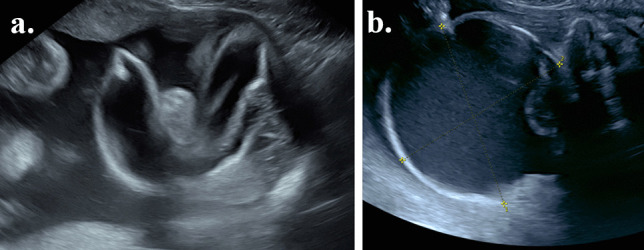
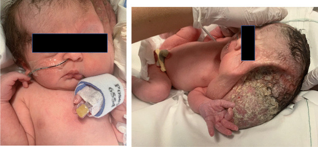
We present the case of a pregnant 32-year-old woman who presented with a giant fetal facial tumour at 22 weeks. The mass, initially 4 × 3.5 × 3 cm in size, was largely cystic with a small solid component. It subsequently increased to 9 × 9 × 10 cm. Significant compression effects on the fetal orbit, temple and infratemporal fossa, with potential compression of the optic nerve, were noted on ultrasound and MRI. The cyst required drainage twice in the pregnancy: firstly to reduce the compression effects and secondly to facilitate caesarean delivery. Postnatally, the baby had significant compression and displacement of the craniofacial skeleton from the mass effect. Postnatal histology revealed a diagnosis of a teratoma. This case highlights the complexities and challenges surrounding the diagnosis and management of a giant fetal facial tumour.

Keywords: Congenital tumour; Fetal anomaly; Fetus; Pregnancy.


Figures

Fig. 1
Fig. 1
Ultrasound at 22 weeks’ gestation. a and b are ultrasound images of the lesion at 22 weeks gestation. The lesion (a) measures 4 × 3.5 × 3 cm in size with a single solid component of 1.02 × 1.03 cm size seen in (b)
Fig. 2
Fig. 2
MRI at 23 weeks gestation. Coronal a and axial b T2-weighted MRI of the fetal head and neck demonstrate a large cystic lesion (arrowhead) in the left infratemporal region with a smaller solid component in its medial aspect. It is causing significant distortion of surrounding structures including displacement of the left orbit (dashed arrow) and ear (arrow)
Fig. 3
Fig. 3
Ultrasound showing the enlarged lesion at 30 + 2 weeks gestation. This shows an ultrasound image of the mass at 30 + 2 weeks gestation. The tumour now measures 9 × 9 × 10 cm
Fig. 4
Fig. 4
Fetal MRI at 31 weeks gestation. Axial T2-weighted fetal MRI demonstrates interval increase in the size of the large cystic mass (arrowhead) with increased mass effect on the left orbit (dotted arrow)
Fig. 5
Fig. 5
Ultrasound images of the lesion before and after drainage. a highlights the collapse of the cystic structure after the removal of 640 ml of light yellow coloured at 33 + 3 weeks gestation. b shows the reaccumulation by 34 + 3 weeks gestation
Fig. 6
Fig. 6
Images of the baby in the immediate postnatal period. This shows the large cystic lesion immediately after caesarean delivery. The cyst re-accumulated after drainage at the time of caesarean delivery
Fig. 7
Fig. 7
Postnatal MRI of the head and neck. Coronal T2-weighted MRI of the head and neck on day 1 of life again demonstrates the large left-sided cystic mass (arrowhead) with a solid component medially (star). There is distortion and displacement of adjacent structures including the left orbit (dashed arrow). Some decrease in the size of the cystic component is noted due to drainage a few hours previously postnatally
Fig. 8
Fig. 8
CT images of the head and neck. Bony 3D reformat of a CT of the head and neck demonstrates the extent of bony remodelling in the left frontal, parietal and temporal bones with extensive bony thinning and a prominent bony ridge (arrowheads) at the margins of the mass

References

    1. Alamo L, Beck-Popovic M, Gudinchet F, Meuli R. Congenital tumors: imaging when life just begins. Insights Imaging. 2011;2(3):297–308. doi: 10.1007/s13244-011-0073-8. - DOI - PMC - PubMed
    1. Feygin T, Khalek N, Moldenhauer JS (2020) Fetal brain, head, and neck tumors: Prenatal imaging and management. Prenat Diagn 40(10):1203–1219 - PubMed
    1. Sun RW, Tuchin VV, Zharov VP, et al. Current status, pitfalls and future directions in the diagnosis and therapy of lymphatic malformation. J Biophotonics. 2018;11(8):e201700124. doi: 10.1002/jbio.201700124. - DOI - PMC - PubMed
    1. Sefidbakht S, Iranpour P, Keshavarz P, et al. Fetal MRI in prenatal diagnosis of encephalocele. J Obstet Gynaecol Can. 2020;42(3):304–307. doi: 10.1016/j.jogc.2019.03.013. - DOI - PubMed
    1. Bagchi A, Hira P, Mittal K, et al. Branchial cleft cysts: a pictorial review. Pol J Radiol. 2018;83:e204–e209. doi: 10.5114/pjr.2018.76278. - DOI - PMC - PubMed

LinkOut - more resources