. 2023 Mar 25;3(1):40.
doi: 10.1038/s43856-023-00270-4.

Fusion peptide is superior to co-expressing subunits for arming oncolytic herpes virus with interleukin 12

Affiliations

Fusion peptide is superior to co-expressing subunits for arming oncolytic herpes virus with interleukin 12

Hiroshi Fukuhara et al. Commun Med (Lond). .

Abstract

Background: G47∆ is a triple-mutated oncolytic herpes simplex virus type 1 (HSV-1) recently approved as a new drug for malignant glioma in Japan. As the next-generation, we develop armed oncolytic HSV-1 using G47∆ as the backbone. Because oncolytic HSV-1 elicits specific antitumor immunity, interleukin 12 (IL-12) can function as an effective payload to enhance the efficacy.

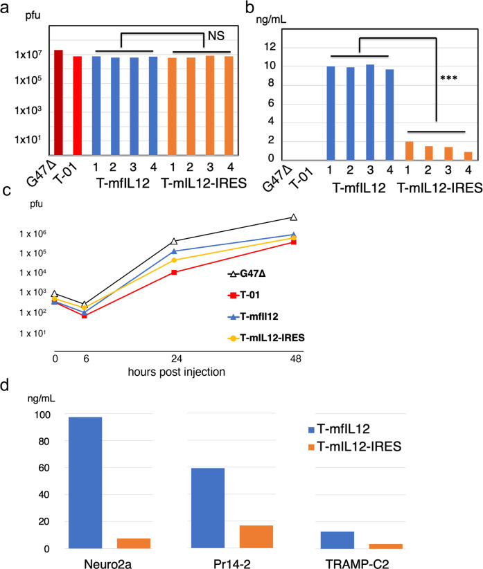
Methods: We evaluate the optimal methods for expressing IL-12 as a payload for G47∆-based oncolytic HSV-1. Two new armed viruses are generated for evaluation by employing different methods to express IL-12: T-mfIL12 expresses murine IL-12 as a fusion peptide, with the genes of two subunits (p35 and p40) linked by bovine elastin motifs, and T-mIL12-IRES co-expresses the subunits, with the two genes separated by an internal ribosome entry site (IRES) sequence.

Results: T-mfIL12 is significantly more efficient in producing IL-12 than T-mIL12-IRES in all cell lines tested, whereas the expression methods do not affect the replication capabilities and cytopathic effects. In two syngeneic mouse subcutaneous tumor models of Neuro2a and TRAMP-C2, T-mfIL12 exhibits a significantly higher efficacy than T-mIL12-IRES when inoculated intratumorally. Furthermore, T-mfIL12 shows a significantly higher intratumoral expression of functional IL-12, causing stronger stimulation of specific antitumor immune responses than T-mIL12-IRES.

Conclusions: The results implicate that a fusion-type expression of IL-12 is a method superior to co-expression of separate subunits, due to higher production of functional IL-12 molecules. This study led to the creation of triple-mutated oncolytic HSV-1 armed with human IL-12 currently used in phase 1/2 trial for malignant melanoma.

Plain language summary

Some viruses, including the herpes virus, can be modified so that they can target and kill cancers. These viruses can be loaded with factors that stimulate the immune system, which can help to eradicate cancer cells. Here, we test different methods of loading a cancer-killing version of the herpes virus with interleukin 12, an immune-stimulating factor. We show that one method, which involves loading the virus with the different parts of interleukin 12 fused together, is superior to another, and leads to improved anti-cancer effects in mouse models. These findings have contributed to the creation of a cancer-killing virus that is currently in clinical trials in patients with melanoma.


Conflict of interest statement

The authors declare no competing interests.

Figures

Fig. 1
Fig. 1. The structures of T-mfIL12 and T-mIL12-IRES.
The boxes (top line) represent inverted repeat sequences flanking the long (UL) and short (US) unique sequences of HSV-1 DNA. Oncolytic HSV-1 uses G47∆ as the backbone and contains 1.0 kb deletions in both copies of the γ34.5 gene, a 0.3 kb deletion in the α47 gene, and a 0.9 kb deletion in the ICP6 gene. The lacZ gene and the cytomegalovirus (CMV) promoter-driven interleukin-12 (IL-12) gene are oriented in opposite directions, and inserted into the deleted ICP6 locus. T-mfIL12 contains a cassette carrying the p35 and p40 genes of murine IL-12 linked by two bovine elastin motifs. Murine IL-12 is expressed as a single fusion peptide and folds to become active. T-mIL12-IRES contains a cassette in which the p40 and p35 genes of murine IL-12 are separated by an internal ribosome entry site (IRES) sequence from the encephalomyocarditis virus (EMCV). The p40 and p35 subunits are efficiently co-expressed to form active murine IL-12. T-01 contains a cassette with no transgene.
Fig. 2
Fig. 2. In vitro replication capabilities and murine IL-12 expressions of T-mfIL12 and T-mIL12-IRES.
a In vitro replication assay. Vero cells were infected with G47∆, T-01, T-mfIL12 candidate (4 clones) or T-mIL12-IRES candidate (4 clones) at a multiplicity of infection (MOI) of 0.01, the progeny virus was recovered 48 h after infection, and the number determined by plaque assay. T-mfIL12 and T-mfIL12-IRES showed no significant difference in replication capability (p = 0.736, t-test). b In vitro murine IL-12 expression. Vero cells were infected with G47∆, T-01, T-mfIL12 candidate (4 clones) or T-mIL12-IRES candidate (4 clones) at an MOI of 1, and the amount of murine interleukin-12 (IL-12) (p70) secreted was determined by enzyme-linked immune-sorbent assay (ELISA). T-mfIL12 expressed a significantly higher amount of p70 IL-12 than T-mIL12-IRES (p < 0.001, t-test). c The time course of viral yields. Vero cells were infected with G47∆, T-01, T-mfIL12 or T-mIL12-IRES in duplicate at an MOI of 0.01, the progeny virus was recovered 0 h, 6 h, 24 h and 48 h after infection, and titrated by plaque assay. The time course for the viral yields of T-mfIL12 was comparable to that of T-mIL12-IRES. d In vitro murine IL-12 expression in murine tumor cell lines. Neuro2a, Pr14-2 or TRAMP-C2 cells were infected with T-mfIL12 or T-mIL12-IRES at an MOI of 1, and the amount of murine IL-12 (p70) secreted was determined by ELISA. T-mfIL12 expressed a higher amount of p70 IL-12 than T-mIL12-IRES in all three cell lines. All assays were performed in duplicate. ***, p < 0.001; NS, not significant.
Fig. 3
Fig. 3. In vitro cytopathic effects of T-mfIL12 and T-mIL12-IRES.
In all three murine cancer cell lines, both T-mfIL12 and T-mIL12-IRES showed cytopathic effects comparable to the control virus T-01 at both multiplicity of infection (MOI) = 0.1 (dotted lines) and MOI = 1 (solid lines). The number of surviving cells was counted daily and expressed as a percentage of mock-infected controls. The results are the means of triplicate wells.
Fig. 4
Fig. 4. In vivo efficacy of T-mfIL12 and T-mIL12-IRES.
a, b and c Assessment by subcutaneous tumor growth inhibition. a Male C57BL/6 mice harboring unilateral subcutaneous TRAMP-C2 tumors (prostate cancer) were treated with intratumoral inoculations with T-01, T-mfIL12 or T-mIL12-IRES (5 × 106 pfu) or mock on days 0 and 3 (n = 6). T-mfIL12 showed the highest efficacy. All three viruses were significantly more efficacious than mock (p < 0.001 for all) (Significance not indicated in the figure). b Female A/J mice harboring bilateral subcutaneous Neuro2a tumors (neuroblastoma) were treated with intratumoral inoculations with T-01, T-mfIL12 or T-mIL12-IRES (5 × 104 pfu) or mock into the left tumors only on days 0 and 3 (n = 9-12). T-mfIL12 exhibited the highest efficacy in both treated and untreated sides. All three viruses were significantly more efficacious than mock in the treated side (p = 0.001 for T-01 and p < 0.001 for both T-mfIL12 and T-mIL12-IRES) and T-mfIL12 and T-mIL12-IRES than mock in the untreated side (p < 0.001 for both viruses) (Significance not indicated in the figure). c NOG mice harboring bilateral subcutaneous Neuro2a tumors were treated with intratumoral inoculations with T-01, T-mfIL12 or T-mIL12-IRES (1 × 106 pfu) or mock into the left tumors only on days 0 and 3 (n = 10). T-01, T-mfIL12 and T-mIL12-IRES showed no difference in efficacy in the treated side. The effect of IL-12 expression was completely abolished for both T-mfIL12 and T-mIL12-IRES in both the treated side and the untreated side. Tumor volume = length × width × height × 0.52. Results represent the mean. Bars, standard error of the mean (SEM). *, p < 0.05; **, p < 0.01; ***, p < 0.001; Two-way ANOVA with Bonferroni’s multiple comparisons test. d and e Assessment by survival. Female A/J mice harboring bilateral subcutaneous Neuro2a tumors were treated with intratumoral inoculations with T-01, T-mfIL12 or T-mIL12-IRES, either 5 × 105 pfu (d) or 5 × 104 pfu (e), or mock into the left tumors on days 0 and 3. Animals were sacrificed when the maximum diameter of one of the bilateral tumors reached 22 mm. Both T-mfIL12 and T-mIL12-IRES significantly prolonged the survival of tumor-bearing mice compared with T-01 at both doses (log-rank test). One or two T-mfIL12-treated mice showed a long-term survival for both doses, but there was no significant difference between T-mfIL12 and T-mIL12-IRES.
Fig. 5
Fig. 5. Immune responses by T-mfIL12 and T-mIL12-IRES.
a Immunohistochemistry. Bilateral subcutaneous Neuro2a tumors were generated in A/J mice, left tumors only were inoculated with T-mfIL12, T-mIL12-IRES, T-01 (2 × 105 pfu) or mock on days 0 and 3, and the tumors were harvested on day 6 (n = 3 per group). An increased infiltration of CD4+ and CD8+ lymphocytes were observed in the tumor for all three viruses, both in the treated and the untreated side, most prominently with T-mfIL12 (Fig. 5a). HSV-1 positive cells were observed in the tumor with all viruses in the treated side, but not in the untreated side. HE, hematoxylin and eosin. Scale bars, 100 μm. b In vivo levels of interleukin-12 (IL-12) and Interferon γ (IFNγ). In the unilateral subcutaneous Neuro2a model, established tumors were inoculated with T-01, T-mfIL12, T-mIL12-IRES (2 × 106 pfu) or mock, sera and tumor samples were collected on days 1, 3 and 6 (n = 3 per group), and the levels of mouse IL-12 and IFNγ were measured by ELISA. The intratumoral IL-12 levels for T-mfIL12 were significantly higher than those for T-mIL12-IRES at all time points (p = 0.018, p = 0.016 and p = 0.046 for days 1, 3 and 6, respectively). The levels of IL-12 detected from the serum were remarkably lower than those in the tumor. Correlating with the intratumoral IL-12, the serum IL-12 level for T-mfIL12 was higher than that for T-mIL12-IRES on day 1 (p = 0.047). The IFNγ levels of T-mfIL12 were significantly higher than those of T-mIL12-IRES both in the tumor and serum on day 1 (p = 0.014 and p = 0.027, tumor and serum, respectively). c Immune responses specific to Neuro2a cells. In the unilateral subcutaneous Neuro2a model, established tumors were inoculated with T-01, T-mfIL12, T-mIL12-IRES (5 × 104 pfu) or mock on days 0 and 3, and the spleen was harvested on day 6. By ELISpot assay, splenocytes from T-mfIL12-treated mice showed a significantly higher number of IFNγ release stimulated by Neuro2a cells than those from T-01- and T-mIL12-IRES- treated ones (p = 0.005 and p = 0.004 vs T-01 and T-mIL12-IRES, respectively). No significant difference in number of IL-4 releasing splenocytes was observed among the three virus-treated groups. The IFNγ or IL-4 releases specific to Neuro2a cells were calculated by subtracting the numbers of SaI/N responding spots from those of Neuro2a responding spots. For b and c, Graphs show the means. Dots represent individual data. Bars, SD. *, p < 0.05; **, p < 0.01; NS, not significant; one-way ANOVA with Tukey’s multiple comparisons.
Fig. 6
Fig. 6. Comparison of in vivo efficacy of T-mfIL12 and direct intratumoral injection with recombinant interleukin-12 (rIL-12).
In the bilateral subcutaneous Neuro2a model, rIL-12, T-01 (5 × 104 pfu) without or with rIL-12, T-mfIL12 (5 × 104 pfu) or mock was inoculated into the left tumors only on days 0 and 4 (n = 10 per group). Three different doses were used for rIL-12; 500 ng (a), 50 ng (b) and 1 ng (c). The dose 50 ng represents the intratumoal IL-12 level treated with T-mfIL12 at 2 × 106 pfu and 1 ng represents that at 5 × 104 pfu, the T-mfIL12 dose used in these experiments. a When the dose of 500 ng was used for rIL-12, rIL-12, T-01, T-01+rIL-12 and T-mfIL12 were all significantly more efficacious than mock in the treated side (p < 0.001 vs mock for all). In the untreated side, rIL-12 alone showed no significant antitumor effect compared with mock, whereas T-01+rIL-12 and T-mfIL12 showed a significantly higher efficacy than mock (p = 0.039 and p < 0.001 vs mock, respectively). Further, in the untreated side, T-mfIL12, but not T-01+rIL-12, was significantly more efficacious than rIL-12 alone (p = 0.003). b Results similar to a were obtained when the dose of 50 ng was used for rIL-12. c When the dose of 1 ng was used for rIL-12, rIL-12 alone showed no significant efficacy in both treated and untreated sides, and T-mfIL12, but not T-01+rIL-12, was significantly more efficacious than rIL-12 alone in the treated side. Tumor volume = length × width × height × 0.52. Results represent the mean. Bars, standard error of the mean (SEM). *, p < 0.05; **, p < 0.01; ***, p < 0.001; ns, not significant; Two-way ANOVA with Bonferroni’s multiple comparisons test.

References

    1. Kirn D, Martuza RL, Zwiebel J. Replication-selective virotherapy for cancer: Biological principles, risk management and future directions. Nat. Med. 2001;7:781–787. doi: 10.1038/89901. - DOI - PubMed
    1. Fukuhara H, Ino Y, Todo T. Oncolytic virus therapy: A new era of cancer treatment at dawn. Cancer Sci. 2016;107:1373–1379. doi: 10.1111/cas.13027. - DOI - PMC - PubMed
    1. Martuza RL, Malick A, Markert JM, Ruffner KL, Coen DM. Experimental therapy of human glioma by means of a genetically engineered virus mutant. Science. 1991;252:854–856. doi: 10.1126/science.1851332. - DOI - PubMed
    1. Coffin R. Interview with Robert Coffin, inventor of T-VEC: the first oncolytic immunotherapy approved for the treatment of cancer. Immunotherapy. 2016;8:103–106. doi: 10.2217/imt.15.116. - DOI - PubMed
    1. Mineta T, Rabkin SD, Yazaki T, Hunter WD, Martuza RL. Attenuated multi-mutated herpes simplex virus-1 for the treatment of malignant gliomas. Nat. Med. 1995;1:938–943. doi: 10.1038/nm0995-938. - DOI - PubMed