. 2023 Jun;199(2):221-230.
doi: 10.1007/s10549-023-06916-0. Epub 2023 Mar 25.

Contrast enhanced mammography in breast cancer surveillance

Affiliations

Contrast enhanced mammography in breast cancer surveillance

Kenneth Elder et al. Breast Cancer Res Treat. 2023 Jun.

Abstract

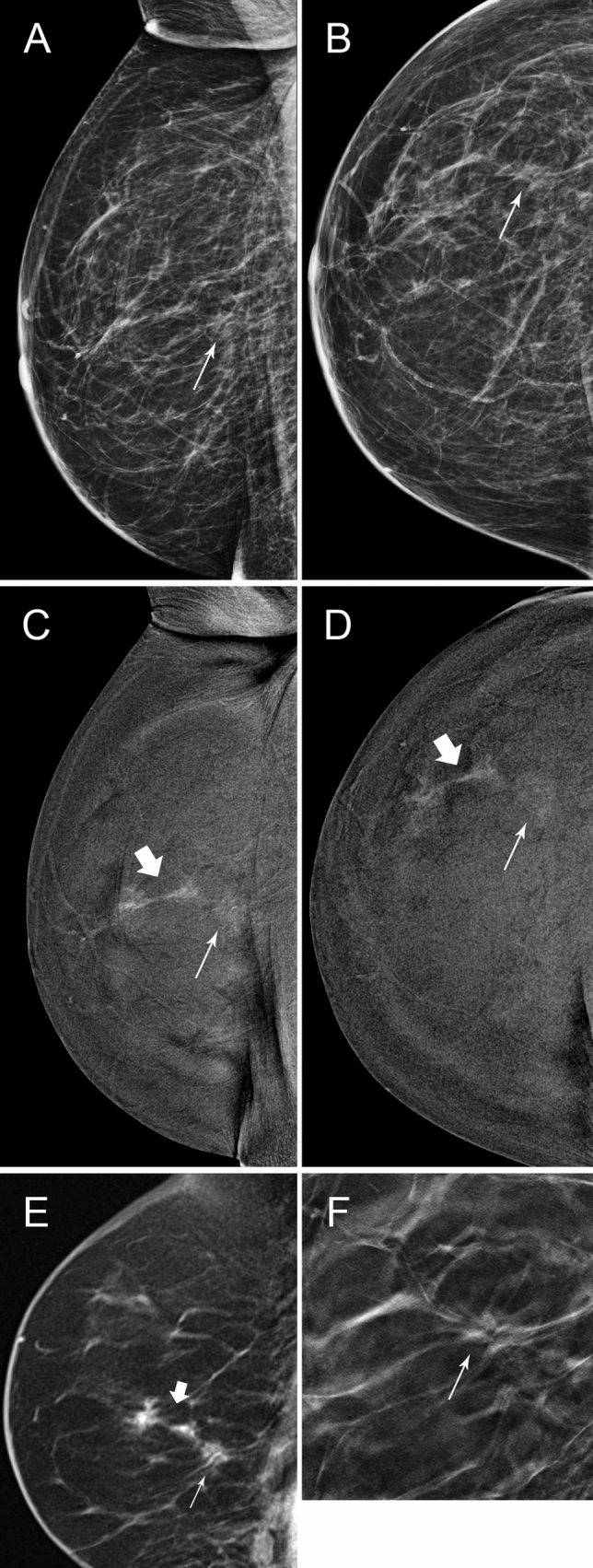
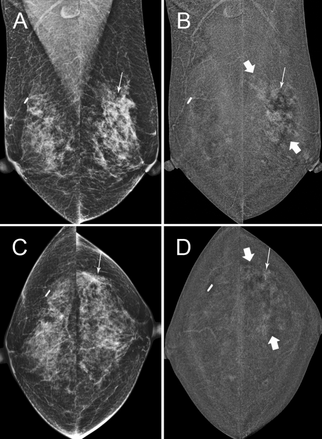
Purpose: Mammography (MG) is the standard imaging in surveillance of women with a personal history of breast cancer or DCIS (PHBC), supplemented with ultrasound. Contrast Enhanced Mammography (CEM) has higher sensitivity than MG and US. We report the performance of CEM compared with MG ± US.

Methods: A retrospective study of patients undergoing their first surveillance CEM in an Australian hospital setting between June 2006 and October 2020. Cases where a patient was recalled for assessment were identified, recording radiology, pathology and treatment details. Blinded re-reading of recalled cases was performed to determine the contribution of contrast. Use of surveillance US across the board was assessed for the period.

Results: 73/1191 (6.1%) patients were recalled. 35 (48%) were true positives (TP), with 26 invasive cancers and 9 cases of DCIS, while 38 (52%) were false positive (FP) with a positive predictive value (PPV) 47.9%. 32/73 were recalled due to MG findings, while 41/73 were only recalled due to Contrast. 14/73 had 'minimal signs' with a lesion identifiable on MG with knowledge of the contrast finding, while 27/73 were visible only with contrast. 41% (17/41) recalled due to contrast were TP. Contrast-only TPs were found with low and high mammographic density (MD). Screening breast US reduced by 55% in the year after CEM was implemented.

Conclusion: Compared to MG, CEM as a single surveillance modality for those with PHBC has higher sensitivity and comparable specificity, identifying additional malignant lesions that are clinically significant. Investigation of interval cancer and subsequent round outcomes is warranted.

Keywords: Breast cancer; CEM; Contrast; Mammography; Surveillance.


Conflict of interest statement

The authors have no relevant financial or non-financial interests to disclose.

Figures

Fig. 1
Fig. 1
A and B, 72 year-old patient, 2D 2019 surveillance images, MD BIRADS B. She had right wide local excision 2009 for a small Gd 3 invasive node-positive cancer. Thin arrow shows a small spiculated density thought to be part of the scar, stable on tomography. F is the CC tomographic image of this density(thin arrow). C and D are C + images from the same surveillance study showing faint enhancement (thin arrow) in the spiculated density, but a further 35 mm of spiculated enhancement anteriorly (thick arrow), not seen on 2D, 3D or targeted ultrasound. E is a sagittal contrast enhanced MR image (thin MIP) at the time of MR guided biopsy showing excellent correlation of CEM & MR images. Right mastectomy showed invasive carcinoma NST, Grade 3 with 2 tumours 22 &14 mm, triple negative, Ki67 20%. SNB negative. This was classified as a minimal signs case on 2D/3D after re-reading
Fig. 2
Fig. 2
A and C, 53 year-old patient, 2D 2019 surveillance images, MD BIRADS C. She had right wide local excision 2015 for 20 mm Intermediate grade DCIS and excision of papilloma with atypia on the left in the upper outer quadrant at the same time. Thin arrow on left is site of scar, stable on tomography. No other abnormalities on 2D,3D or ultrasound on the left and no calcifications. B and D are C + images from the same surveillance study showing extensive non-mass enhancement (thick arrows) throughout the left upper outer quadrant and mild BPE bilaterally. Stereotactic biopsy targeted to the enhancing scar (thin arrow) in the left upper outer quadrant was performed. Mastectomy showed 9 cm intermediate grade DCIS without invasion. SNB negative. This is an example of a contrast-only lesion
Fig. 3
Fig. 3
A and B, 45 year-old patient, 2D 2019 surveillance images, MD BIRADS B. She had right wide local excision 2017 for 12 mm invasive carcinoma NST with negative SNB. There were no abnormalities on 2D/3D. C and D are the C + images from the same surveillance study and show two small foci of enhancement (thin arrows) just superolateral to the nipple. Targeted ultrasound, E and F, showed two subtle hypoechoic lesions at 12.30 o’clock 2 cm(E, thin arrow) from nipple and 12.30 o’clock 5 cm(F thick arrow) from nipple, thought to correlate. US guided biopsies were performed and tissue markers were placed. G and H, post biopsy CEM shows that only one of the 2 lesions was correctly localised (thin arrow). It showed invasive carcinoma. The other biopsy contained only normal breast tissue on histopathology(thick arrow). Left wide local excision contained two foci of invasive carcinoma grade1, 4 mm & 1 mm and 25 mm of high grade DCIS. Post biopsy CEM is an excellent method of confirming that the correct lesion has been sampled. Ultrasound can present several “likely” lesions especially when the contrast lesion is small. In these instances MR or CEM guided biopsy, if available is more reliable and expeditious

References

    1. Houssami N, Abraham LA, Miglioretti DL, Sickles EA, Kerlikowske K, Buist DSM, et al. Accuracy and outcomes of screening mammography in women with a personal history of early-stage breast cancer. JAMA. 2011;305:790–799. doi: 10.1001/jama.2011.188. - DOI - PMC - PubMed
    1. Lam DL, Houssami N, Lee JM. Imaging surveillance after primary breast cancer treatment. Am J Roentgenol. 2017;208:676–686. doi: 10.2214/ajr.16.16300. - DOI - PMC - PubMed
    1. Consortium F the BCS, Buist DSM, Abraham LA, Barlow WE, Krishnaraj A, Holdridge RC, et al. Diagnosis of second breast cancer events after initial diagnosis of early stage breast cancer. Breast Cancer Res Tr 2010; 124:863–873.. - PMC - PubMed
    1. Bernardi D, Macaskill P, Pellegrini M, Valentini M, Fantò C, Ostillio L, et al. Breast cancer screening with tomosynthesis (3D mammography) with acquired or synthetic 2D mammography compared with 2D mammography alone (STORM-2): a population-based prospective study. Lancet Oncol. 2016;17:1105–1113. doi: 10.1016/s1470-2045(16)30101-2. - DOI - PubMed
    1. Ciatto S, Houssami N, Bernardi D, Caumo F, Pellegrini M, Brunelli S, et al. Integration of 3D digital mammography with tomosynthesis for population breast-cancer screening (STORM): a prospective comparison study. Lancet Oncol. 2013;14:583–589. doi: 10.1016/s1470-2045(13)70134-7. - DOI - PubMed

MeSH terms