. 2023 Dec;478(12):2795-2811.
doi: 10.1007/s11010-023-04700-8. Epub 2023 Mar 26.

Biochemical study of the effect of mesenchymal stem cells-derived exosome versus L-Dopa in experimentally induced Parkinson's disease in rats

Affiliations

Biochemical study of the effect of mesenchymal stem cells-derived exosome versus L-Dopa in experimentally induced Parkinson's disease in rats

Asmaa S Mohamed et al. Mol Cell Biochem. 2023 Dec.

Abstract

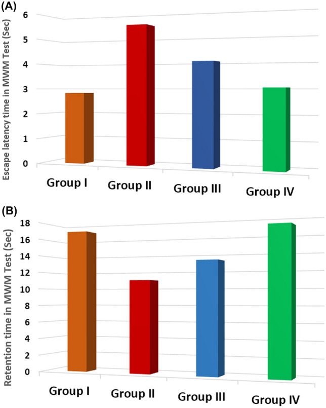
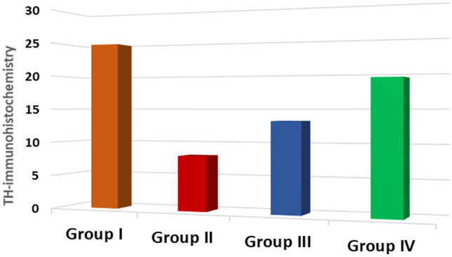
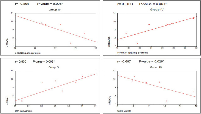
Parkinson's disease (PD) is a chronic and ongoing neurological condition. Unfortunately, as the dopaminergic terminals continue to deteriorate, the effectiveness of anti-Parkinson therapy decreases. This study aimed to examine the effects of BM-MSCs-derived exosomes in rats induced with Parkinson's disease. The goal was to determine their potential for neurogenic repair and functional restoration. Forty male albino rats were divided into four groups: control (group I), PD (group II), PD-L-Dopa (group III), and PD-exosome (group IV). Motor tests, histopathological examinations, and immunohistochemistry for tyrosine hydroxylase were performed on brain tissue. The levels of α-synuclein, DJ-1, PARKIN, circRNA.2837, and microRNA-34b were measured in brain homogenates. Rotenone induced motor deficits and neuronal alterations. Groups (III) and (IV) showed improvement in motor function, histopathology, α-synuclein, PARKIN, and DJ-1 compared to group (II). Group (IV) showed improvement in microRNA-34b and circRNA.2837 compared to groups (III) and (II). MSC-derived exosomes showed a greater suppression of neurodegenerative disease (ND) compared to L-Dopa in Parkinson's patients.

Keywords: DJ-1; Exosome; PARKIN; Parkinson’ disease (PD); circRNA.2837; microRNA-34b.


Conflict of interest statement

The authors have no relevant financial or non-financial interests to disclose.

Figures

Fig. 1
Fig. 1
Flowchart of experimental design
Fig. 2
Fig. 2
Comparison regarding turn time (A) and total time (B) (Sec) of vertical pole test in different studied groups along the course of the study
Fig. 3
Fig. 3
Comparison regarding escape latency time (A) and retention time (B) (Sec) of Morris water maze test in different studied groups along the course of the study
Fig. 4
Fig. 4
MSCs in culture. MSCs were identified through their morphology as they look spindle-shaped fibroblast-like cells, as well as their colony-forming unit (CFU). Scale bar = 100 μm
Fig. 5
Fig. 5
MSCs characterization by flow cytometry analysis. MSCs are negative for surface marker CD45 (0.4%), but positive for CD105 (97%)
Fig. 6
Fig. 6
Exosome characterization by TEM and Western Blotting. A Exosomes were characterized through TEM by their cup-shaped spheroidal morphology and their size was about 100 nm, scale bar = 100 nm. B Western blotting analysis shows a positive result for surface marker CD81 and CD63, which are characteristics for exosomes [(Note: β-actin in B) is the reference protein (internal control)]
Fig. 7
Fig. 7
Histopathological sections in the SN of different studied groups (H&E). AD H&E × 100, scale bar = 100 μm]. A Section in the SN of group (I) showing normal neurons of variable sizes and shapes. The glial cells are smaller and darker than the neurons and have dense nuclei. B Section in the SN of group (II) showing neuronal loss with increased glial cells (arrows). C Section in the SN of group (III) showing neurons, glial cells, and dilated blood vessels. D Section in the SN of group (IV) showing neurons of different shape and size and glial cells. E–H H&E × 400, scale bar = 50 μm]. E section in the SN of group (I) showing neurons with large pale nuclei, prominent nucleoli and lightly stained cytoplasm (black arrow). The glial cells are smaller and darker have dense nuclei. F section in the SN of group (II) showing neurons having nuclei with peripheral condensation of the chromatin (black arrow). Some of them have typical hyaline cytoplasmic inclusions—Lewy bodies (blue arrow). The neurons are surrounded by perineuronal spaces. Other deeply stained shrunken neurons (S) are observed. G section in the SN of group (III) showing some neurons having rounded nuclei with nucleoli (blue arrow). Other neurons (black arrow) have nuclei with peripheral condensation of the chromatin, while Lewy body is seen in some neurons (yellow arrow). H section in the SN of group (IV) showing neurons having large pale nuclei, prominent nucleoli, and lightly stained cytoplasm (black arrow) admixed with smaller glial cells
Fig. 8
Fig. 8
Tyrosine hydroxylase (TH)-immunostained sections in different studied groups. A–D IHC × 100, scale bar = 100 μm]. A TH-immunostained section in the SN of group (I) showing strong positive TH immunoreactivity in neurons. B TH-immunostained section in the SN of group (II) showing few mild positive neurons. C TH-immunostained section in the SN of group (III) showing moderate TH immunoreactivity of the neurons. D TH-immunostained section in the SN of group (VI) showing strong positive TH-stained neurons. E–H IHC × 400, scale bar = 50 μm]. E TH-immunostained section in the SN of group (I) showing strong positive TH-stained neurons (arrows) and the surrounding dense TH-immunoreactive neuropil. F TH-immunostained section in the SN of group (II) showing few mild positive TH-stained neurons (arrows) and weak positivity of the neuropil. G TH-immunostained section in the SN of group (III) showing moderate TH immunoreactivity of the neurons. H TH-immunostained section in the SN of group (VI) showing strong positive TH-stained neurons (arrows) and the surrounding neuropil
Fig. 9
Fig. 9
Comparison regarding TH-positive cells in the SN among the studied groups
Fig. 10
Fig. 10
Comparison regarding brain tissue α-SYN, Parkin, and DJ-1 levels among the studied groups
Fig. 11
Fig. 11
Comparison regarding brain tissue miRNA-34b and circRNA 2837 expression levels (fold change) among the studied groups
Fig. 12
Fig. 12
Correlation between the studied parameters in group II (PD group)
Fig. 13
Fig. 13
Correlation between the studied parameters in group IV (PD-MSCs-derived exosomes group)

Similar articles

Cited by

References

    1. Aarsland D, Batzu L, Halliday GM, et al. Parkinson disease-associated cognitive impairment. Nat Rev Dis Primers. 2021;7:47. doi: 10.1038/s41572-021-00280-3. - DOI - PubMed
    1. d’Angelo M, Cimini A, Castelli V. Insights into the effects of mesenchymal stem cell-derived secretome in Parkinson’s disease. Int J Mol Sci. 2020;21(15):5241. doi: 10.3390/ijms21155241. - DOI - PMC - PubMed
    1. Merimi M, El-Majzoub R, Lagneaux L, Moussa Agha D, et al. The therapeutic potential of mesenchymal stromal cells for regenerative medicine: current knowledge and future understandings. Front Cell Dev Biol. 2021;9:661532. doi: 10.3389/fcell.2021.661532. - DOI - PMC - PubMed
    1. Wang M, Yu F, Ding H, Wang Y, et al. Emerging function and clinical values of exosomal MicroRNAs in cancer. Mol Ther Nucleic Acids. 2019;16:791–804. doi: 10.1016/j.omtn.2019.04.027. - DOI - PMC - PubMed
    1. Roy B, Lee E, Li T, Rampersaud M. Role of microRNAs in neurodegeneration: from disease cause to tools of biomarker discovery and therapeutics. Genes. 2022;13:425. doi: 10.3390/genes13030425. - DOI - PMC - PubMed