Esophageal Epithelium and Lamina Propria Are Unevenly Involved in Eosinophilic Esophagitis
- PMID: 36967100
- PMCID: PMC10518022
- DOI: 10.1016/j.cgh.2023.03.014
Esophageal Epithelium and Lamina Propria Are Unevenly Involved in Eosinophilic Esophagitis
Abstract
Background & aims: The nature of the involvement of esophageal tissue in eosinophilic esophagitis (EoE) is unclear. We estimated the intrabiopsy site agreements of the EoE Histologic Scoring System (EoEHSS) scores for the grade (degree) and stage (extent) of involvement of the esophageal epithelial and lamina propria and examined if the EoE activity status influenced the intrabiopsy site agreement.
Methods: Demographic, clinical, and EoEHSS scores collected as part of the prospective Outcome Measures for Eosinophilic Gastrointestinal Diseases Across Ages study were analyzed. A weighted Cohen's kappa agreement coefficient (k) was used to calculate the pairwise agreements for proximal:distal, proximal:middle, and middle:distal esophageal biopsy sites, separately for grade and stage scores, for each of the 8 components of EoEHSS. A k > 0.75 was considered uniform involvement. Inactive EoE was defined as fewer than 15 eosinophils per high-powered field.
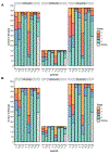
Results: EoEHSS scores from 1263 esophageal biopsy specimens were analyzed. The k for the stage of involvement of the dilated intercellular spaces across all 3 sites in inactive EoE was consistently greater than 0.75 (range, 0.87-0.99). The k for lamina propria fibrosis was greater than 0.75 across some of the biopsy sites but not across all 3. Otherwise, the k for all other features, for both grade and stage, irrespective of the disease activity status, was 0.75 or less (range, 0.00-0.74).
Conclusions: Except for the extent of involvement of dilated intercellular spaces in inactive EoE, the remaining epithelial features and lamina propria are involved unevenly across biopsy sites in EoE, irrespective of the disease activity status. This study enhances our understanding of the effects of EoE on esophageal tissue pathology.
Keywords: Eosinophilic Esophagitis; Epithelial Alterations; Histology Scoring System; Lamina Propria Fibrosis; Peak Eosinophil Counts.
Copyright © 2023 AGA Institute. Published by Elsevier Inc. All rights reserved.
Figures


References
-
- Liacouras CA, Furuta GT, Hirano I, et al. Eosinophilic esophagitis: Updated consensus recommendations for children and adults. Journal of Allergy and Clinical Immunology 2011;128:3–20.e6. - PubMed
Publication types
MeSH terms
Supplementary concepts
Grants and funding
LinkOut - more resources
Full Text Sources
Medical

