. 2023 Apr;26(3):216-221.
doi: 10.1080/17518423.2023.2193626. Epub 2023 Mar 26.

Anodal Contralesional tDCS Enhances CST Excitability Bilaterally in an Adolescent with Hemiparetic Cerebral Palsy: A Brief Report

Affiliations

Anodal Contralesional tDCS Enhances CST Excitability Bilaterally in an Adolescent with Hemiparetic Cerebral Palsy: A Brief Report

Rodrigo G Delatorre et al. Dev Neurorehabil. 2023 Apr.

Abstract

Hemiparetic cerebral palsy (HCP), weakness on one side of the body typically caused by perinatal stroke, is characterized by lifelong motor impairments related to alterations in the corticospinal tract (CST). CST reorganization could be a useful biomarker to guide applications of neuromodulatory interventions, such as transcranial direct current stimulation (tDCS), to improve the effectiveness of rehabilitation therapies. We evaluated an adolescent with HCP and CST reorganization who demonstrated persistent heightened CST excitability in both upper limbs following anodal contralesional tDCS. The results support further investigation of targeted tDCS as an adjuvant therapy to traditional neurorehabilitation for upper limb function.

Keywords: Brain Excitability; Hemiparesis; Motor Evoked Potential; Perinatal Stroke; Transcranial Direct Current Stimulation.


Conflict of interest statement

Declaration of Interest: Author Dr. Tim Feyma receives compensation as a member of the speaker bureau for PTC Therapeutics. Other authors report no conflict of interest.

Figures

Figure 1.
Figure 1.
A) Coronal and B) Axial views of participant’s T1-weighted MRI scan. Images are presented in radiological orientation (left side of the image displays the right hemisphere).
Figure 2.
Figure 2.
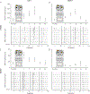
TMS assessment results displayed for intervention participant for A) Left Hand B) right Hand C) Left Wrist D) Right Wrist. Line graphs display MEP amplitudes (calculated from peak to peak, Mean +/− Standard Deviation) at each time point; Waterfall plots illustrate a subset of 10/20 trials (odd-numbered) at Pre-tDCS, Post 0, Post 30, and Post 60 assessments. The TMS pulse was delivered at time 0, with plots displaying EMG recordings from 50 msec before the TMS pulse to 100 msec after the TMS pulse. No MEPs were recorded at the baseline assessment (pre-tDCS) in the Right ED. Between Pre-tDCS and Post 0 assessments, the participant received 1.5 mA anodal tDCS to the right hemisphere while participating in the TrackTest fine motor activity. The diagram depicts an M1-SO montage wherein the left (lesioned) hemisphere displays the cathode placed on the forehead, and the right (non-lesioned) hemisphere displays the anode placed over the motor hotspot. Sample TrackTest traces are shown from the participant’s pre-test trials, completed with his left hand.

References

    1. Kirton A, Deveber G. Life after perinatal stroke. Stroke. Nov 2013;44(11):3265–71. doi:10.1161/STROKEAHA.113.000739 - DOI - PubMed
    1. Ganesan V, Hogan A, Shack N, Gordon A, Isaacs E, Kirkham FJ. Outcome after ischaemic stroke in childhood. Dev Med Child Neurol. Jul 2000;42(7):455–61. doi:10.1017/s0012162200000852 - DOI - PubMed
    1. Martin JH. The corticospinal system: from development to motor control. Neuroscientist. Apr 2005;11(2):161–73. doi:10.1177/1073858404270843 - DOI - PubMed
    1. Eyre JA, Taylor JP, Villagra F, Smith M, Miller S. Evidence of activity-dependent withdrawal of corticospinal projections during human development. Neurology. Nov 13 2001;57(9):1543–54. doi:10.1212/wnl.57.9.1543 - DOI - PubMed
    1. Carr LJ, Harrison LM, Evans AL, Stephens JA. Patterns of central motor reorganization in hemiplegic cerebral palsy. Brain. Oct 1993;116 ( Pt 5):1223–47. doi:10.1093/brain/116.5.1223 - DOI - PubMed