. 2023 Mar 9;9(3):e14362.
doi: 10.1016/j.heliyon.2023.e14362. eCollection 2023 Mar.

Gold nanoparticles enhances radiosensitivity in glioma cells by inhibiting TRAF6/NF-κB induced CCL2 expression

Affiliations

Gold nanoparticles enhances radiosensitivity in glioma cells by inhibiting TRAF6/NF-κB induced CCL2 expression

Hao Wang et al. Heliyon. .

Abstract

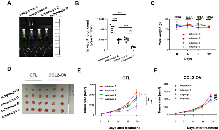
Gliomas are inherently difficult to treat by radiotherapy because glioma cells become radioresistant over time. However, combining radiotherapy with a radiosensitizer could be an effective strategy to mitigate the radioresistance of glioma cells. Gold nanoparticles (AuNPs) have emerged as a promising nanomaterial for cancer therapy, but little is known about whether AuNPs and X-ray radiation have cytotoxic synergistic effects against tumors. In this study, we found that the combination of AuNPs and X-ray irradiation significantly reduced the viabilities, as well as the migration and invasion, of glioma cells. Mechanistically, we observed that the AuNPs inhibited radiation-induced CCL2 expression by inhibiting the TRAF6/NF-κB pathway, which likely manifested the synergistic therapeutic effect between the AuNPs and X-ray radiation. The AuNPs also re-sensitized radioresistant glioma cells by inhibiting CCL2 expression. These results were also observed in another tumor cell line with a different molecular pattern, indicating that the underlying mechanism may be ubiquitous through cancer cells. Lastly, using the glioma mouse model, we observed that AuNPs significantly reduced tumor growth in the presence of X-ray radiation compared to radiotherapy alone.

Keywords: Glioma; Gold nanoparticles; Migration and invasion radio-resistance; Synergistic effect.


Conflict of interest statement

The authors declare no competing interests.

Figures

Fig. 1
Fig. 1
Identification of constructed IR resistant glioma cells U251-R. (A) Cell viabilities (%) expressed by the IR against U251 and U251-R cells at dose ranges from 0 to 4 Gy for 24 h (student t-test for differences determination, n = 3 for each group). (B) Cell apoptosis rates (%) expressed by the IR against U251 and U251-R cell lines at dose 4 Gy for 24 h (student t-test for differences determination, n = 3 for each group). (C) Migration rates (%) and invasion rates (%) expressed by the IR against U251 and U251-R cell lines at dose 4 Gy for 24 h (student t-test for differences determination, n = 3 for each group). The migration and invasion ability were measured with transwell assay. The representative morphology of migration and invasion cells were shown in Supplementary Fig. 1A in the corresponding group. CTL: control group; IR: X-ray irradiation. *: P < 0.05; **: P < 0.01; ***: P < 0.001; NS: not significant.
Fig. 2
Fig. 2
AuNPs and IR had a synergistic anti-tumor effect against glioma cells. (A) Cell viabilities (%) expressed by the AuNPs against U251, U251-R and NHA cells at dose ranges from 0 to 150 mg/L (student t-test for differences determination, n = 3 for each group). (B) Migration rates (%) and invasion rates (%) expressed by the AuNPs at 150 mg/L against U251 and U251-R cell lines (student t-test for differences determination, n = 3 for each group). The migration and invasion ability were measured with transwell assay. The representative morphology of migration and invasion cells were shown in Supplementary Fig. 1A in the corresponding group. (C) Cell viabilities (%) expressed by the AuNPs at 150 mg/L, IR at 4 Gy and Comb against U251 (student t-test for differences determination, n = 3 for each group). (D) Migration rates (%) and invasion rates (%) expressed by the AuNPs at 150 mg/L, IR at 4 Gy and Comb against U251 (student t-test for differences determination, n = 3 for each group). The migration and invasion ability were measured with transwell assay. The representative morphology of migration and invasion cells were shown in Supplementary Fig. 1A in the corresponding group. CTL: control group; IR: X-ray irradiation; Comb: defined as AuNPs at 75 mg/L (half concentration) + IR at 2 Gy (half concentration) treatment. *: P < 0.05; **: P < 0.01; ***: P < 0.001; NS: not significant.
Fig. 3
Fig. 3
AuNPs exerts synergistic effect with IR by reversing the IR induced CCL2 production in glioma cells. (A) Cultured U251 cells were treated with IR at 4 Gy, together with or without AuNPs at 150 mg/L for 24 h, and the CCL2 productions were determined by ELISA assay in the supernatant proteins (student t-test for differences determination, n = 3 for each group). (B) Cell viabilities (%) expressed by the AuNPs at 150 mg/L, IR at 4 Gy and Comb against wide-type U251 (control) and CCL2 stable over-expressed U251 (CCL-OV) (student t-test for differences determination, n = 3 for each group). (C) Migration rates (%) and invasion rates (%) expressed by the AuNPs at 150 mg/L, IR at 4 Gy and Comb against wide-type U251 (control) and CCL2 stable over-expressed U251 (CCL-OV) (student t-test for differences determination, n = 3 for each group). The migration and invasion ability were measured with transwell assay. The representative morphology of migration and invasion cells were shown in Supplementary Fig. 1A in the corresponding group. CTL: control group; IR: X-ray irradiation; Comb: defined as AuNPs at 75 mg/L (half concentration) + IR at 2 Gy (half concentration) treatment. *: P < 0.05; **: P < 0.01; ***: P < 0.001; NS: not significant.
Fig. 4
Fig. 4
AuNPs inhibits TRAF6/NF-κB signaling activation in glioma cells. (A) Cultured U251 cells were pre-transfected with NF-κB reporter plasmid and phRL-TK plasmid (internal control) for 24 h, and then treated with IR at 4 Gy, together with or without AuNPs at 150 mg/L for another 24 h. The activation of the NF-κB promoter was measured by a dual-luciferase reporter gene assay (student t-test for differences determination, n = 3 for each group). (B) Cultured U251 cells were pre-transfected with NF-κB reporter plasmid and phRL-TK plasmid (internal control), together with TRAF6, TAK1, or IKKβ expression plasmid for 24 h, and then treated with or without AuNPs at 150 mg/L for another 24 h. The activation of the NF-κB promoter was measured by a dual-luciferase reporter gene assay (student t-test for differences determination, n = 3 for each group). (C–D) Cultured U251 cells were treated with or without AuNPs at 150 mg/L for 24 h, and (C) the phosphorylation levels of p65 and TRAF6 total protein levels were determined by western blotting assay; (D) the mRNA levels of p65 and TRAF6 were determined by qRT-PCR assay (student t-test for differences determination, n = 3 for each group). (E) Lysates from certain treated U251 cells transiently co-transfected with Flag-TRAF6 and HA-UB (K48) or HA-UB (K63) plasmids were subjected to immunoprecipitation with anti-Flag antibody followed by Western blot analysis with anti-HA antibody. The full scan of the gel was shown in Supplementary Fig. 5. CTL: control group; IR: X-ray irradiation; UB: ubiquitin. *: P < 0.05; **: P < 0.01; ***: P < 0.001; NS: not significant.
Fig. 5
Fig. 5
TRAF6/NF-κB/CCL2 pathway play the key role in promoting glioma cell resistant to IR. (A) Cultured U251 cells and U251-R cells were transient transfected with TRAF6 (TRAF6-OV), together with or without 10 mM NF-κB inhibitor (BAY-117082) for 48 h, and the CCL2 productions were determined by ELISA assay in the supernatant proteins (student t-test for differences determination, n = 3 for each group). (B) Cultured U251-R cells were transient transfected with CCL siRNA (CCL-SI) for 24 h, and then were treated with IR at certain doses for another 24 h, and the cell viabilities (%) were determined (student t-test for differences determination, n = 3 for each group). (C) Cultured U251-R cells were transient transfected with CCL siRNA (CCL-SI) for 24 h, and then were treated with IR at 4 Gy for another 24 h, and the migration rates (%) and invasion rates (%) were determined (student t-test for differences determination, n = 3 for each group). The migration and invasion ability were measured with transwell assay. The representative morphology of migration and invasion cells were shown in Supplementary Fig. 1A in the corresponding group. *: P < 0.05; **: P < 0.01; ***: P < 0.001; NS: not significant.
Fig. 6
Fig. 6
AuNPs sensitize the radio-resistant glioma cells to IR by restricting CCL2 productions. (A) Cell viabilities (%) expressed by the AuNPs at 150 mg/L, IR at 4 Gy and Comb against wide-type U251-R (control) and CCL2 stable over-expressed U251-R (CCL-OV) (student t-test for differences determination, n = 3 for each group). (B) Migration rates (%) and invasion rates (%) expressed by the AuNPs at 150 mg/L, IR at 4 Gy and Comb against wide-type U251-R (control) and CCL2 stable over-expressed U251-R (CCL-OV) (student t-test for differences determination, n = 3 for each group). The migration and invasion ability were measured with transwell assay. The representative morphology of migration and invasion cells were shown in Supplementary Fig. 1A in the corresponding group. CTL: control group; IR: X-ray irradiation; Comb: defined as AuNPs at 25 mg/L (half concentration) + IR at 4Gy (half concentration) treatment.*: P < 0.05; **: P < 0.01; ***: P < 0.001; NS: not significant.
Fig. 7
Fig. 7
In vivo effect of AuNPs on glioma growth after IR therapy. (A–C) C57BL/6 mice were used for brain tumor models construction. Mice were categorized into indicated subgroups. (A) Representative IVIS images showing tumor presence in each group 12 days after treatment. (B) Photon counts obtained from tumors resulting from GL261-luc cells in each group 12 days after treatment (student t-test for differences determination, n = 8 for each group). (C) The weight of mice were measured in each group from 0 to 12 days after treatment (student t-test for differences determination, n = 8 for each group). (D–F) widetype U251 cells (Control) or CCL2 stable overexpressed U251 cells (CCL2-OV) were injected into nude mice to construct xenograft mice model. Mice were categorized into indicated subgroups. (D) Representative images of tumors in nude mice xenografted in each group at the endpoint time (28 days after treatment). (E–F) Tumor volumes in (E) control mice and (F) CCL2-OV mice under certain treatment were measured every 7 days after treatment until endpoint time (28 days after treatment) (student t-test for differences determination, n = 3 for each group). subgroups A: PBS group (control); subgroups B: single AuNPs treatment group; subgroups C: PBS + X-ray treatment group; subgroups D: AuNPs + X-ray treatment group. *: P < 0.05; **: P < 0.01; ***: P < 0.001; NS: not significant. NSA: In the four groups of mice, there was no statistical difference between any two groups.

Similar articles

Cited by

References

    1. Ostrom Q.T., Cote D.J., Ascha M., Kruchko C., Barnholtz-Sloan J. Adult glioma incidence and survival by race or ethnicity in the United States from 2000 to 2014. JAMA Oncol. 2018;4:1254–1262. doi: 10.1001/jamaoncol.2018.1789. - DOI - PMC - PubMed
    1. van Nifterik K.A., Van Den Berg J., Slotman B.J., Van Rijn J. Anti-tumour effects by a trimodal combination of temozolomide, meloxicam and X-rays in cultures of human glioma cells. Int. J. Radiation Biol. 2011;87:192–201. doi: 10.3109/09553002.2010.519423. - DOI - PubMed
    1. Du H.Q., Wang Y., Jiang Y., Wang C.H., Zhou T., Liu H.Y., et al. Silencing of the TPM1 gene induces radioresistance of glioma U251 cells. Oncol. Rep. 2015;33:2807–2814. doi: 10.3892/or.2015.3906. - DOI - PubMed
    1. Liu C., Wang L., Qiu H., Dong Q., Feng Y., Li D., et al. Combined strategy of radioactive 125I seeds and salinomycin for enhanced glioma chemo-radiotherapy: evidences for ROS-mediated apoptosis and signaling crosstalk. Neurochem. Res. 2018;43:1317–1327. doi: 10.1007/s11064-018-2547-2. - DOI - PubMed
    1. Zhao J., Liu P., Ma J., Li D., Yang H., Chen W., et al. Enhancement of radiosensitization by silver nanoparticles functionalized with polyethylene glycol and aptamer As1411 for glioma irradiation therapy. Int. J. Nanomed. 2019;14:9483–9496. doi: 10.2147/IJN.S224160. - DOI - PMC - PubMed

LinkOut - more resources